INFOGRAPHIC – Target measurements for water quality

Water is a main nutrient and carrier for vaccines, medicine – including antibiotics, but also for pathogens
Chemistry


Microbiology


Water is a main nutrient and carrier for vaccines, medicine – including antibiotics, but also for pathogens
At the recent EW Nutrition Poultry Academy in Jakarta, Indonesia, Dr Steve Leeson, Professor Emeritus, University of Guelph, Canada, opened his presentation by stating that “it is obvious that any nutrient deficiency will impact bird health, but not so obvious is that nutrition per se can positively impact immunity and health in an otherwise healthy and high-producing bird.”
Modern high-performing broilers are characterized by extremely high feed intake. This puts a lot of stress on the physiology of the entire gastrointestinal tract, but particularly so on the absorptive epithelial cells of the small intestine. Any organism requires a nutrient source for survival and reproduction. Dr Leeson asked “can we significantly reduce nutrient supply to pathogens, while sustaining bird productivity?”
He reminded the audience that no cellular function comes for free: so there is always a “cost”. A general conclusion is that 10% of nutrients can be used for immune function during disease challenge, and always get priority. Therefore, you don’t want to overstimulate the immune system, which in extreme situations leads to an inflammatory response. In his presentation, Dr Leeson considered factors determining gut health and nutritional tools which are available to support gut health.
Gut pathogens impact the bird and/or the consumer. Clostridia and E. coli are the major concerns regarding bird health and productivity, whereas Salmonella and Campylobacter are major pathogens important for human health.
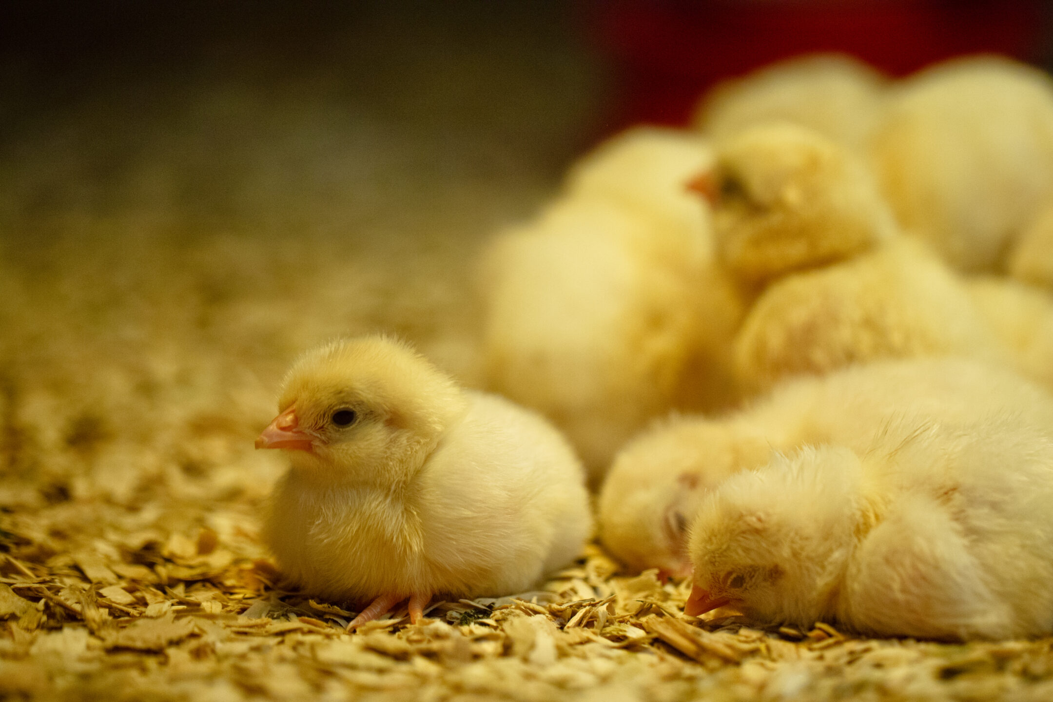
The chick hatches with a gut virtually devoid of microbes, so early colonizers tend to predominate quite quickly. Microbial species present on the hatching tray, during delivery and during the first few days at the farm will likely dictate early gut colonization. In some instances, the chick’s microflora may be established by the time it gets to the farm, so the probiotic faces more of a challenge to establish itself as the predominant species.
Gut villi development matures at around 10-15 days of age. The broiler pre-starter diet therefore is a target for feed additives that positively impact gut structure and development.
Fast growth rate and high egg output are negatively correlated with immune response. Consequently, nutrient-dense diets are not optimal for immunity. With bacteria, it’s a numbers game – but these numbers quickly multiply. The first 7 days are important, therefore probiotics must be established early. Consider the role of targeted feed additives, such as butyrate, phytogenics, antioxidants, PUFAs etc.
Also, maximize feed particle size – the limit is usually pellet quality. Mitigate nutrient transition at any diet change. Review the supply of trace minerals, as there is a trend to lower levels of organic minerals. With all the factors that weigh into production performance, any support that can be rallied through nutrition needs to be considered.
***
EW Nutrition’s Poultry Academy took place in Jakarta and Manila in early September 2023. Dr. Steve Leeson, an expert in Poultry Nutrition & Production with nearly 50 years’ experience in the industry, was the distinguished keynote speaker.
Dr. Leeson had his Ph.D. in Poultry Nutrition in 1974 from the University of Nottingham. Over a span of 38 years, he was a Professor in the Department of Animal &Poultry Science at the University of Guelph, Canada. Since 2014, he has been Professor Emeritus at the same University. As an eminent author, he has more than 400 papers in refereed journals and 6 books on various aspects of Poultry Nutrition & Management. He also won the American Feed Manufacturer’s Association Nutrition Research Award (1981), the Canadian Society of Animal Science Fellowship Award (2001), and Novus Lifetime Achievement Award in Poultry Nutrition (2011).
By Dr. Inge Heinzl, Editor, EW Nutrition
Salmonellosis is third among foodborne diseases leading to death (Ferrari, 2019). More than 91,000 human cases of Salmonellosis are reported by the EU each year, generating overall costs of up to €3 billion a year (EFSA, 2023), 10-20% of which are attributed to pork consumption (Soumet, 2022). The annual costs arising from the resulting human health losses in 2010 were about €90 million (FCC Consortium, 2010). Take the example of Ireland, where a high prevalence of Salmonella in lymph nodes still shows a severe issue pre-slaughter and a big challenge for slaughterhouses to stick to the process hygiene requirements (Deane, 2022).
Several governments already have monitoring programs in place, and the farms are categorized according to the salmonella contamination of their pigs. In some countries, e.g., Denmark, an economic penalty of 2% of the carcass value must be paid if the farm has level 2 (intermediate seroprevalence) and 4-8% if the level is 3. Other countries, e.g., Germany, the UK, Ireland, or the Netherlands, use quality assurance schemes. The farmers can only sell their carcasses under this label if their farm has a certain level.
Salmonellas are rod-shaped gram-negative bacteria of the family of enterobacteria that use flagella for their movement. They were named after the American vet Daniel Elmer Salmon. The genus of Salmonella consists of two species (S. bongori and S. enterica with seven subspecies) with in total more than 2500 serovars (see Figure 1). The effects of the different serovars can range from asymptomatic carriage to severe invasive systemic disease (Gal-Mor, 2014). All Salmonella serovars generally can cause disease in humans; the rosa-marked ones already showed infections.
 Figure 1: the genus of Salmonella with Salmonella serovars relevant for pigs (according to Bonardi, 2017: Salmonella in the pork production chain and its impact on human health in the European Union)
Within the group of Salmonella, some serovars can only reside in one or few species, e.g., S. enterica spp. enterica Serovar Dublin (S. Dublin) in bovines (Waldron, 2018) or S. Cholerasuis in pigs (Chiu, 2004). An infection in humans with these pathogens is often invasive and life-threatening (WHO, 2018). On the contrary, serovars like S. Typhimurium and S. Enteritidis are not host-specific and can cause disease in various species.
The serotypes S. Typhi and S. Paratyphi A, B, or C are highly adapted to humans and only for them pathogenic; they are responsible for the occurrence of typhus.
Serovars occurring in pigs and relevant for humans are, for example, S. Typhimurium (Hendriksen, 2004), S. Serotype 4,[5],12:I (Hauser et al., 2010), S. Cholerasuis (Chiu, 2004), S. Derby (Gonzalez-Santamarina, 2021), S. Agona (Brenner Michael, 2006) and S. Rissen (Elbediwi, 2021).
The way of transmission to humans depends on the serovar:
Human-specific and, therefore, only in humans and higher primates residing serovars S. Typhi and Paratyphi A, B, or C (typhoidal) are excreted via feces or urine. Therefore, any food or water contaminated with the feces or urine of infected people can transmit this disease (Government of South Australia, 2023). Typhoid and paratyphoid Salmonellosis occur endemic in developing countries with the lack of clean water and, therefore, inadequate hygiene (Gal-Mor, 2014).
Serovars which can cause disease in humans and animals (non-typhoidal), can be transmitted by
– animal products such as milk, eggs, meat
– contact with infected persons/animals (pigs, cows, pets, reptiles…) or
– other feces- or urine-contaminated products such as sprouts, vegetables, fruits….
Farm animals take salmonellas from their fellows, contaminated feed or water, rodents, or pests.
In the case of typhoid or paratyphoid Salmonellosis, the onset of illness is gradual. People can suffer from sustained high fever, unwellness, severe headache, and decreased appetite, but also from an enlarged spleen irritating the abdomen and dry cough.
A study conducted in Thailand with children suffering from enteric fever caused by the typhoid serovars S. Typhi and Paratyphi showed a sudden onset of fever and gastrointestinal issues (diarrhea), rose spots, bronchitis, and pneumonia (Thisyakorn et al., 1987)
The non-typhoid Salmonellosis is typically characterized by an acute onset of fever, nausea, abdominal pain with diarrhea, and sometimes vomiting (WHO, 2018). However, 5% of the persons – children with underlying conditions, e.g., babies, or people who have AIDS, malignancies, inflammatory bowel disease, gastrointestinal illness caused by non-typhoid serovars, and hemolytic anemia, or receiving an immunosuppressive therapy can be susceptible to bacteremia. Additionally, serovars like S. Cholerasuis or S. Dublin are apt to develop bacteremia by entering the bloodstream with little or no involvement of the gut (Chiu, 1999). In these cases, consequences can be septic arthritis, pneumonia, peritonitis, cutaneous abscess, mycotic aneurysm, and sometimes death (Chen et al., 2007; Chiu, 2004, Wang et al., 1996).
In pigs, S. Cholerasuis causes high fever, purple discolorations of the skin, and thereinafter diarrhea. The mortality rate in pigs suffering from this type of Salmonellosis is high. Barrows orally challenged with S. Typhimurium showed elevated rectal temperature by 12h, remaining elevated until the end of the study. Feed intake decreased with a peak at 48h after the challenge and remained up to 120h after the challenge. Daily gain reduced during the following two weeks after infection. A higher plasma cortisol level and a lower IGF-I level could also be noticed. All these effects indicate significant changes in the endocrine stress and the somatotropic axis, also without significant alterations in the systemic pro-inflammatory mediators (Balaji et al., 2000)
There are three main steps to keep the contamination of pork as low as possible:
To answer this question, we must look at how the pathogen can be transported to the farm. According to the Code of Practice for the Prevention and Control of Salmonella on Pig Farms (Ministry of Agriculture, Fisheries and Food and the Scottish Executive Rural Affairs Department), there are several possibilities to infiltrate the pathogen into the farm:
To minimize/prevent the entry of Salmonella into the livestock, bought-in animals must come from reputable breeding farms with a salmonella monitoring system in place. As possible carrier animals are more likely to excrete Salmonella when stressed; they should be kept in isolation after purchasing. Additionally, the animals must go through a disinfectant foot bath before entering the farm.
Generally, the production site must be kept clean and as unattractive as possible for all these animals. Rests of feed must be removed, and dead animals and afterbirths must be promptly and carefully disposed of. A well-planned baiting and trapping policy should be in place to effectively control rodents.
In any case, the number of persons entering the hog house must be kept as low as possible. Farmworkers should be trained in the principles of hygiene. They should wear adequate clothing (waterproof boots and protective overalls) that can be easily cleaned/laundered and disinfected. The clothes/shoes should always be used only at this site. Thorough hand washing and the disinfection of the boots when entering and leaving the pig unit are a must.
If visits are necessary, the visitors should take the same measures as the farm workers. And, of course, they should not have had contact with another pig farm during the last 48 hours.
Farm equipment should not be shared with other farms. If this cannot be avoided, it must be cleaned and disinfected before re-entering the farm. Also, the vehicles for the transport of the animals must be cleaned and disinfected as soon as possible after usage, as contaminated transporters always pose the risk of infection.
To get high feed quality, the feed should be purchased from feed mills/sources with a well-functioning bacterial control to guarantee the absence of Salmonella. It is essential that birds, domestic and wild animals cannot enter the feed stores.
It is also advised to keep dry feed dry as possibly contaminating Salmonella can multiply in such humid conditions. Additionally, all feed bins and delivery pipes for dry and wet feed must be consciously cleaned, and the damp feed pipes also disinfected.
The change from pellets to mash could be helpful as the pellets facilitate Salmonella colonization by stimulating the secretion of mucins (Hedemann et al., 2005).
For sanitation of the feed, we offer organic acids (Acidomix product range) or mixtures of organic acids and formaldehyde in countries where formaldehyde products are allowed (Formycine) to decrease the pathogenic load of the feed materials. In vitro trials show the effectiveness of the products:
For the in vitro trial with Formycine, autoclaved feed samples were inoculated with Salmonella enteritidis serovar Typhimurium DSM 19587 strain to reach a Salmonella contamination of 106 CFU/g of feed. After incubating at room temperature for three hours, Formycine Liquido was added to the contaminated feed samples at 0, 500, 1000, and 2000 ppm. The control and inoculated feed samples were further incubated at room temperature, and Salmonella counts (CFU/g) were carried out at 24, 48, 72 hours and on day 15. The limit of Salmonella detection was set at 100 CFU/g (102). Results are shown in figure 2.
 Fig. 2: Effect of treatment time and different inclusion levels of Formycine Liquido on the Salmonella count in feed
As important as uncontaminated feed is clean water for drinking. It can be achieved by taking the water from a main or a bacteriologically controlled water borehole. Regular cleaning/disinfection of the tanks, pipes, and drinkers is essential.
Straw material containing feces of other animals (rodents, pets) always carries the risk of Salmonella contamination. Also, wet or moldy bedding is not recommended because it is an additional challenge for the animal. To optimize the quality of bedding, the straw should be bought from reliable and as few as possible sources. The material must be stored dry and as far as practicable from the pig buildings (Ministry of Agriculture, Fisheries and Food & Scottish Executive Rural Affairs Department, 2000).
For the control of Salmonella in swine herds, vaccination is an effective tool. De Ridder et al. (2013) showed that an attenuated vaccine reduced the transmission of Salmonella Typhimurium in pigs. The vaccination with an attenuated S. Typhimurium strain, followed by a booster vaccination with inactivated S. Cholerasuis, showed better effects than an inactivated S. Cholerasuis vaccine alone (Alborali et al., 2017). Bearson et al. (2017) could delimitate transmission through less shedding and protect the animals against systemic disease.
To achieve the best effects, the producer must understand the diversity of Salmonella serovars to choose the most promising vaccination strategy (FSIS, 2023).
If there are already cases of Salmonella on the farm, infected animals must be separated from the rest of the herd. Small batch sizes are beneficial, as well as not mixing different litters after weaning. If feasible, separate units for different production phases with an all-in/all-out system could break the reinfection cycle and help reduce Salmonella contamination on the farm. And also in this case, vaccination is helpful.
An effective tool is acidifying the feed with organic acids, as Salmonella doesn’t like acid conditions. A trial was conducted with Acidomix AFG and Acidomix AFL to show their effects against Salmonella. For the test, 105 CFU/g of Salmonella enterica ser. Typhimurium was added to feed containing 1000 ppm, 2000 ppm, and 3000 ppm of Acidomix AFG or AFL. The stomach and intestine were simulated in vitro by adjusting the pH with HCl and NaHCO3 as follows:
Stomach 2.8
Intestine 6.8-7.0
After the respective incubation, the microorganisms were recovered from feed and plated on an appropriate medium for CFU counting. The results are shown in figures 3 and 4.

Figures 3 + 4: Effects of different concentrations of Acidomix AFG and Acidomix AFL against Salmonella enterica ser. Typhimurium in feed
Plant compounds or phytomolecules can also be used against Salmonella in pigs. Some examples of phytomolecules to be used are Piperine, Allicin, Eugenol, and Carvacrol. Eugenol, e.g., increases the permeability of the Salmonella membrane, disrupts the cytoplasmic membrane, and inhibits the production of bacterial virulence factors (Keita et al., 2022; Mak et al., 2019). Thymol and Carvacrol interact with the cell membrane by H bonding, also resulting in a higher permeability.
An already published in vitro trial conducted with our product Ventar D also showed excellent effects against Salmonella while sparing the beneficial gut flora. A further trial once more demonstrated the susceptibility of Salmonella to Ventar D. It showed that Ventar D controls Salmonella by suppressing their motility and, at higher concentrations, inactivating the cells (see figures 5 + 6):
 Figure 5: S. enterica motility test: on the left side – control; on the right side – motility medium containing.750 µg/mL of Ventar
 Fig 6 . Disk diffusion assay employing S. enterica. upper left side – disk containing 10 µL of Ventar; upper right – 5 µL; lower left – control; lower right – 1µL.
In addition to the direct Salmonella-reducing effect, essential oils / secondary plant compounds / phytomolecules improve digestive enzyme activity and digestion, leading to increased nutrient absorption and better feed conversion (Windisch et al., 2008).
In general, the slaughterhouse personnel is responsible for adequate hygiene management to prevent contamination of carcasses and meat. However, also the farmer can make his contribution to maintain the risk of contamination in the slaughterhouse as low as possible. A study by Vieira-Pinto (2006) revealed that one Salmonella-positive pig can contaminate several other carcasses.
According to a trial conducted by Hurd et al. (2002), infection and, therefore, “contamination” of other pigs can rapidly occur, meaning that cross-contamination is a topic during transport to the slaughterhouse and in the lairages when the pigs come together with animals from other farms. The stress to which the pigs are exposed influences physiological and biochemical processes. The microbiome and animal’s immunity are affected, leading to higher excretion of Salmonella during transport and in the lairages. So, the animals should not be stressed during loading and unloading or transportation. The trailer poses a further risk of infection if it was not cleaned and disinfected before. So, reliable people who treat the animals well and keep their trailers clean should be chosen for transportation.
At least in the EU, pig producers have the big duty to keep Salmonella low in their herds; otherwise, they will have financial losses. They are not only responsible for their farm, but also the slaughterhouses count on them. Besides the standard strict hygiene management and vaccination, farmers can use products provided by the industry to sanitize feed but also to support their animals directly with phytomolecules acting against pathogens and supporting gut health.
All these measures together should be a solution to the immense challenge of Salmonella, to protect people and prevent economic losses.
References:
Alborali, Giovanni Loris, Jessica Ruggeri, Michele Pesciaroli, Nicola Martinelli, Barbara Chirullo, Serena Ammendola, Andrea Battistoni, Maria Cristina Ossiprandi, Attilio Corradi, and Paolo Pasquali. “Prime-Boost Vaccination with Attenuated Salmonella Typhimurium Δznuabc and Inactivated Salmonella Choleraesuis Is Protective against Salmonella Choleraesuis Challenge Infection in Piglets.” BMC Veterinary Research 13, no. 1 (2017): 284. https://doi.org/10.1186/s12917-017-1202-5.
Balaji, R, K J Wright, C M Hill, S S Dritz, E L Knoppel, and J E Minton. “Acute Phase Responses of Pigs Challenged Orally with Salmonella Typhimurium.” Journal of Animal Science 78, no. 7 (2000): 1885. https://doi.org/10.2527/2000.7871885x.
Bearson, Bradley L, Shawn M. Bearson, Brian W Brunelle, Darrell O Bayles, In Soo Lee, and Jalusa D Kich. “Salmonella Diva Vaccine Reduces Disease, Colonization, and Shedding Due to Virulent S. Typhimurium Infection in Swine.” Journal of Medical Microbiology 66, no. 5 (2017): 651–61. https://doi.org/10.1099/jmm.0.000482.
Brenner Michael, G, M Cardoso, and S Schwarz. “Molecular Analysis of Salmonella Enterica Subsp. Enterica Serovar Agona Isolated from Slaughter Pigs.” Veterinary Microbiology 112, no. 1 (2006): 43–52. https://doi.org/10.1016/j.vetmic.2005.10.011.
Chen, P.-L., C.-M. Chang, C.-J. Wu, N.-Y. Ko, N.-Y. Lee, H.-C. Lee, H.-I. Shih, C.-C. Lee, R.-R. Wang, and W.-C. Ko. “Extraintestinal Focal Infections in Adults with Non-typhoid Salmonella Bacteraemia: Predisposing Factors and Clinical Outcome.” Journal of Internal Medicine 261, no. 1 (2007): 91–100. https://doi.org/10.1111/j.1365-2796.2006.01748.x.
Chiu, Cheng-Hsun, Lin-Hui Su, and Chishih Chu. “Salmonella EntericaSerotype Choleraesuis: Epidemiology, Pathogenesis, Clinical Disease, and Treatment.” Clinical Microbiology Reviews 17, no. 2 (2004): 311–22. https://doi.org/10.1128/cmr.17.2.311-322.2004.
De Ridder, L., D. Maes, J. Dewulf, F. Pasmans, F. Boyen, F. Haesebrouck, E. Méroc, P. Butaye, and Y. Van der Stede. “Evaluation of Three Intervention Strategies to Reduce the Transmission of Salmonella Typhimurium in Pigs.” The Veterinary Journal 197, no. 3 (2013): 613–18. https://doi.org/10.1016/j.tvjl.2013.03.026.
Deane, Annette, Declan Murphy, Finola C. Leonard, William Byrne, Tracey Clegg, Gillian Madigan, Margaret Griffin, John Egan, and Deirdre M. Prendergast. “Prevalence of Salmonella spp. in Slaughter Pigs and Carcasses in Irish Abattoirs and Their Antimicrobial Resistance.” Irish Veterinary Journal 75, no. 1 (2022). https://doi.org/10.1186/s13620-022-00211-y.
Edel, W., M. Schothorst, P. A. Guinée, and E. H. Kampelmacher. “Effect of Feeding Pellets on the Prevention and Sanitation of Salmonella Infections in Fattening Pigs1.” Zentralblatt für Veterinärmedizin Reihe B 17, no. 7 (2010): 730–38. https://doi.org/10.1111/j.1439-0450.1970.tb01571.x.
EFSA. “Salmonella.” European Food Safety Authority. Accessed August 7, 2023. https://www.efsa.europa.eu/en/topics/topic/salmonella.
Elbediwi, Mohammed, Daiwei Shi, Silpak Biswas, Xuebin Xu, and Min Yue. “Changing Patterns of Salmonella Enterica Serovar Rissen from Humans, Food Animals, and Animal-Derived Foods in China, 1995–2019.” Frontiers in Microbiology 12 (2021). https://doi.org/10.3389/fmicb.2021.702909.
Elnekave, Ehud, Samuel Hong, Alison E Mather, Dave Boxrud, Angela J Taylor, Victoria Lappi, Timothy J Johnson, et al. “Salmonella Enterica Serotype 4,[5],12:I:- In Swine in the United States Midwest: An Emerging Multidrug-Resistant Clade.” Clinical Infectious Diseases 66, no. 6 (2018): 877–85. https://doi.org/10.1093/cid/cix909.
FCC Consortium. “Final Report – Food Safety.” European Commission, 2010. https://food.ec.europa.eu/system/files/2016-10/biosafety_food-borne-disease_salmonella_fattening-pigs_slaughthouse-analysis-costs.pdf.
Ferrari, Rafaela G., Denes K. Rosario, Adelino Cunha-Neto, Sérgio B. Mano, Eduardo E. Figueiredo, and Carlos A. Conte-Junior. “Worldwide Epidemiology of Salmonella serovars in Animal-Based Foods: A Meta-Analysis.” Applied and Environmental Microbiology 85, no. 14 (2019). https://doi.org/10.1128/aem.00591-19.
“FSIS Guideline to Control Salmonella in Swine Slaughter and Pork Processing Establishments.” FSIS Guideline to Control Salmonella in Swine Slaughter and Pork Processing Establishments | Food Safety and Inspection Service. Accessed August 14, 2023. https://www.fsis.usda.gov/guidelines/2023-0003.
Gal-Mor, Ohad, Erin C. Boyle, and Guntram A. Grassl. “Same Species, Different Diseases: How and Why Typhoidal and Non-Typhoidal Salmonella Enterica Serovars Differ.” Frontiers in Microbiology 5 (2014). https://doi.org/10.3389/fmicb.2014.00391.
González-Santamarina, Belén, Silvia García-Soto, Helmut Hotzel, Diana Meemken, Reinhard Fries, and Herbert Tomaso. “Salmonella Derby: A Comparative Genomic Analysis of Strains from Germany.” Frontiers in Microbiology 12 (2021). https://doi.org/10.3389/fmicb.2021.591929.
Government of South Australia. Typhoid and paratyphoid – including symptoms, treatment, and prevention, April 3, 2022. https://www.sahealth.sa.gov.au/wps/wcm/connect/public+content/sa+health+internet/conditions/infectious+diseases/typhoid+and+paratyphoid/typhoid+and+paratyphoid+-+including+symptoms+treatment+and+prevention.
Hauser, Elisabeth, Erhard Tietze, Reiner Helmuth, Ernst Junker, Kathrin Blank, Rita Prager, Wolfgang Rabsch, Bernd Appel, Angelika Fruth, and Burkhard Malorny. “Pork Contaminated with Salmonella Enterica Serovar 4,[5],12:I:−, an Emerging Health Risk for Humans.” Applied and Environmental Microbiology 76, no. 14 (2010): 4601–10. https://doi.org/10.1128/aem.02991-09.
Health and Wellbeing; address=11 Hindmarsh Square, Adelaide scheme=AGLSTERMS.AglsAgent; corporateName=Department for. “Sa Health.” Typhoid and paratyphoid – including symptoms, treatment, and prevention, April 3, 2022. https://www.sahealth.sa.gov.au/wps/wcm/connect/public+content/sa+health+internet/conditions/infectious+diseases/typhoid+and+paratyphoid/typhoid+and+paratyphoid+-+including+symptoms+treatment+and+prevention.
Hedemann, M. S., L. L. Mikkelsen, P. J. Naughton, and B. B. Jensen. “Effect of Feed Particle Size and Feed Processing on Morphological Characteristics in the Small and Large Intestine of Pigs and on Adhesion of Salmonella Enterica Serovar Typhimurium DT12 in the Ileum in Vitro1.” Journal of Animal Science 83, no. 7 (2005): 1554–62. https://doi.org/10.2527/2005.8371554x.
Hendriksen, Susan W.M., Karin Orsel, Jaap A. Wagenaar, Angelika Miko, and Engeline van Duijkeren. “Animal-to-Human Transmission ofSalmonellaTyphimurium DT104A Variant.” Emerging Infectious Diseases 10, no. 12 (2004): 2225–27. https://doi.org/10.3201/eid1012.040286.
Keita, Kadiatou, Charles Darkoh, and Florence Okafor. “Secondary Plant Metabolites as Potent Drug Candidates against Antimicrobial-Resistant Pathogens.” SN Applied Sciences 4, no. 8 (2022). https://doi.org/10.1007/s42452-022-05084-y.
Ministry of Agriculture, Fisheries and Food, and Scottish Executive Rural Affairs Department. “Salmonella on Pig Farms – Code of Practice for the Prevention and Control Of.” ReadkonG.com, 2000. https://www.readkong.com/page/code-of-practice-for-the-prevention-and-control-of-5160969.
Morrow, W.E. Morgan, and Julie Funk. Ms. Salmonella as a Foodborne Pathogen in Pork. North Carolina State University Animal Science, n.d.
Soumet, C., A. Kerouanton, A. Bridier, N. Rose, M. Denis, I. Attig, N. Haddache, and C. Fablet. Report, Salmonella excretion level in pig farms and impact of quaternary ammonium compounds based disinfectants on Escherichia coli antibiotic resistance § (2022).
Thisyakorn, Usa. “Typhoid and Paratyphoid Fever in 192 Hospitalized Children in Thailand.” Archives of Pediatrics & Adolescent Medicine 141, no. 8 (1987): 862. https://doi.org/10.1001/archpedi.1987.04460080048025.
Ung, Aymeric, Amrish Y. Baidjoe, Dieter Van Cauteren, Nizar Fawal, Laetitia Fabre, Caroline Guerrisi, Kostas Danis, et al. “Disentangling a Complex Nationwide Salmonella Dublin Outbreak Associated with Raw-Milk Cheese Consumption, France, 2015 to 2016.” Eurosurveillance 24, no. 3 (2019). https://doi.org/10.2807/1560-7917.es.2019.24.3.1700703.
Vieira-Pinto, M, R Tenreiro, and C Martins. “Unveiling Contamination Sources and Dissemination Routes of Salmonella Sp. in Pigs at a Portuguese Slaughterhouse through Macrorestriction Profiling by Pulsed-Field Gel Electrophoresis.” International Journal of Food Microbiology 110, no. 1 (2006): 77–84. https://doi.org/10.1016/j.ijfoodmicro.2006.01.046.
Waldron, P. “Keeping Cows and Humans Safe from Salmonella Dublin.” Cornell University College of Veterinary Medicine, December 25, 2018. https://www.vet.cornell.edu/news/20181218/keeping-cows-and-humans-safe-salmonella-dublin.
Wang, J.-H., Y.-C. Liu, M.-Y. Yen, J.-H. Wang, Y.-S. Chen, S.-R. Wann, and D.-L. Cheng. “Mycotic Aneurysm Due to Non-Typhi Salmonella: Report of 16 Cases.” Clinical Infectious Diseases 23, no. 4 (1996): 743–47. https://doi.org/10.1093/clinids/23.4.743.
WHO. “Salmonella (Non-Typhoidal).” World Health Organization, February 20, 2018. https://www.who.int/news-room/fact-sheets/detail/salmonella-(non-typhoidal).
Windisch, W., K. Schedle, C. Plitzner, and A. Kroismayr. “Use of Phytogenic Products as Feed Additives for Swine and Poultry1.” Journal of Animal Science 86, no. suppl_14 (2008). https://doi.org/10.2527/jas.2007-0459.
Windisch, W., K. Schedle, C. Plitzner, and A. Kroismayr. “Use of Phytogenic Products as Feed Additives for Swine and Poultry1.” Journal of Animal Science 86, no. suppl_14 (2008). https://doi.org/10.2527/jas.2007-0459.
By Dr Merideth Parke BVSc, Regional Technical Manager Swine, EW Nutrition
We care for our animals, and antibiotics are a crucial component in the management of disease due to susceptible pathogens, supporting animal health and welfare. However, the administration of antibiotics in pig farming has become a common practice to prevent bacterial infections, reduce economic losses, and increase productivity.
All antibiotic applications have collateral consequences of significance, bringing a deeper consideration to their non-essential application. This article aims to challenge the choice to administer antibiotics by exploring the broader impact that antibiotics have on animal and human health, economies, and the environment.
Antibiotics do not specifically target pathogenic bacteria. By impacting beneficial microorganisms, they disrupt the natural balance of microbial communities within animals. They reduce the microbiota diversity and abundance of all susceptible bacteria – beneficial and pathogenic ones… many of which play crucial roles in digestion, brain function, the immune system, and respiratory and overall health. Resulting microbiota imbalances may present themselves in animals showing health performance changes associated with non-target systems, including the nasal, respiratory, or gut microbiome 7, 8, 14. The gut-respiratory microbiome axis is well-established in mammals. Gut microbiota health, diversity, and nutrient supply directly impact respiratory health and function13. In pigs specifically, the modulation of the gut microbiome is being considered as an additional tool in the control of respiratory diseases such as PRRS due to the link between the digestion of nutrients, systemic immunity, and response to pulmonary infections11.
The collateral effect of antibiotic administration disrupting not only the microbial communities throughout the animal but also linked body systems needs to be considered significant in the context of optimal animal health, welfare, and productivity.
The consideration of the pathogenesis of individual bacteria is critical to mitigate potential for direct collateral effects associated with antibiotic administration. For example, in cases of toxin producing bacteria, when animals are medicated either orally or parenterally, mortality may increase due to the associated release of toxins when large numbers of toxin producing bacteria are killed quickly2.
Numerous animal studies have investigated the modulatory role of intestinal microbes on the gut-brain axis. One identified mechanism seen with antibiotic-induced changes in fecal microbiota is the decreased concentrations of hypothalamic neurotransmitter precursors, 5-hydroxytryptamine (serotonin), and dopamine5. Neurotransmitters are essential for communication between the nerve cells. Animals with oral antibiotic-induced microbiota depletion have been shown to experience changes in brain function, such as spatial memory deficits and depressive-like behaviors.
Anaerobic treatment technology is well accepted as a feasible management process for swine farm wastewater due to its relatively low cost with the benefit of bioenergy production. Additionally, the much smaller volume of sludge remaining after anaerobic processing further eases the safe disposal and decreases the risk associated with the disposal of swine waste containing residual antibiotics4.
The excretion of antibiotics in animal waste, and the resulting presence of antibiotics in wastewater, can impact the success of anaerobic treatment technologies, which already could be demonstrated by several studies 6, 11. The degree to which antibiotics affect this process will vary by type, combination, and concentration. Furthermore, the presence of antibiotics within the anaerobic system may result in a population shift towards less sensitive microbes or the development of strains with antibiotic-resistant genes1, 12.
Regulatory authorities specify detailed withdrawal periods after antibiotic treatment. However, residues of antibiotics and their metabolites may persist in animal tissues, such as meat and milk, even after this period. These residues can enter the human food chain if not adequately monitored and controlled.
Prolonged exposure to low levels of antibiotics through the consumption of animal products may contribute to the emergence of antibiotic-resistant bacteria in humans, posing a significant public health risk.
As mentioned, the administration of antibiotics to livestock can result in the release of these compounds into the environment. Antibiotics can enter the soil, waterways, and surrounding ecosystems through excretions from treated animals, inappropriate disposal of manure, and runoff from agricultural fields. Once in the environment, antibiotics can contribute to the selection and spread of antibiotic-resistant bacteria in natural bacterial communities. This contamination poses a potential risk to wildlife, including birds, fish, and other aquatic organisms, as well as the broader ecological balance of affected ecosystems.
One of the widely researched concerns associated with antibiotic use in livestock is the development of antibiotic resistance. The development of AMR does not require prolonged antibiotic use and, along with other collateral effects, also occurs when antibiotics are used within recommended therapeutic or preventive applications.
Gene mutations can supply bacteria with abilities that make them resistant to certain antibiotics (e.g., a mechanism to destroy or discharge the antibiotic). This resistance can be transferred to other microorganisms, as seen with the effect of carbadox on Escherichia coli5 and Salmonella enterica2 and the carbadox and metronidazole effect on Brachyspira hyodysenteriae15. Additionally, there is an indication that the zinc resistance of Staphylococcus of animal origin is associated with the methicillin resistance coming from humans3.
Consequently, the effectiveness of antibiotics in treating infections in target animals becomes compromised, and the risk of exposure to resistant pathogens for in-contact animals and across species increases, including humans.
To successfully minimize the collateral effects of antibiotic administration in livestock, a unified strategy with support from all stakeholders in the production system is essential. The European Innovation Partnership – Agriculture9 concisely summarizes such a process as requiring…
In general, implementing responsible antibiotic stewardship practices is paramount. This includes limiting antibiotic use to the treatment of diagnosed infections with an effective antibiotic, and eliminating their use as growth promotors or for prophylactic purposes.
While antibiotics play a crucial role in ensuring the health and welfare of livestock, their extensive administration in the agricultural industry has collateral effects that cannot be ignored. The development of antibiotic resistance, environmental contamination, disruption of microbial communities, and the potential transfer of antibiotic residues to food pose significant challenges.
Adopting responsible antibiotic stewardship practices, including veterinary oversight, disease prevention programs, optimal animal husbandry practices, and alternatives to antibiotics, can strike a balance between animal health, efficient productive performance, and environmental and human health concerns.
The collaboration of stakeholders, including farmers, veterinarians, policymakers, industry and consumers, is essential in implementing and supporting these measures to create a sustainable and resilient livestock industry.
References
By Madalina Diaconu, Product Manager Pretect D, EW Nutrition and
With costs of over 14 billion USD per year (Blake, 2020), coccidiosis is one of the most devastating enteric challenges in the poultry industry. With regard to costs, subclinical forms of coccidiosis account for the majority of production losses, as damage to intestinal cells results in lower body weight, higher feed conversion rates, lack of flock uniformity, and failures in skin pigmentation. This challenge can only be tackled, if we understand the basics of coccidiosis control in poultry and what options producers have to manage coccidiosis risks.
Good farm management, litter management, and coccidiosis control programs such as shuttle and rotation programs form the basis for preventing clinical coccidiosis. More successful strategies include disease monitoring, strategic use of coccidiostats, and increasingly coccidiosis vaccines. However, the intrinsic properties of coccidia make these parasites often frustrating to control. Acquired resistance to available coccidiostats is the most difficult and challenging factor to overcome.
Optimally, coccidiosis control programs are developed based on the farm history and the severity of infection. The coccidiostats traditionally used were chemicals and ionophores, with ionophores being polyether antibiotics. To prevent the development of resistance, the coccidiostats were used in shuttle or rotation programs, at which in the rotation program, the anticoccidial changes from flock to flock, and in the shuttle program within one production cycle (Chapman, 1997).
The control strategies, however, are not 100% effective. The reason for that is a lack of diversity in available drug molecules and the overuse of some molecules within programs. An additional lack of sufficient coccidiosis monitoring and rigorous financial optimization often leads to cost-saving but only marginally effective solutions. At first glance, they seem effective, but in reality, they promote resistance, the development of subclinical coccidiosis, expressed in a worsened feed conversion rate, and possibly also clinical coccidiosis.
Changing coccidiosis control strategies has two main drivers: the global interest in mitigating antimicrobial resistance and the consumer’s demand for antibiotic-free meat production.
Already in the late 1990s, due to the fear of growing antimicrobial resistance, the EU withdrew the authorization for Avoparcin, Bacitracin zinc, Spiramycin, Virginiamycin, and Tylosin phosphate, typical growth promoters, to “help decrease resistance to antibiotics used in medical therapy”. However, ionophores, being also antibiotics, were left untouched: The regulation (EC) No 1831/2003 [13]of the European Parliament and the Council of 22 September 2003 clearly distinguished between coccidiostats and antibiotic growth promoters. Unlike the antibiotic growth promoters, whose primary action site is the gut microflora, coccidiostats only have a secondary and residual activity against the gut microflora. Furthermore, the Commission declared in 2022 that the use of coccidiostats would not presently be ruled out “even if of antibiotic origin” (MEMO/02/66, 2022) as “hygienic precautions and adaptive husbandry measures are not sufficient to keep poultry free of coccidiosis” and that “modern poultry husbandry is currently only practicable if coccidiosis can be prevented by inhibiting or killing parasites during their development”. In other words, the Commission acknowledged that ionophores were only still authorized because it believed there were no other means of controlling coccidiosis in profitable poultry production.
Due to consumers’ demand for antibiotic-reduced or, even better, antibiotic-free meat production, intensified industrial research to fight coccidiosis with natural solutions has shown success. Knowledge, research, and technological developments are now at the stage of offering solutions that can be an effective part of the coccidia control program and open up opportunities to make poultry production even more sustainable by reducing drug dependency.
Producers from other countries have already reacted. Different from the handling of ionophores regime in the EU, where they are allowed as feed additives, in the United States, coccidiostats belonging to the polyether-ionophore class are not permitted in NAE (No Antibiotics Ever) and RWE (Raised Without Antibiotics) programs. Instead of using ionophores, coccidiosis is controlled with a veterinary-led combination of live vaccines, synthetic compounds, phytomolecules, and farm management. This approach can be successful, as demonstrated by the fact that over 50% of broiler meat production in the US is NAE. Another example is Australia, where the two leading retail store chains also exclude chemical coccidiostats from broiler production. In certain European countries, e.g., Norway, the focus is increasingly on banning ionophores.
In the beginning, the transition from conventional to NAE production can be difficult. There is the possibility to leave out the ionophores and manage the control program only with chemicals of different modes of action. More effective, however, is a combination of vaccination and chemicals (bio-shuttle program) or the combination of phytomolecules with vaccination and/or chemicals (Gaydos, 2022).
When it is decided that natural solutions shall be used to control coccidiosis, some things about vaccination must be known:
As the availability of vaccines is limited and the application costs are relatively high, the industry has been researching supportive measures or products and discovered phytochemicals as the best choice. Effective phytochemical substances have antimicrobial and antiparasitic properties and enhance protective immunity in poultry infected by coccidiosis. They can be used in rotation with vaccination, to curtail vaccination reactions of (non-attenuated) wild strain vaccines, or in combination with chemical coccidiostats in a shuttle program.
In a recent review paper (El-Shall et al., 2022), natural herbal products and their extracts have been described to effectively reduce oocyst output by inhibiting Eimeria species’ invasion, replication, and development in chicken gut tissues. Phenolic compounds in herbal extracts cause coccidia cell death and lower oocyst counts. Additionally, herbal additives offer benefits such as reducing intestinal lipid peroxidation, facilitating epithelial repair, and decreasing Eimeria-induced intestinal permeability.
Various phytochemical remedies are shown in this simplified adaptation of a table from El-Shall et al. (2022), indicating the effects exerted on poultry in connection to coccidia infection.
| Bioactive compound | Effect |
| Saponins | Inhibition of coccidia: By binding to membrane cholesterol, the saponins disturb the lipids in the parasite cell membrane. The impact on the enzymatic activity and metabolism leads to cell death, which then induces a toxic effect in mature enterocytes in the intestinal mucosa. As a result, sporozoite-infected cells are released before the protozoa reach the merozoite phase.Support for the chicken: Saponins enhance non-specific immunity and increase productive performance (higher daily gain and improved FCR, lower mortality rate). They decrease fecal oocyst shedding and reduce ammonia production. |
| Tannins | Inhibition of coccidia: Tannins penetrate the coccidia oocyst wall and inactivate the endogenous enzymes responsible for sporulation.Support for the chicken: Additionally, they enhance anticoccidial antibodies’ activity by increasing cellular and humoral immunity. |
| Flavonoids and terpenoids | Inhibition of coccidia: They inhibit the invasion and replication of different species of coccidia.Support for the chicken: They bind to the mannose receptor on macrophages and stimulate them to produce inflammatory cytokines such as IL-1 through IL-6 and TNF. Higher weight gain and lower fecal oocyst output are an indication of suppression of coccidiosis. |
| Artemisinin | Inhibition of coccidia: Its impact on calcium homeostasis compromises the oocyst wall formation and leads to a defective cell wall and, in the end, to the death of the oocyst. Enhancing the production of ROS directly inhibits sporulation and also wall formation and, therefore, affects the Eimeria life cycle.Support for the chicken: Reduction of oocyst shedding |
| Leaf powder of Artemisia annua | Support for the chicken: Protection from pathological symptoms and mortality associated with Eimeria tenella infection. Reduced lesion score and fecal oocyst output. The leaf powder was more efficient than the essential oil, which could be due to a lack of Artemisinin in the oil, and to the greater antioxidant ability of A. annua leaves than the oil. |
| Phenols | Inhibition of coccidia: Phenols change the cytoplasmic membrane’s permeability for cations (H+ and K+), impairing essential processes in the cell. The resulting leakage of cellular constituents leads to water unbalance, collapse of the membrane potential, inhibition of ATP synthesis, and, finally, cell death. Due to their toxic effect on the upper layer of mature enterocytes of the intestinal mucosa, they accelerate the natural renewal process, and, therefore, sporozoite-infected cells are shed before the coccidia reaches the merozoite phase. |
Table 1: Bioactive compounds and their anticoccidial effect exerted in poultry
Due to still rising antimicrobial resistance, consumers push for meat production without antimicrobial usage. Phytomolecules, as a natural solution, create opportunities to make poultry production more sustainable by reducing dependency on harmful drugs. With their advent, there is hope that antibiotic resistance can be held in check without affecting the profitability of poultry farming.
By Lea Poppe, Regional Technical Manager On-Farm Solutions Europe, and Dr. Inge Heinzl, Editor
One of the ten global public health threats is antimicrobial resistance (AMR). Jim O’Neill predicted 10 million people dying from AMR annually by 2050 (O’Neill, 2016). The following article will show the causes of antimicrobial resistance and how antibodies from the egg could help mitigate the problem of AMR.
Antimicrobial substances are used to prevent and cure diseases in humans, animals, and plants and include antibiotics, antivirals, antiparasitics, and antifungals. The use of these medicines does not always happen consciously, partially due to ignorance and partially for economic reasons.
Let’s take the example of neonatal calf diarrhea, one of the most common diseases with a high economic impact. Calf diarrhea can be caused by a wide range of bacteria, viruses, or parasites. This infectious form can be a complication of non-infectious diarrhea caused by dietary, psychological, and environmental stress (Uetake, 2012). The pathogens causing diarrhea in calves can vary with the region. In Switzerland and the UK, e.g., rotaviruses and cryptosporidia are the most common pathogens, whereas, in Germany, E. coli is also one of the leading causes. To minimize the occurrence of AMR, it is always crucial to know which pathogen is behind the disease.
However, in several countries, consumers request reduced or even no usage of antibiotics (“No Antibiotics Ever” – NAE), and animal producers must react.
Bacteria, viruses, parasites, and fungi that no longer respond to antimicrobial therapy are classified as resistant. The drugs become ineffective and, therefore, the treatment of disease inefficient or even impossible. All the different usages mentioned before offer the possibility that resistant bacteria/microorganisms will occur and proliferate. Due to global trade and the mobility of people, drug-resistant pathogens are spreading rapidly throughout the world, and common diseases cannot be treated anymore with existing antimicrobial medicines like antibiotics. Standard surgeries can become a risk, and, in the worst case, humans die from diseases once considered treatable. If new antibiotics are developed, their long-term efficacy again depends on their correct and limited use.
There have already been different approaches to fighting AMR. As examples, the annually published MARAN Report compiled in the Netherlands, the EU ban on antibiotic growth promoters in 2006, “No antibiotics ever (NAE) programs” in the US, or the annually published “Antimicrobial resistance surveillance in Europe” can be mentioned. One of the latest approaches is an advisory “One Health High-Level Expert Panel” (OHHLEP) founded by the Food and Agriculture Organization of the United Nations (FAO), the World Organization for Animal Health (OIE), the United Nations Environment Program (UNEP), and the World Health Organization (WHO) in May 2021. As AMR has many causes and, consequently, many players are involved in its reduction, the OHHLEP wants to improve communication and collaboration between all sectors and stakeholders. The goal is to design and implement programs, policies, legislations, and research to improve human, animal, and environmental health, which are closely linked. Approaches like those mentioned help reduce the spread of resistant pathogens and, with this, remain able to treat diseases in humans, animals, and plants.
On top of the pure health benefits, reducing AMR improves food security and safety and contributes to achieving the Sustainable Development Goals (e.g., zero hunger, good health and well-being, and clean water).
Young animals like calves, lambs, and piglets do not receive immunological equipment in the womb and need a passive immune transfer by maternal colostrum. Accordingly, optimal colostrum management is the first way to protect newborn animals from infection, confirmed by the general discussion on the Failure of Passive Transfer: various studies suggest that calves with poor immunoglobulin supply suffer from diarrhea more frequently than calves with adequate supply.
Especially during the immunological gap when the maternal immunoglobulins are decreasing and the own immunocompetence is still not fully developed, it is crucial to have a look at housing, stress triggers, biosecurity, and the diet to reduce the risk of infectious diseases and the need for treatments.
Also, if newborn animals receive enough colostrum in time and if everything goes optimally, the animals suffer from two immunity gaps: the first one occurs just after birth before the first intake of colostrum, and the second one occurs when the maternal antibodies decrease, and the immune system of the young animal is still not developed completely. These immunity gaps raise the question of whether something else can be done to support newborns during their first days of life.
The answer was provided by Felix Klemperer (1893), a German internist researching immunity. He found that hens coming in contact with pathogens produce antibodies against these agents and transfer them to the egg. It is unimportant if the pathogens are relevant for chickens or other animals. In the egg, the immunoglobulins usually serve as an immune starter kit for the chick.
Technology enables us today to produce a high-value product based on egg powder containing natural egg immunoglobulins (IgY – immunoglobulins from the yolk). These egg antibodies mainly act in the gut. There, they recognize and tie up, for example, diarrhea-causing pathogens and, in this way, render them ineffective.
The efficacy of egg antibodies was demonstrated in different studies (Kellner et al., 1994; Erhard et al., 1996; Ikemori et al., 1997; Yokoyama et al., 1992; Marquart, 1999; Yokoyama et al., 1997) for piglets and calves.
One trial conducted in Germany showed promising results concerning the reduction of mortality in the farrowing unit. For the trial, 96 sows and their litters were divided into three groups with 32 sows each. Two of the groups orally received a product containing egg immunoglobulins, the EP -1 + 3 group on days 1 and 3 and the EP – 1 + 2 + 3 group on the first three days. The third group served as a control. Regardless of the frequency of application, the egg powder product was very supportive and significantly reduced mortality compared to the control group. The measure resulted in 2 additionally weaned piglets than in the control group.

IgY-based products were also tested in calves to demonstrate their efficacy. In a field trial conducted on a Portuguese dairy farm with 12 calves per group, an IgY-containing oral application was compared to a control group without supplementation. The test product was applied on the day of birth and the two consecutive days. Key observation parameters during a two-week observation period were diarrhea incidence, onset, duration, and antibiotic treatments, the standard procedure on the trial farm in case of diarrhea. On-farm tests to check for the pathogenic cause of diarrhea were not part of the farm’s standards.

In this trial, 10 of 12 calves in the control group suffered from diarrhea, but in the trial group, only 5 calves. Total diarrhea and antibiotic treatment duration in the control group was 37 days (average 3.08 days/animal), and in the trial group, only 7 days (average 0.58 days/animal). Additionally, diarrhea in calves of the Globigen Calf Paste group started later, so the animals already had the chance to develop an at least minimally working immune system.
The supplement served as an effective tool to support calves during their first days of life and to reduce antibiotic treatments dramatically.
Antimicrobial reduction is one of the biggest tasks for global animal production. It must be done without impacting animal health and parameters like growth performance and general cost-efficacy. This overall demand can be supported with a holistic approach considering biosecurity, stress reduction, and nutritional support. Feed supplements such as egg immunoglobulins are commercial options showing great results and benefits in the field and making global animal production take the right direction in the future.
References upon request.
By Tingting Fan, Regional Technical Manager Poultry, EW Nutrition
Chicken coccidiosis is a common and important disease in poultry production, with an incidence of infection as high as 50-70%. The mortality rates are around 20-30% or higher in highly severe cases. In addition to losses due to mortality, producers lose money due to poor growth as well as decreased meat yield and quality. Additionally, the birds get more susceptible to secondary infections, e.g., necrotic enteritis (Moore, 2016).
The costs caused by coccidiosis in poultry are about 13 billion US $ (Blake, 2020). These costs globally divide into 1 billion costs for prophylaxis/treatment and 12 billion due to performance losses. Until now, only 5% of the prophylaxis costs have been created by natural solutions. That means that there is still a high potential to be tapped.
For a long time, ionophores fitting the classical definition of antibiotics and chemicals were used in coccidia-fighting programs – and contributed to the development of antimicrobial resistance (Nesse et al., 2015). Nowadays, the combination with vaccination in rotation or shuttle programs has reduced this danger, but there is still potential. Meanwhile, some natural solutions are available that can be integrated into coccidiosis-fighting programs. However, producers using natural solutions are still a minority.
For thousands of years, plants have been used in human and veterinary medicine. Before the discovery of antibiotics in 1928, diseases were fought with plants. To regain the effectiveness of antibiotics, using natural solutions for prophylaxis should be once more standard, and the use of antibiotics is the treatment only for critical cases.
The pathogenic mechanism of coccidia or Eimeria spp. is mainly the massive destruction of host intestinal cells when it reproduces, resulting in severe damage to the intestinal mucosa. On the one hand, the damaged gut wall loses its capability for effective digestion and absorption of nutrients, leading to worse feed conversion and lower weight gain.
On the other hand, this damage reduces the chicken’s immunity and paves the way for other infections, such as necrotic enteritis, and raises mortality.
Table 1:The seven most known Eimeria species in broilers and their main site of occurrence
| Eimeria species | Predilection site |
| E. tenella | Ceca |
| E. acervulina | Duodenum and prox. jejunum |
| E. maxima | Central jejunum |
| E. mitis | Distal jejunum and ileum |
| E. necatrix | Central jejunum and ceca |
| E. brunetti | Ileum, entrance of the ceca and rectum |
| E. praecox | Duodenum and prox. jejunum |
Concerning their pathogenicity, for poultry, the Eimeria species must be ordered in the following way: E. necatrix> E. tenella > E. brunetti > E. maxima > E. acervulina > Eimeria mitis, and Eimeria praecox.
Thanks to its bi-layered wall with a robust structure, the oocysts of coccidia are extremely resilient. They can survive 4 to 9 months in the litter or soil and are resistant to common disinfectants. Farm personnel and visitors are also important vectors, so good biosecurity practices can reduce the number of oocysts contaminating the premises and help prevent clinical out-brakes. Coccidiosis control in poultry should focus on “prevention” rather than “treatment”, combining biosecurity practices, feed additives, and/or vaccination.
To prevent coccidia infections, one of the most critical points is hygiene. Biosecurity practices are crucial and include cleaning and disinfection of the poultry houses and their surroundings, pest control and prevention, restriction, control, and management of the entry of personnel, visitors, vehicles, and equipment, among others.
Coccidia oocysts are ubiquitous and survive for a long time, and even effective cleaning and disinfection cannot completely remove them. After a severe outbreak, it is recommended to take drastic biosecurity measures such as flame or caustic soda disinfection to prevent further spread of the disease.
When there are birds in the house, it must be paid attention that the litter is not excessively humid. Litter moisture should be maintained around 25%; turning and replacing moist litter are the best practices to follow. For keeping the litter dry, adequate ventilation and appropriate stocking density are beneficial.
To avoid unnecessary stress and gut health issues, the birds must be fed according to their requirements with high-quality feed so that the animals build up good immunity and resilience.
Anticoccidial drugs were the first means of preventing and controlling coccidiosis in chickens and once achieved very good results. Since Sulfaquinoxaline was found to be effective in the 1850s, about fifty other drugs have been developed for the prevention and control of coccidiosis. Generally, the anticoccidials used for years to prevent the disease can be divided into ionophores and chemicals.
Ionophores, produced as by-products of bacterial fermentation, are technically antibiotics. The great benefits of ionophores are that they kill the parasite before it can infect the bird and thus prevent damage to the host cells. Eimeria species also take a long time to develop resistance to ionophores (Chapman, 2015). Well-established ionophores are products that contain monensin, lasalocid, salinomycin, narasin, or maduramycin; the trade names are Coban/Monensin, Avatec, Coxisstac, Monteban, and Cygro.
Chemicals, these molecules, are produced by chemical synthesis. They differ from each other and ionophores as each one has a unique mode of action against coccidia. In general, they act by interfering with one or more stages of the life cycle of Eimeria, e.g., supplying fake nutrients (Amprolium, Vit. B1) to the parasite, starving them out. The active components here are nicarbazin, amprolium, zoalene, decoquinate, clopidol, robenidine and diclazuril, and the respective trade names Nicarb, Amprol, Zoamix, Deccox, Coyden, Robenz and Clinacox. Eimeria species develop resistance to these chemical molecules; therefore, they must be used carefully and with strict planning. However, cross-resistance does not develop, making them highly valuable in rotation programs.
Vaccination against coccidiosis is accepted by many farmers as a good solution to control coccidiosis in chickens. Vaccination aims to replace resistant field strains with vaccine strains, which are sensitive to anticoccidials. Currently, commercial chicken vaccines are available in natural and attenuated strains; research to obtain safer and more efficient vaccines is also ongoing.
Non-attenuated vaccines are less expensive and make for good immunity, but as they may mildly damage the intestinal epithelium, the risk of necrotic enteritis can increase. On the contrary, attenuated strains – usually “precocious” strains with shorter reproduction cycles, cause less intestinal damage and thus have a lower risk of provoking bacterial or necrotic enteritis. The immunity is like after normal infections; however, you have a controlled epidemiology, fewer coccidiosis outbreaks, and an improved uniformity of the flock.
Phytomolecules-based natural anticoccidials saponins and tannins are natural components that can also help control coccidiosis (e.g., Pretect D, EW Nutrition GmbH). These ingredients act in different ways: the tannins improve the intestinal barrier function locally and systemically. The saponins directly impact the oocysts by preventing their growth, interacting with the cholesterol in the cell membrane (triterpenoid saponin), or hindering further sporulation and causing cell death by causing pores in the cell membrane of the parasite. Altogether, Pretect D promotes the beneficial microbial population and reduces the harmful one, improves the gut barrier function, reduces mucosal inflammation, inhibits growth and replication of Eimeria, preventing their lesions, and fosters birds’ immune response against Eimeria spp.
To prove Pretect D’s effectiveness in the reduction of coccidiosis, several trials were conducted. One of the trials was carried out in Poland with 360.000 broilers in commercial conditions. The animals were divided into ten houses, and two cycles were tested. Half of the birds served as control and received Narasin and Nicarbazin in the starter and grower I diet and salinomycin in the grower II diet. The other half also were fed Narasin and Nicarbazin in the starter and grower I diet, but Pretect D @1kg/t in grower II and 0.5kg/t in the finisher diet. The results are shown in figure 1: The application of Pretect D in the grower II and finisher diet decreased the number of oocysts in the droppings more than the application of salinomycin and, therefore, reduced the spreading of coccidiosis. In addition, the performance of the broilers receiving Pretect D was nothing short of the control’s performance showing Pretect as an optimal completion in shuttle or rotation programs (see more HERE).
 Figure 1: Reduction of oocysts in the droppings by Pretect D
Coccidiosis is a challenge aggravated by our current high level of production. Tools such as ionophores, chemicals, but also vaccines, and natural products are available to fight coccidiosis. However, due to the high probability of resistance development, these tools must be used carefully and in structured programs. The phytomolecules-based product Pretect D gives the possibility to reduce antimicrobial resistance as part of programs against coccidiosis.
References upon request
By Dr. Ajay Bhoyar, Global Technical Manager Poultry, EW Nutrition
Gut health is a critical challenge in antibiotics-free (ABF) production as it plays a vital role in the overall health and well-being of animals. Antibiotics have long been used as a means of preventing and treating diseases in animals, but their overuse has led to the development of antibiotic-resistant bacteria. As a result, many farmers and producers are shifting towards antibiotics-free production methods. This shift presents a significant challenge as maintaining gut health without antibiotics can be difficult. It is, however, not impossible.
One of the main challenges in antibiotics-free production is the prevention of bacterial infections in the gut. The gut microbiome plays a crucial role in the immune system and overall health of animals. When the balance of microbes in the gut is disrupted (dysbiosis), it can lead to poor nutrient absorption which subsequently results in reduced live bird performance including feed efficiency and weight gain in broiler chicken. In the absence of antibiotics, farmers and producers must rely on other methods to maintain a healthy gut microbiome.

The trend in recent years has been for poultry producers to reduce their use of antibiotics to promote public health and improve the sustainability of their operations. This has been driven by concerns about the development of antibiotic-resistant bacteria and the potential impact on human health, as well as by consumer demand for meat produced without antibiotics. Many countries now have regulations in place that limit the use of antibiotics in food and animal production.
Phasing out AGPs will likely lead to changes in the microbial profile of the intestinal tract. It is hoped that strategies such as infectious disease prevention programs and using non-antibiotic alternatives minimize possible negative consequences of antibiotic removal on poultry flocks (Yegani and Korver, 2008).
A healthy gastrointestinal system is important for poultry to achieve its maximum production potential. Gut health in poultry refers to the overall well-being and functioning of the gastrointestinal tract in birds. This includes the balance of beneficial bacteria, the integrity of the gut lining, and the ability to digest and absorb nutrients. Gut health is important for maintaining the overall health and well-being of the birds. A healthy gut helps to improve feed efficiency, nutrient absorption, and the overall immunity of the birds.
The gut is host to more than 640 different species of bacteria and 20+ different hormones. It digests and absorbs the vast majority of nutrients and makes up for nearly a quarter of body energy expenditure. It is also the largest immune organ in the body (Kraehenbuhl and Neutra, 1992). Consequently, ‘gut health’ is highly complex and encompasses the macro and micro-structural integrity of the gut, the balance of the microflora, and the status of the immune system (Chot, 2009).
The gut is a critical component of the immune system, as it is the first line of defense against pathogens that enter the body through the digestive system. Chickens have a specialized immune system in the gut, known as gut-associated lymphoid tissue (GALT), which helps to identify and respond to potential pathogens. The GALT includes Peyer’s Patches, which are clusters of immune cells located in the gut wall, as well as the gut-associated lymphocytes (GALs) that are found throughout the gut. These immune cells are responsible for recognizing and responding to pathogens that enter the gut.
The gut-mediated immune response in chickens involves several different mechanisms, including the activation of immune cells, the production of antibodies, and the release of inflammatory mediators. The GALT and GALs play a crucial role in this response by identifying and responding to pathogens, as well as activating other immune cells to help fight off the infection.
The gut microbiome also plays a critical role in gut-mediated immunity in chickens. The gut microbiome is made up of a highly varied community of microorganisms, and these microorganisms can have a significant impact on the immune response. For example, certain beneficial bacteria can help to stimulate the immune response and protect the gut from pathogens.
Overall, the gut microbiome, GALT, and GALs all work together to create an environment that is hostile to pathogens while supporting the growth and health of beneficial microorganisms.
Dysbiosis is an imbalance in the gut microbiota because of an intestinal disruption. Dysbacteriosis can lead to wet litter and caking issues. Prolonged contact with the caked litter can lead to pododermatitis (feet ulceration) and hock-burn, resulting in welfare issues as well as degradation of the carcass (Bailey, 2010). Apart from these issues, the major economic impact comes from reduced growth rates, FCR, and increased veterinary treatment costs. Coccidiosis infection and other enteric diseases can be aggravated when dysbiosis is prevalent. Generally, animals with dysbiosis have high concentrations of Clostridium that generate more toxins, leading to necrotic enteritis.
 Fig.1: Dysbiosis – a result of challenging animal’s microbiome. Source: Charisse Petersen and June L. Round. 2014
It is believed that both non-infectious and infectious factors can play a role in dysbacteriosis (DeGussem, 2007). Any changes in feed and feed raw materials, as well as the physical quality of feed, influence the balance of the gut microbiota. There are some risk periods during poultry production when the bird will be challenged, for example during feed change, vaccination, handling, transportation, etc. During these periods, the gut microbiota can fluctuate and, in some cases, if management is sub-optimal, dysbacteriosis can occur.
Infectious agents that potentially play a role in dysbacteriosis include mycotoxins, Eimeria spp., Clostridium perfringens, and other bacteria producing toxic metabolites.
The factors affecting broiler gut health can be summarized as follows:
 Fig. 2. Key factors affecting broilers’ gut health
Two key approaches for managing gut health in poultry without the use of antibiotics are outstandingly successful.
Ensuring the birds have access to clean water, high-quality feed, and a stress-free environment is crucial for ABF poultry production. A balanced diet in terms of protein, energy, and essential vitamins and minerals is essential for maintaining gut health.
The environment in which birds have kept plays a major role in maintaining gut health. Proper sanitation and ventilation, as well as the right temperature and humidity, are crucial to prevent the spread of disease and infection. There is no alternative to the strict implementation of stringent biosecurity measures to prevent the spread of disease.
Early detection and treatment of diseases can help to prevent them from becoming more serious problems affecting the profitability of ABF production. It is important to keep a close eye on birds for signs of disease, such as diarrhea, reduced water, and feed consumption.
Another approach to maintaining gut health in antibiotics-free poultry production is using gut health-supporting feed additives. A variety of gut health-supporting feed additives including phytochemicals/essential oils, organic acids, probiotics, prebiotics, exogenous enzymes, etc. in combination or alone are used in animal production. Particularly, phytogenic feed additives (PFAs) have gained interest as cost-effective feed additives with already well-established effects on improving broiler chickens’ intestinal health.
Plant secondary metabolites and essential oils (generically called phytogenics, phytochemicals, or phytomolecules) are biologically active compounds that have recently garnered interest as feed additives in poultry production, due to their capacity to improve feed efficiency by enhancing the production of digestive secretions and nutrient absorption. This helps reduce the pathogenic load in the gut, exert antioxidant properties and decrease the microbial burden on the animal’s immune status (Abdelli et al. 2021).
Phytochemicals are naturally occurring compounds found in plants. Many phytomolecules have been found to have antimicrobial properties, meaning they can inhibit the growth or kill microorganisms such as bacteria, viruses, and fungi. Examples of phytomolecules with antimicrobial properties include compounds found in garlic, thyme, and tea tree oil. Essential oils (EOs) are raw plant extracts (flowers, leaves, roots, fruit, etc.) whereas phytomolecules are active ingredients of essential oils or other plant materials. A phytomolecule is clearly defined as one active compound. Essential oils (EOs) are important aromatic components of herbs and spices and are used as natural alternatives for replacing antibiotic growth promoters (AGPs) in poultry feed. The beneficial effects of EOs include appetite stimulation, improvement of enzyme secretion related to food digestion, and immune response activation (Krishan and Narang, 2014).
A wide variety of herbs and spices (thyme, oregano, cinnamon, rosemary, marjoram, yarrow, garlic, ginger, green tea, black cumin, and coriander, among others), as well as EOs (from thyme, oregano, cinnamon, garlic, anise, rosemary, citruses, clove, ginger), have been used in poultry, individually or mixed, for their potential application as AGP alternatives (Gadde et al., 2017).
 Fig. 3: Phytomolecule-based feed additive outperforms AGPs with improved broiler performance (42 Days field study)
One of the primary modes of action of EOs is related to their antimicrobial effects which allow for controlling potential pathogens (Mohammadi and Kim, 2018).
| Phytomolecule blend | Clostridium perfringens | Enterococcus caecorum | Enterococcus hirae | Escherichia coli | Salmonella typhimurium | Staphylococcus aureus |
| Ventar D | 1250 | 2500 | 5000 | 2500 | 5000 | 2500 |
Fig. 4: Effectivity of phytomolecule-based feed additive (Ventar D) against enteropathogenic bacteria (MIC value in PPM)
Phytomolecules have been shown to have anti-inflammatory properties. These compounds include flavonoids, polyphenols, carotenoids, and terpenes, among others. One of the ways in which phytomolecules exhibit anti-inflammatory effects is through their ability to inhibit the activity of pro-inflammatory enzymes and molecules. For example, polyphenols have been shown to inhibit the activity of nuclear factor-kappa B (NF-kB), a transcription factor that plays a key role in regulating inflammation.
Phytomolecules also have antioxidant properties, which can help to protect cells from damage caused by reactive oxygen species (ROS) and other reactive molecules that can contribute to inflammation. Plant extracts are also proposed to be used as antioxidants in animal feed, protecting animals from oxidative damage caused by free radicals. The presence of phenolic OH groups in thymol, carvacrol, and other plant extracts act as hydrogen donors to the peroxy radicals produced during the first step in lipid oxidation, thus retarding the hydroxyl peroxide formation (Farag et al., 1989, Djeridane et al., 2006). Thymol and carvacrol are reported to inhibit lipid peroxidation (Hashemipour et.al. 2013) and have strong antioxidant activity (Yanishlieva et al., 1999).
Overall, the anti-inflammatory effects of phytomolecules are thought to be due to a combination of their ability to inhibit the activity of pro-inflammatory enzymes and molecules, their antioxidant properties, and their ability to modulate the immune system. Plant extracts (i.e. carvacrol, cinnamaldehyde, eugenol. etc.) inhibit the production of pro-inflammatory cytokines and chemokines from endotoxin-stimulated immune cells and epithelial cells (Lang et al., 2004, Lee et al., 2005, Liu et al., 2020). It has been indicated that anti-inflammatory activities may be partially mediated by blocking the NF-κB activation pathway (Lee et al., 2005).
 Fig. 5: Anti-inflammatory effect of phytomolecule-based feed additive (Ventar D) – the reduced activity of inflammatory cytokines
Several phytogenic compounds have also been shown to be largely absorbed in the upper GIT, meaning that without proper protection, the majority would not reach the lower gut where they would exert their major functions (Abdelli et al. 2021). The benefits of supplementing the broiler diet with a mixture of encapsulated EOs were higher than the tested PFA in powdered, non-protected form (Hafeez et al. 2016). Novel delivery technologies have been developed to protect PFAs from the degradation and oxidation process during feed processing and storage, ease the handling, allow a slower release, and target the lower GIT (Starčević et al. 2014). The specific protection techniques used during the commercial production of an EO/phytomolecule blend are crucial in delivering on all the objectives with remarkable consistency.

Fig. 6: Pelleting stability of phytomolecule – based feed additive (Ventar D) at high temperature and longer conditioning time
Removal of antibiotics in poultry production can be challenging for controlling mortality and maintaining the production performance of the birds. Phytogenic feed additives have been shown to improve production performance of chicken due to their antimicrobial, anti-inflammatory, antioxidant, and digestive properties. Possible mechanisms behind improved nutrient digestibility by phytogenic feed additives (PFAs) supplementation could be attributed to the ability of these feed additives to stimulate appetite, saliva secretion, intestinal mucus production, bile acid secretion, and activity of digestive enzymes such as trypsin and amylase as well as to positively affect the intestinal morphology (Oso et al. 2019). EOs are perceived as growth promoters in poultry diets, with strong antimicrobial and anticoccidial activities (Zahi et al., 2018). PFAs have positive effects on body weight gain and FCR in chickens (Khattak et al. 2014, Zhang et el. 2009).

Fig. 7: Phytomolecule-based feed additive improved broiler FCR and mortality in field trial
In conclusion, managing gut health is a significant challenge in ABF broiler production that must be addressed to achieve optimal performance and welfare of the birds. The use of antibiotics as a preventative measure in broiler production has been widely used, but with the increasing demand for antibiotic-free products, alternative methods to maintain gut health must be implemented. These include using gut health-supporting feed additives, and proper management practices such as implementing biosecurity measures, maintaining optimal environmental conditions, providing adequate space and ventilation, and reducing stress. However, it is essential to note that there is no one-size-fits-all solution for gut health management in ABF broiler production. It is important to continuously monitor and assess their flock’s gut health and make adjustments as necessary. Additionally, research and development in this field should be encouraged to identify new and innovative ways to maintain gut health in ABF broiler production.
Overall, managing gut health is a critical challenge that requires a multi-faceted approach and ongoing monitoring and management. By implementing the appropriate strategies and utilizing new technologies, poultry operators can ensure the health and well-being of their flocks while meeting the growing demand for antibiotic-free products sustainably.
References:
Abdelli N, Solà-Oriol D, Pérez JF. Phytogenic Feed Additives in Poultry: Achievements, Prospective and Challenges. Animals (Basel). 2021 Dec 6;11(12):3471.
Bailey R. A. 2010. Intestinal microbiota and the pathogenesis of dysbacteriosis in broiler chickens. PhD thesis submitted to the University of East Anglia. Institute of Food Research, United Kingdom
Choct M. Managing gut health through nutrition. British Poultry Science Volume 50, Number 1 (January 2009), pp. 9—15.
De Gussem M, “Coccidiosis in Poultry: Review on Diagnosis, Control, Prevention and Interaction with Overall Gut Health,” Proceedings of the 16th European Symposium on Poultry Nutrition, Strasbourg, 26-30 August, 2007, pp. 253-261.H.J. Dorman, S.G. Deans. Antimicrobial agents from plants: antibacterial activity of plant volatile oils. J Appl Microbiol, 88 (2000), pp. 308-316
Djeridane A., M. Yousfi M, Nadjemi B, Boutassouna D., Stocker P., Vidal N. Antioxidant activity of some Algerian medicinal plants extracts containing phenolic compounds. Food Chem, 97 (2006), pp. 654-660
Farag R. S., Daw Z.Y., Hewedi F.M., El-Baroty G.S.A. Antimicrobial activity of some Egyptian spice essential oils. J Food Prot, 52 (1989), pp. 665-667
Gadde U., Kim W.H., Oh S.T., Lillehoj H.S. Alternatives to antibiotics for maximizing growth performance and feed efficiency in poultry: A review. Anim. Health Res. Rev. 2017;18:26–45.
Guo, F.C., Kwakkel, R.P., Williams, B.A., Li, W.K., Li, H.S., Luo, J.Y., Li, X.P., Wei, Y.X., Yan, Z.T. and Verstegen, M.W.A., 2004. Effects of mushroom and herb polysaccharides, as alternatives for an antibiotic, on growth performance of broilers. British Poultry Science, 45(5), pp.684-694.
Hafeez A., Männer K., Schieder C., Zentek J. Effect of supplementation of phytogenic feed additives (powdered vs. encapsulated) on performance and nutrient digestibility in broiler chickens. Poult. Sci. 2016;95:622–629.
Hammer K.A., Carson C.F., Riley T.V. Antimicrobial activity of essential oils and other plant extracts. J Appl Microbiol, 86 (1999), pp. 985-990
Hashemipour H, Kermanshahi H, Golian A, Veldkamp T. Effect of thymol and carvacrol feed supplementation on performance, antioxidant enzyme activities, fatty acid composition, digestive enzyme activities, and immune response in broiler chickens. Poultry Science. Volume 92. Issue 8. 2013, Pp 2059-2069,
Khattak F., Ronchi A., Castelli P., Sparks N. Effects of natural blend of essential oil on growth performance, blood biochemistry, cecal morphology, and carcass quality of broiler chickens. Poult. Sci. 2014;93:132–137
Kraehenbuhl, J.P. & Neutra, M.R. (1992) Molecular and cellular basis of immune protection of mucosal surfaces. Physiology Reviews, 72: 853–879.Krishan and Narang J. Adv. Vet. Anim. Res., 1(4): 156-162, December 2014
Lang A., Lahav M., Sakhnini E, Barshack I., Fidder H. H., Avidan B. Allicin inhibits spontaneous and TNF-alpha induced secretion of proinflammatory cytokines and chemokines from intestinal epithelial cells. Clin Nutr, 23 (2004), pp. 1199-1208
Lee S.H., Lee S.Y., Son D.J., Lee H., Yoo H.S., Song S. Inhibitory effect of 2′-hydroxycinnamaldehyde on nitric oxide production through inhibition of NF-kappa B activation in RAW 264.7 cells Biochem Pharmacol, 69 (2005), pp. 791-799
Liu, S., Song, M., Yun, W., Lee, J., Kim, H. and Cho, J., 2020. Effect of carvacrol essential oils on growth performance and intestinal barrier function in broilers with lipopolysaccharide challenge. Animal Production Science, 60(4), pp.545-552.
Mitsch, P., Zitterl-Eglseer, K., Köhler, B., Gabler, C., Losa, R. and Zimpernik, I., 2004. The effect of two different blends of essential oil components on the proliferation of Clostridium perfringens in the intestines of broiler chickens. Poultry science, 83(4), pp.669-675.
Mohammadi Gheisar M., Kim I.H. Phytobiotics in poultry and swine nutrition—A review. Ital. J. Anim. Sci. 2018;17:92–99.
Oso A.O., Suganthi R.U., Reddy G.B.M., Malik P.K., Thirumalaisamy G., Awachat V.B., Selvaraju S., Arangasamy A., Bhatta R. Effect of dietary supplementation with phytogenic blend on growth performance, apparent ileal digestibility of nutrients, intestinal morphology, and cecal microflora of broiler chickens. Poult. Sci. 2019;98:4755–4766
Oviedo-Rondón, Edgar O., et al. “Ileal and caecal microbial populations in broilers given specific essential oil blends and probiotics in two consecutive grow-outs.” Avian Biology Research 3.4 (2010): 157-169.
Petersen C. and June L. Round. Defining dysbiosis and its influence on host immunity and disease. Cellular Microbiology (2014)16(7), 1024–1033
Starčević K., Krstulović L., Brozić D., Maurić M., Stojević Z., Mikulec Ž., Bajić M., Mašek T. Production performance, meat composition and oxidative susceptibility in broiler chicken fed with different phenolic compounds. J. Sci. Food Agric. 2014;95:1172–1178.
Yanishlieva, N.V., Marinova, E.M., Gordon, M.H. and Raneva, V.G., 1999. Antioxidant activity and mechanism of action of thymol and carvacrol in two lipid systems. Food Chemistry, 64(1), pp.59-66.
Yegani, M. and Korver, D.R., 2008. Factors affecting intestinal health in poultry. Poultry science, 87(10), pp.2052-2063.
Zhai, H., H. Liu, Shikui Wang, Jinlong Wu and Anna-Maria Kluenter. “Potential of essential oils for poultry and pigs.” Animal Nutrition 4 (2018): 179 – 186.
Zhang G.F., Yang Z.B., Wang Y., Yang W.R., Jiang S.Z., Gai G.S. Effects of ginger root (Zingiber officinale) processed to different particle sizes on growth performance, antioxidant status, and serum metabolites of broiler chickens. Poult. Sci. 2009;88:2159–2166.
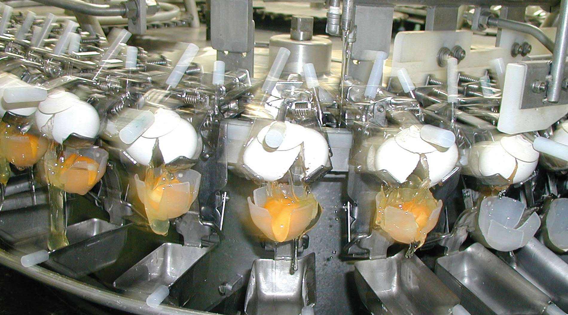
By Dr. Inge Heinzl, Editor, EW Nutrition
For a long time now, IgY technology has been used to provide clear benefits in diagnostics, human medicine, and animal production. To give you a deeper insight into this topic, in the following, we will show you some steps of production, the benefits, and the applications of IgY.
IgY (immunoglobulin of the yolk) are immunoglobulins that hens produce to protect their chicks during the first weeks of life against occurring pathogens. They are the equivalent of immunoglobulin G in the colostrum of mammalians. IgY are an entirely natural product; every egg sold in the supermarket contains IgY.
IgY develops in the hen against the pathogens with which the hens are confronted. Thereby, it does not matter if these pathogens are relevant for the hens. They also produce antibodies against, e. g., bovine, porcine, or human-specific pathogens. This fact was already noticed by Vaillard (1891). He saw that the intraperitoneal injection of tetanus bacteria raised immunity against tetanus bacteria in hens’ serum.

A short time later, Klemperer (1892) documented that the serum antibodies were also transferred into the egg. For this purpose, he did a similar trial with hens but collected the eggs. He fed mice a solution containing the egg yolk, and afterward, he infected them with tetanus. All mice with a higher dosage of egg yolk remained healthy, the others receiving a low dosage or no egg yolk died.
The “usual” production of antibodies in mammals includes pain and stress-causing procedures such as immunization, bleeding, and sacrifice. The only stress factor in producing egg antibodies is the hyper-immunization with the pathogen or parts of it; the rest -collecting the eggs- is non-invasive (Ikemori et al., 1993). The European Centre for the Validation of Alternative Methods (ECVAM) ), one of Europe’s health and consumer protection institutes, strongly recommends egg immunoglobulins as an alternative to mammalian antibodies (Schade et al., 1996).
IgY production is also advantageous in terms of quantitative and qualitative output. Usually, one egg (with 15 mL of yolk) contains about 100-150 mg IgY (Pereira et al., 2019). Assuming that a hen lays about 300 eggs per year, one bird can produce between 30 and 45 g IgY in this period. After the isolation of the IgY from the egg yolk and the extraction from the remaining proteins, a final purification step that includes chromatography could achieve IgY with >90 % purity (Morgan et al., 2021).
The targeted confrontation of the animal with specific pathogens or antigens leads to the production of specific antibodies. In a field trial with piglets, Kellner et al. (1994) compared three groups of piglets suffering from diarrhea on day 1 of the test. One group received egg powder originating from hens hyperimmunized with diarrhea-causing pathogens, the second group egg powder from regular eggs, and the third didn’t receive any egg powder. The following results they achieved in one of two farms. The trial shows that, after applying egg powder with selected antibodies, the animals completely recovered within three days. In the group receiving egg powder of regular eggs, still, 9.1% suffered from severe diarrhea and in the control group without any egg powder, only 27.3 % recovered.

Figure 1: Comparison of eggs originating from regular and hyperimmunized hens
A process must meet specific requirements to be suitable for industrial production. In the case of IgY production, the crucial preconditions are that…
Industrially produced IgY can target selected pathogens, e.g., enteric bacteria or viruses, respiratory pathogens, SARS-COV-2, etc. As the antibodies act not only in birds but also in other animals, such as mammals including humans, they can be used to prevent disease or support persons/animals in the case of illness. IgY is safe for animals and humans.
Concerning the economic benefits of IgY production, it can be said that it is a cost-effective method due to the high concentration of IgY in the egg yolk and the relatively simple process of the purification of the antibodies. Additionally, feeding and handling are easier and more cost-effective for hens than for many other animals.
There are different methods of IgY production. One possibility is to hyperimmunize the hens simultaneously with multiple antigens. This method seems to be convenient but does not deliver standardized products concerning the content of immunoglobulins.
The other possibility is the immunization of different groups of hens, each with one antigen (e.g., Rotavirus, Salmonella, E. coli). The content of immunoglobulins is determined, and the different egg powders are mixed. The result is an IgY product with standardized amounts of specific immunoglobulins.
There are different application areas for IgY or IgY products. In human medicine, egg immunoglobulins can be used against the toxin of rattlesnakes or scorpions, or Streptococcus mutans bacteria, causing dental caries (Gassmann and Hübscher, 1992) Egg immunoglobulins are important for diagnostic tests such as radioimmunoassay (RIA) and enzyme-linked immunoassay (ELISA).
A further application area is animal nutrition. Young animals, such as calves or piglets, but also young dogs or cats, are born with immature immune systems. If they, additionally, are deprived of maternal colostrum in adequate quantity and/or quality, they suffer from immunity gaps during their first weeks of life and are susceptible to pathogens in their environment.
Antibiotics have been used prophylactically for a long time to protect young animals in this critical phase. With increasing antibiotic resistance, this procedure is not allowed anymore.
Products based on egg immunoglobulins against enteric pathogens, e.g., support young animals against newborn or weaning diarrhea (e.g., Yokoyama et al., 1992; Ikemori et al., 1992; Ikemori et al., 1997, Yokoyama et al., 1998).
IgY technology is an animal-friendly technology with high output. Its various applications make IgY a helpful tool for human medicine as well as animal production. To get the best results, attention must be paid to quality, meaning, amongst others the standardization of the products.
IgY is an optimal tool to help young animals such as calves and piglets cope with pathogenic challenges in early life. Consequently, IgY technology enables us to limit (preventive) antimicrobial use in critical periods of animal rearing and, therefore, reduce antimicrobial resistance.
References:
Gassmann, M., and U. Hübscher. “Use of Polyclonal Antibodies from Egg Yolk of Immunised Chickens .” ALTEX – Alternatives to animal experimentation 9, no. 1 (1992): 5–14.
Ikemori, Yutaka, Masahiko Kuroki, Robert C. Peralta, Hideaki Yokoyama, and Yoshikatsu Kodama. “Protection of Neonatal Calves against Fatal Enteric Colibacillosis by Administration of Egg Yolk Powder from Hens Immunized with K99-Piliated Enterotoxigenic Escherichia Coli.” Amer. J. Vet. Res. 53, no. 11 (1992): 2005–8. https://doi.org/PMID: 1466492.
Ikemori, Yutaka, Masashi Ohta, Kouji Umeda, Faustino C. Icatlo, Masahiko Kuroki, Hideaki Yokoyama, and Yoshikatsu Kodama. “Passive Protection of Neonatal Calves against Bovine Coronavirus-Induced Diarrhea by Administration of Egg Yolk or Colostrum Antibody Powder.” Veterinary Microbiology 58, no. 2-4 (1997): 105–11. https://doi.org/10.1016/s0378-1135(97)00144-2.
Ikemori, Yutaka, Robert C. Peralta, Masahiko Kuroki, Hideaki Yokoyama, and Yoshikatsu Kodama. “Research Note: Avidity of Chicken Yolk Antibodies to Enterotoxigenic Escherichia Coli Fimbriae.” Poultry Science 72, no. 12 (1993): 2361–65. https://doi.org/10.3382/ps.0722361.
Kellner, J., M.H. Erhard, M. Renner, and U. Lösch. “Therapeutischer Einsatz Von Spezifischen Eiantikörpern Bei Saugferkeldurchfall – Ein Feldversuch.” Tierärztliche Umschau 49, no. 1 (January 1, 1994): 31–34.
Klemperer, Felix. “Ueber Natürliche Immunität Und Ihre Verwerthung Für Die Immunisirungstherapie.” Archiv für Experimentelle Pathologie und Pharmakologie 31, no. 4-5 (1893): 356–82. https://doi.org/10.1007/bf01832882.
Pereira, E.P.V., M.F. van Tilburg, E.O.P.T. Florean, and M.I.F. Guedes. “Egg Yolk Antibodies (Igy) and Their Applications in Human and Veterinary Health: A Review.” International Immunopharmacology 73 (2019): 293–303. https://doi.org/10.1016/j.intimp.2019.05.015.
Schade, R., C. Staak, C. Hendriksen, M. Erhard, H. Hugl, G. Koch, A. Larsson, et al. “The Production of Avian (Egg Yolk) Antibodies: IgY,” 1996. https://www.researchgate.net/publication/281466059_The_production_of_avian_egg_yolk_antibodies_IgY_The_report_and_recommendations_of_ECVAM_workshop_21.
Schade, R., C. Staak, C. Hendriksen, M. Erhard, H. Hugl, G. Koch, A. Larsson, et al. “The Production of Avian (Egg Yolk) Antibodies: IgY. The Report and Recommendations of ECVAM Workshop 21.” ATLA (Alternatives to Laboratory Animals) 24 (1996): 925–34. https://doi.org/https://www.researchgate.net/publication/281466059_The_production_of_avian_egg_yolk_antibodies_IgY_The_report_and_recommendations_of_ECVAM_workshop_21.
Yokoyama, H, R C Peralta, R Diaz, S Sendo, Y Ikemori, and Y Kodama. “Passive Protective Effect of Chicken Egg Yolk Immunoglobulins against Experimental Enterotoxigenic Escherichia Coli Infection in Neonatal Piglets.” Infection and Immunity 60, no. 3 (1992): 998–1007. https://doi.org/10.1128/iai.60.3.998-1007.1992.
Yokoyama, Hideaki, Robert C. Peralta, Kouji Umeda, Tomomi Hashi, Faustino C. Icatlo, Masahiko Kuroki, Yutaka Ikemori, and Yoshikatsu Kodama. “Prevention of Fatal Salmonelosis in Neonatal Calves, Using Orally Administered Chicken Egg Yolk Salmonella-Specific Antibodies.” Amer. J. Vet. Res. 59, no. 4 (1998): 416–20. https://doi.org/PMID: 9563623.
Yokoyama, Hideaki, Robert C. Peralta, Sadako Sendo, Yutaka Ikemori, and Yoshikatsu Kodama. “Detection of Passage and Absorption of Chicken Egg Yolk Immunoglobulins in the Gastrointestinal Tract of Pigs by Use of Enzyme-Linked Immunosorbent Assay and Fluorescent Antibody Testing.” American Journal of Veterinary Research 54, no. 6 (1993): 867–72. https://doi.org/PMID: 8323054.
Zhang, Xiao-Ying, Ricardo S. Vieira-Pires, Patricia M. Morgan, Rüdiger Schade, Xiao-Ying Zhang, Rao Wu, Shikun Ge, and Álvaro Ferreira Júnior. “Immunization of Hens.” Essay. In IGY-Technology: Production and Application of Egg Yolk Antibodies. Basic Knowledge for a Successful Practice., 116–34. Cham, Switzerland: Springer Nature, 2021.
Zhang, Xiao-Ying, Ricardo S. Vieira-Pires, Patricia M. Morgan, Schade Rüdiger, Patricia M. Morgan, Marga G. Freire, Ana Paula M. Tavares, Antonysamy Michael, and Xiao-Ying Zhang. “Extraction and Purification of IgY .” Essay. In IGY-Technology: Basic Knowledge for a Successful Practice, 135–60. Cham: Springer International Publishing AG, 2021.
Zhang, Xiao-Ying, Ricardo S. Vieira-Pires, Patricia M. Morgan, Schade Rüdiger, Patricia M. Morgan, Xiao-Ying Zhang, Antonysamy Michael, Ana Paula M. Tavares, and Marga G. Freire. “Extraction and Purification of IgY (Chapter 11).” Essay. In IGY-Technology: Basic Knowledge for a Successful Practice, 135–60. Cham: Springer International Publishing AG, 2021.

By Dr. Inge Heinzl, Editor, EW Nutrition
According to the American Medical Association, antimicrobial resistance is one of the main threats to public health nowadays. More than 2 million people are infected with bacteria resistant to different types of antibiotics every year (Marquardt and Suzhen, 2018). Prof Dame Sally Davies (2012), Chief Medical Officer for England, mentions that antibiotics are losing their effectiveness at alarming rates. Bacteria are finding ways to survive the antibiotics, so these molecules no longer work. O’Neill (2016) predicted in his report that 10 million people a year could be dying by 2050 due to antimicrobial resistance.
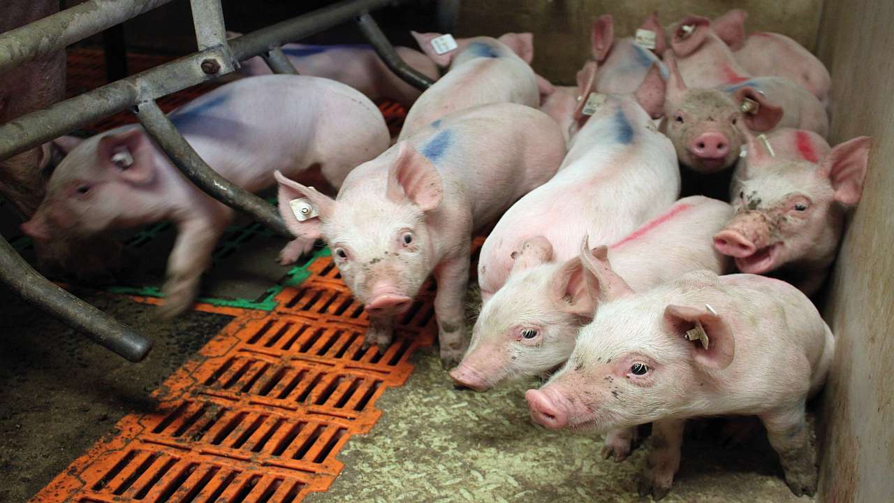
Antimicrobial resistance is a natural process but this is accelerated by inappropriate prescribing of antimicrobials, poor infection control practices and the unnecessary use of antimicrobials in agriculture (Barber and Sutherland, 2017).
Resistance to specific antibiotics occurs through mutations that enable the bacteria to withstand an antibiotic treatment. One mechanism is the production of enzymes degrading or altering the antibiotic, rendering them harmless. The elimination of entrances for antibiotics or the development of pumps discharging them is another possibility. A further option is the elimination of the targets the antibiotic would attack.
So-called “resistance genes” are responsible for resistance. These genes can be transferred from one bacterium to another and also from beneficial bacteria to harmful ones. When antibiotics are used, “normal” bacteria are killed; the resistant ones survive and have all possibilities to proliferate. The Dutch Government has been tracking resistant bacteria in poultry flocks for the last two decades. A clear correlation between antibiotic use and the percentage of resistance could be observed. The good thing: according to the 2020 MARAN report (De Greeff et al., 2020), by reducing the use of antibiotics, the occurrence of resistances can be pushed back.
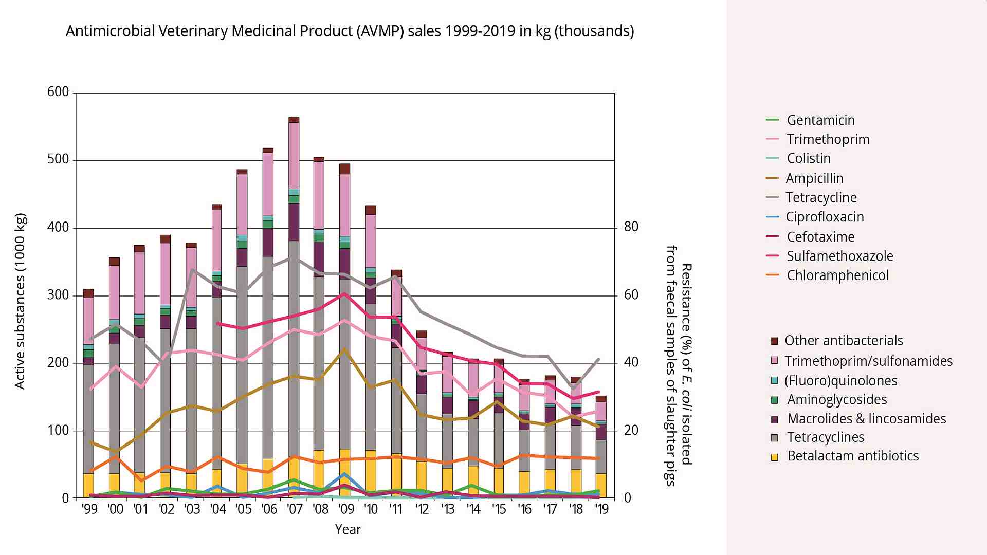
Figure 1. Sales of antibiotics from 1999 to 2016 and the development of resistances (MARAN report, 2018)
In pig production, antibiotics are often used in stressful situations such as weaning or moving. Antibiotics decrease the pathogenic pressure in animals and help them overcome these critical periods. Disadvantage: Antibiotics do not differentiate between good and bad but between susceptible and resistant. Therefore, also the beneficial gut flora gets destroyed through antibiotic treatment, and resistance is spread.
After the ban of antibiotic growth promoters in Europe in 2006, the US has also made considerable efforts to reduce the use of antibiotics.
When antibiotics are taken out of livestock production, measures in different areas must be implemented to keep performance and profitability high. Without supporting the animals by other means, they will get sick and even die in acute cases. Subclinical disease forms reduce their feed intake, and growth performance consequently decreases. According to literature, losses due to decreased average weight gain can be up to $40 per pig (Hao et al., 2014).
To support pigs, especially during the afore-mentioned critical periods, alternatives focusing on the maintenance of gut health and, therefore, also overall health must be chosen. This goal can only be achieved by balancing the intestinal flora with reducing pathogenic bacteria occurrence.
Phytomolecules are produced by plants to defend themselves against predators or pathogens. Farmers use the substances in animal feeds to support digestion, improve palatability, but also to reduce pathogenic pressure (Baser and Buchbauer, 2010).
In animal feeding, different application forms are available:
A trial conducted at the Federal University of Lavras (Brazil) evaluated if phytomolecules as a regular diet component can deliver the same effects on growth performance as AGPs in pig production.
For the trial, 108 castrated newborn male pigs were allocated to 3 groups (control, AGP (antibiotic growth promoters), and Activo). Pigs were weaned at 23 days of age with an average weight of 6.3 kg. They were fed a 3-phase diet (nursery, growing, and finishing). The inclusion rates of the additives (antibiotics and phytomolecules-based product – Activo) are shown in table 1.
On days 0, 1, and 2 of the experiment, the animals were challenged by applying a solution containing 107 CFU of E. coli K88, producing the toxins LT, Sta, and bST. Additionally, during the two last days before the growing phase, the animals were exposed to 5h of heat stress, using infrared lamps and closed windows. The parameters weight gain, final weight, FCR, and gut flora composition in the cecum were evaluated.
| Phase | Control | AGP | Activo | |
| Nursery | 0-7 days | — | Gentamycin 2.7kg/t | 0.4kg/t |
| 8-42 days | — | Haloquinol 0.2kg/t | 0.3kg/t | |
| Growing | 42-52 days | — | Tylosin 0.45kg/t | 0.4kg/t |
| 53-87 days | — | Enramycin 0.125kg/t | 0.2kg/t | |
| Finishing | 88-97 days | — | Tylosin 0.45kg/t | 0.4kg/t |
| 98-126 days | — | Enramycin 0.063kg/t | 0.2kg/t | |
Table 1. Inclusion rate of the additives in the feed
AGP: Antibiotic growth promoter; Activo: product based on phytomolecules, microencapsulated (EW Nutrition)
The results of this trial are shown in figure 2.
Concerning growth performance, the group fed the phytomolecules-based product Activo showed a 4.36 kg higher final weight after 126 days than the group provided AGPs (p=0.11), resulting in a 3.28 kg higher weight gain (p=0.21) and a 13 points better feed conversion.
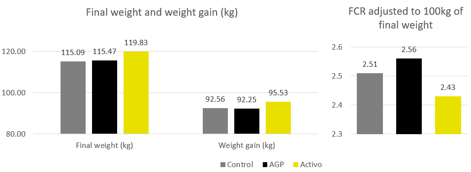
Figure 2. Data of growth performance including final weight, weight gain and FCR adjusted to 100kg
The evaluation of some bacteria naturally occurring in the gut flora showed that, in contrast to the antibiotic prophylaxes, Activo has no negative impact on E. coli, Lactobacillus and Bifidobacterium. However, the antibiotic group showed a slight decrease in the population of Lactobacilli (Figure 3).
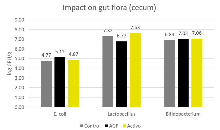
Figure 3. Impact of antibiotics and phytomolecules (Activo) on the composition of the gut flora
This trial shows Activo increasing growth performance and feed conversion without any negative impact on gut flora. The addition of phytomolecules (Activo) to the feed is documented as optimal long-term support instead of antibiotic growth promoters.
In a trial conducted in the USA, a product containing phytomolecules and organic acids (Activo Liquid, EW Nutrition) was compared to an antibiotic for controlling bacterial diseases in US pig production (Mecadox). For the trial, a total of 360 weanling pigs, about 19 days old and weighing 5.70 kg, were divided into four groups. Each group consists of 9 pens with 10 animals per pen. All groups were fed a 3-phase diet.
To the different trial groups, the following products were added (table 2):
| Feeding valid for all groups | Group / Product | Inclusion rate and period of application | |
| 3-phase feeding after weaning: | Mecadox | 50 g/t of feed during the whole period | |
| Phase I (days 0-7): | 23 % CP, 5.4 % CF | Activo Liquid 3 | 375 ml/1000 L of water for 3 days post-weaning |
| Phase II (days 8-21): | 21 % CP, 4.1 % CF | Activo Liquid 5 | 375 ml/1000 L of water for 5 days post-weaning |
| Phase III (days 22-42): | 19 % CP, 4.4 % CF | Activo Liquid 7 | 375 ml/1000 L of water for 7 days post-weaning |
These performance parameters were evaluated: live weight, daily gain, daily feed intake, feed:gain ratios, and mortality.
Table 2. Feeding and inclusion of the additives
The results of the trial are shown in figure 4. Concerning growth, no significant differences could be seen between the groups, only numerical differences. Live weight in the antibiotic group was 25.95 kg, and in the Activo Liquid groups, it ranges from 25.77 kg (shortest period of application) to 26.20 kg (see below). This resulted in calculated values for an average daily gain of 473 g in the Mecadox fed animals and 463 to 487g in the Activo Liquid groups. Due to a lower feed intake per kg of weight gain, all groups fed Activo Liquid showed a significantly (p=0.05) better feed conversion than the Mecadox group.
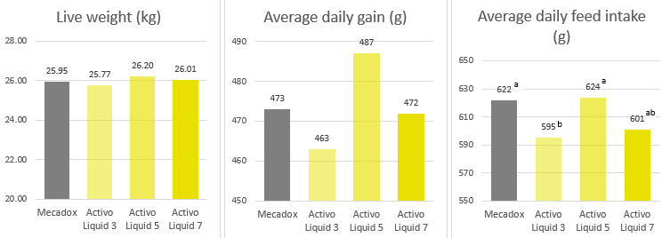
Figure 4. Live weight in the groups fed the antibiotic Mecadox and the phytomolecules-based product Activo Liquid for different periods
Average daily gain in the different trial groups
Average daily feed intake in the different trial groups (P=0.05)
Concerning mortality, the group fed Activo Liquid for 5 days showed the lowest mortality rate of 1.1% (figure 5).
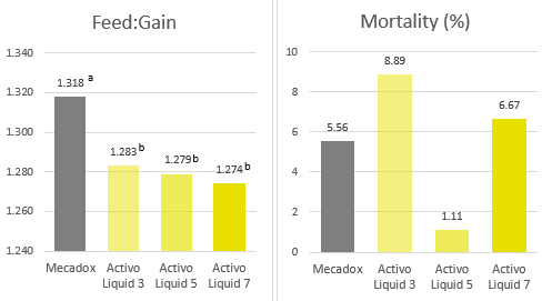
Figure 5. Feed:gain ratio in the different trial groups (P=0.05) & Mortality rates
Considering all parameters, the group fed Activo Liquid for five days provided the best results: numerically the lowest mortality rate, highest daily gain, and one of the two lowest feed:gain ratios. This trial concludes that Activo Liquid with an application period of five days can safely replace antibiotic growth promoters in the diet. Therefore, Activo Liquid is an interesting tool to additionally support pigs during critical periods of life.
The trials conducted with two types of phytomolecules-based products show that phytomolecules efficiently support pigs to achieve their genetic potential. A basic supply of these substances within the feed yields results similar to those of animals receiving antibiotic growth promoters (AGPs). In challenging situations like weaning, additional short-term supply is recommended, which can be done with liquid products via the waterline.
With this strategy, the use of antibiotic growth promoters and, therefore, antibiotics in general can be drastically reduced. This approach can help decrease antimicrobial resistance and, not to be forgotten, accommodates final customers’ requests for the lower usage of antibiotics in livestock.
Barber, Sarah, and Nikki Sutherland. “O’Neill Review into Antibiotic Resistance.” House of Commons Library, March 6, 2017. https://commonslibrary.parliament.uk/research-briefings/cdp-2017-0074/.
Baser, Kemal Hüsnü Can, and Gerhard Buchbauer. Handbook of Essential Oils: Science, Technology, and Applications. Boca Raton, FL: Taylor & Francis distributor, 2010.
Davies, Dame Sally. “Antibiotic Resistance ‘Big Threat to Health’.” BBC News. BBC, November 16, 2012. https://www.bbc.co.uk/news/health-20354536.
De Greeff, S.C., A.F. Schoffelen, and C.M. Verduin. “MARAN Reports.” WUR. National Institute for Public Health and the Environment – Ministery of Health, Welfare and Sport, June 2020. https://www.wur.nl/en/Research-Results/Research-Institutes/Bioveterinary-Research/In-the-spotlight/Antibiotic-resistance/MARAN-reports.htm.
Hao, Haihong, Guyue Cheng, Zahid Iqbal, Xiaohui Ai, Hafiz I. Hussain, Lingli Huang, Menghong Dai, Yulian Wang, Zhenli Liu, and Zonghui Yuan. “Benefits and Risks of Antimicrobial Use in Food-Producing Animals.” Frontiers in Microbiology 5, no. Art. 288 (2014): 1–11. https://doi.org/10.3389/fmicb.2014.00288.
Marquardt, Ronald R, and Suzhen Li. “Antimicrobial Resistance in Livestock: Advances and Alternatives to Antibiotics.” Animal Frontiers 8, no. 2 (2018): 30–37. https://doi.org/10.1093/af/vfy001.
O’Neill, J. “Tackling Drug-Resistant Infections Globally.” Review on Antimicrobial Resistance. Wellcome Trust / HM Government, May 19, 2016. https://amr-review.org/sites/default/files/160518_Final%20paper_with%20cover.pdf.