No revision of the Feed Additives law, says the European Commission

IMG

The authorization and marketing of feed additives in the European Union is currently governed by Feed Additives Regulation (EC) No 1831/2003, which came into effect in 2004. In 2021, the European Commission formalized an initiative to revise it, stating as reasons both the focus brought by the Farm to Fork Strategy, as well as inherent complexities in phrasing, process, and more. Representatives of the EC’s responsible unit, DG SANTE Unit G5, have now confirmed to EW Nutrition that, following consultations and analysis, the revision of the legislation on the authorisation of feed additives will not happen under the current Commission’s mandate.

The revision was initially deemed necessary on several grounds:

  • Not enough focus on sustainable animal farming
  • Lack of flexibility in promoting technical and scientific innovation
  • A lengthy authorization process
  • Unnecessary administrative burden
  • Ineffective imports control leading to unfair competition between EU and non-EU operators
  • Dependency on imports from third countries for some additives (e.g., vitamins)
  • Restrictions on the circulation of feed additives only intended for export
  • Insufficient legal clarity and consistency for a few aspects of the Regulation, e.g. use of certain additives in drinking water or labelling provisions for worker safety provisions in various complementary but unclear Regulations
  • Extensive, unnecessary labeling regulations that create physical and administrative burdens


Near the end of the two-year assessment process, however, the response of European governmental, supra-national, and non-governmental bodies appears to have been lukewarm. Overall, the conclusion of the EC unit overseeing the process was that “while a review of the framework would be useful, it does not appear necessary, considering the possibilities already granted by the existing legal framework.” In other words, applicants will have to use the existing mechanisms for applications, with no prospect for change in the near future.

Other strategies and regulations have also fallen through the cracks. For instance, the EU Animal Health Strategy 2007-2013 has not been updated in 10 years and there are no plans to renew the initiative. This is likely because the Green Deal and the flurry of new or upcoming regulations related to it are expected to supplant the framework for protein production in the European Union.

As the mandate of the current EC ends in 2024, there is a slim chance that the feed additive authorization process might be made less cumbersome once a new commission takes over.


Ketosis: the most critical metabolic disease in dairy cows

Kuehe Stal

Judith Schmidt, Product Manager On-Farm Solutions

Improvements in genetics, nutrition, and management continue to enhance dairy cows’ performance. However, being high-performance athletes comes at a cost, putting an extremely high burden on the animals’ energy metabolism. Especially around calving and during the first eight weeks of lactation, dairy cows can experience many stress factors: subclinical hypocalcemia, abomasum displacements, herd composition changes, or lameness. The more stress factors put the cows’ organism under pressure, the more likely they will become sick. A common consequence of stress is the occurrence of metabolic diseases, especially ketosis.

Both in terms of animal health and economic aspects, ketosis is probably the most critical dairy cow disease when also considering the correlated diseases. In this article, we explore the causes and consequences of ketosis and highlight prevention strategies that keep this issue under control.

Ketosis: causes and consequences

How ketosis develops

A restricted feed intake capacity and/or reduced energy concentration in the ration lead to a deficit in the animal’s energy balance. This situation occurs, for instance, at calving when the mother animal focuses her resources on the calf and its care. To compensate for the energy deficit, body fat is broken down for energy production. This process creates free fatty acids that accumulate in the liver and are partially converted into ketone bodies. These ketone bodies are a “transport medium” for energy, which various organs can use as an alternative energy source.

The problem arises when the deficiency lasts too long: more and more body fat is broken down, more and more fatty acids reach the liver, which leads to a fatty liver, and too high an amount of ketone bodies is formed and released into the blood. The ketone bodies in the blood inhibit appetite, resulting in less feed consumption and an energy deficit – the vicious cycle of ketosis begins.

Subclinical ketosis

Subclinical ketosis is defined as the stage of the disease at which an increased level of ketone bodies can be detected in the blood, urine, and milk. Furthermore, signs of hypoglycemia, increased levels of non-esterified fatty acid, and decreased hepatic gluconeogenesis can be seen in the blood. These conditions are typically not detected because there are no clinical signs.

Subclinical ketosis is a problem as it does not cause visible symptoms but leads to an increased incidence of subsequent diseases such as lab stomach displacement, clinical ketosis, and uterine inflammation. In addition, there may be loss of milk and fertility problems. Subclinically ill animals cannot be identified by the farmer by observation alone. Therefore, subclinical ketosis must be detected at an early stage to be able to act at the right time: prophylaxis instead of therapy.

There are several test possibilities to find out if an animal suffers from ketosis:

  1. Milk: Milk test for ketosis detection has been available for many years. The results are to be obtained based on a color gamut. In contrast to blood analysis, the milk test does not evaluate exact values but shows a color change of the contained indicator. However, an increased milk cell content of the feeding of poorly fermented silages with a high butyric acid content significantly influences the result. The test often does not adequately reflect the actual conditions.
  2. Urine: Another possibility is the examination of urine samples. Urine can be obtained spontaneously or with the help of a catheter. The results can also be read on a color scale of the urine test stripes. Like the milk test, the urine test only distinguishes different concentration ranges, but these are more finely graded than in the milk tests.
  3. Blood: The most accurate but also most complex and expensive method is a blood test. It has the advantage that not only ketone bodies but also other parameters such as free fatty acids, minerals, and liver enzymes can be analyzed. In addition, the blood analysis results are evaluated in numbers and are more comparable than the color changes of test stripes. A good alternative is a rapid test by using a rapid test device, which is also used for measuring human blood sugar. A result is displayed with a drop of blood on a test strip within a few seconds.

Clinical ketosis

Depending on why there are elevated ketone body levels in the blood, we distinguish between primary and secondary clinical ketosis. For the primary form of clinical ketosis, the energy deficit itself (due to high performance and/or incorrect feeding) causes the condition. This form mainly occurs in susceptible, high-yielding dairy cows between the second and seventh weeks of lactation (Vicente et al., 2014). Secondary ketosis is caused indirectly by other diseases disease. A cow suffering from, for example, a claw disease might no longer consume a performance-based feed ration, leading to an energy deficit.

Typical symptoms

Typical of metabolic diseases, ketosis leads to a broad spectrum of symptoms. The classic symptoms at the beginning of the disease are a loss of appetite and decreased milk performance. As the disease develops, motor skills may be affected, and the excrement’s consistency becomes firmer and darker in color. The respiratory rate of sick animals increases, and they show dyspnea. Dyspnea is the medical description for breathing difficulties. Affected animals suffer from air shortage, which can occur in different situations. Due to the excretion of ketone bodies via the mucous membranes, the animals’ breath smells more or less strongly of acetone (Robinson and Williamson, 1977).

In addition, the animals undergo rapid and severe weight loss, and their general body conditions deteriorate noticeably. Furthermore, cows suffering from ketosis show increased milk fat content or an increased milk fat/protein quotient. Clinical symptoms include reduced general well-being, apathy, blindness, staggering, persistent “absent-minded” licking of the environment or overexcitability, muscle tremors, and aggressiveness (Andersson, 1984).

Effects on animal health and performance

Even in its subclinical form – if untreated – ketosis will engender health risks and reduced performance, negatively impacting milk yield and cows’ fertility. For clinical cases, typical effects include infertility, udder and hoof problems, and a fatty liver. Ketosis during early lactation is usually associated with fatty liver disease. In severe cases, the liver becomes enlarged and more fragile. It then no longer performs its detoxification function, toxic compounds increase, and the central nervous system is damaged. Anorexia or even a total loss of consciousness, the so-called hepatic coma, might ensue, ending in a complete liver function failure.

Direct economic costs range from high veterinary costs to the total loss of the dairy cow, i.e., approximately € 600 to € 1.000 per cow. Moreover, producers face indirect costs from secondary diseases such as fatty liver disease, increased postpartum behavior such as uterine infections, abomasum dislocations, or claw diseases.

­­Ketosis prevention: feeding and targeted supplementation

Feeding strategy

As part of the preparatory feeding, both dry and pregnant cows should receive rations that lead to an optimal (and not maximum) body condition at the time of calving. Animals with a poorer nutritional status do not have enough body fat reserves to compensate for lack of energy in the first phase of lactation. In more cases, animals have a too high BCS, leading to a risk of difficult births, and the cows have too little appetite at the beginning of lactation. These cows tend to show an excessive mobilization of fat reserves and develop a fatty liver. So prevention of ketosis of the current lactation starts with preventing a too-high BCS in the middle of the previous lactation.

The aim of feeding measures is to keep the lactating cow’s discrepancy between nutrient requirements and nutrient uptake as low as possible when the genetically determined performance potential is exhausted. For this reason, the ration must have a certain minimum energy density (high-quality forage and appropriate concentrate supplements). Also, anything that prevents the cows from ingesting the maximum amount of dry matter should be avoided.

Ket-o-Vital bolus for metabolic support

Another important preventive measure is the specific support of the calving cow’s liver, rumen, and immune system. EW Nutrition’s Ket-o-Vital Bolus was explicitly designed to reduce the risk of ketosis. It contains fast-available glucogenic substances, positively influencing the cow’s energy metabolism. Another advantage the bolus offers is the slow release of the contained cobalt, selenium, niacin, and active yeast:

  • Cobalt is a trace element important to form cobalamin, the so-called vitamin B12. It is essential for blood formation and the functioning of the nervous system.
  • Selenium protects cells from oxidative damage and ensures an intact immune defense;
  • Niacin is a B vitamin that intervenes in energy metabolism and prevents fatty liver syndrome;
  • And active yeast supports rumen health, preventing rumen acidosis and increasing feed intake.

The application of the Ket-o-Vital Bolus is profitable and straightforward. Only one bolus per application is required.

Ketosis control: be one step ahead

High-performance dairy cows are at risk of ketosis, which results in involuntary culling, poor health, and performance losses. Advanced feed management practices combined with the targeted use of the Ket-o-Vital bolus offer a solution for preventing this debilitating disease. The bolus protects the cows from clinical and subclinical ketosis, reduces metabolic disorders, increases appetite, and improves health – leading to a quick recovery and ensuring profitable production.

References

Vicente, Fernando, María Luisa Rodríguez, Adela Martínez-Fernández, Ana Soldado, Alejandro Argamentería, Mario Peláez, and Begoña de la Roza-Delgado. “Subclinical ketosis on dairy cows in transition period in farms with contrasting butyric acid contents in silages.” The Scientific World Journal 2014 (November 25, 2014): 1–4. https://doi.org/10.1155/2014/279614.

Andersson, L. “Concentrations of blood and milk ketone bodies, blood isopropanol and plasma glucose in dairy cows in relation to the degree of hyperketonaemia and clinical signs*.” Zentralblatt für Veterinärmedizin Reihe A 31, no. 1-10 (1984): 683–93. https://doi.org/10.1111/j.1439-0442.1984.tb01327.x.

Robinson, A. M., and D. H. Williamson. “Effects of acetoacetate administration on glucose metabolism in mammary gland of fed lactating rats.” Biochemical Journal 164, no. 3 (1977): 749–52. https://doi.org/10.1042/bj1640749.


Respiratory disease – the biggest problem in horses

MG

Author: Judith Schmidt, Product Manager On-Farm Solutions

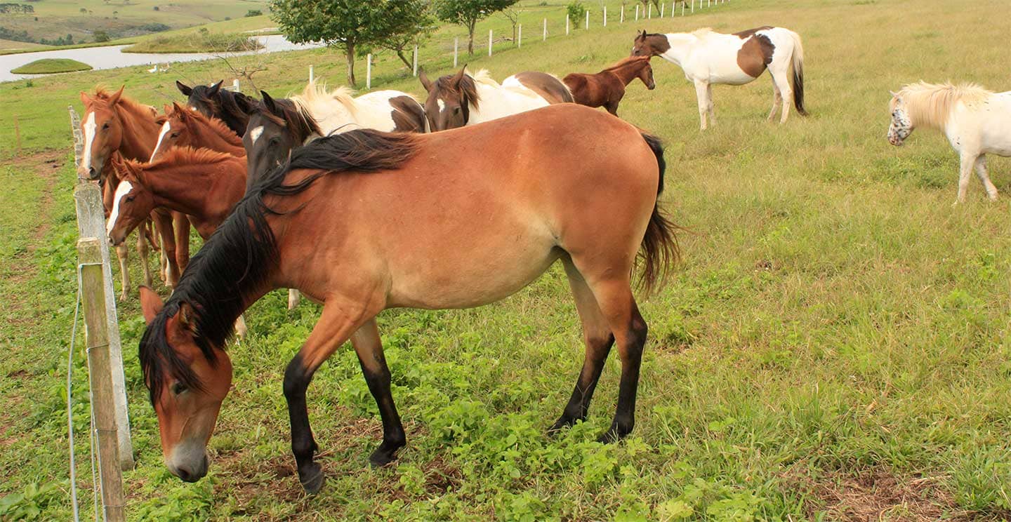
The respiratory tract in horses is prone to various problems, ranging from allergic reactions and inflammation to infections. Through early diagnosis, appropriate treatment, and preventive measures, horse owners can help maintain the respiratory health of their horses and promote their well-being and performance.

Respiratory diseases are a constant topic of suffering and irritation among horse owners. According to a study published in 2005, respiratory diseases account for about 40 % of all equine internal diseases recorded worldwide (Thein 2005).

The high-performance organ: the horse’s lung

The respiratory tract of our horses is a high-performance system with a large exchange surface between the inside of the body and the environment. The lungs enable the so-called gas exchange, i.e., the transfer of oxygen from the air into the horse´s bloodstream. Only when this gas exchange functions properly can the horse supply its muscles with sufficient oxygen.

Even at rest, about 50 to 80 liters of air per minute enter the lungs of a 600 kg horse. With increasing load, this value can rise up to 2.000 liters per minute at maximum load. If a horse is healthy, it breathes calmly and slowly and takes eight to sixteen deep breaths per minute.

In order to protect the lungs as best as possible from harmful influences, the entire respiratory tract is equipped with a special mucous membrane. When irritated by pathogens or foreign bodies, for example, this mucous membrane forms more mucous and transports it towards the mouth cavity with the help of the finest cilia. In this way, most harmful particles are usually intercepted quickly, reliably and, above all, effectively and, if necessary, coughed up before they can even reach the alveoli and cause damage there.

Horse Pixabay

The most common causes of respiratory diseases in horses

Chronic obstructive bronchitis

Chronic obstructive bronchitis is better known as COB or equine asthma. COB is more common in horses that are regularly kept in dusty or poorly ventilated environments, such as cramped stables or pastures with high levels of mold. Inhalation of dust particles and allergens can cause inflammation of the respiratory tract, resulting in coughing, increased mucus expectoration and breathing difficulties. The clinical picture of COB can vary greatly. From occasional poor performance in show horses to chronic coughing with purulent nasal discharge or significant weight loss.

Tracheitis

Another common respiratory disease in horses is tracheitis. This disease is often caused by bacterial or viral infections. Young horses, older horses or those with a weakened immune system are particularly susceptible to tracheitis. Besides infections, irritating factors such as dust, smoke or chemicals can also irritate the mucous membrane of the trachea and trigger inflammation.

Hay fever

Hay fever, also know as allergic respiratory disease or allergic rhinitis, is a common condition that can also affect horses. Like humans, it is an allergic reaction to certain pollens, molds or other environmental allergens that are suspended in the air. Common signs include sneezing, a runny nose and itchy eyes. However, some horses may also suffer from coughing or respiratory symptoms. Hay fever in horses can occur seasonally, depending on the pollen seasons. Depending on the region and season, the symptoms may be more serve during spring, summer or autumn.

Asthma

Asthma in horses, also known as equine asthma or heaves, is a chronic respiratory disease that occurs mainly in horses. It is similar to in many ways to asthma in humans. The main cause of this disease is hypersensitivity of the respiratory tract to dust, allergens or mold spores in the horse´s environment.


Respiratory distress or harmless rattling?

Horse owners know it – the four-legged friends have an impressive range of breathing sounds. But which are harmless, such as the exited trumpeting through the nostrils during a fright? And which ones could be symptoms of a respiratory disease?

Diagnosing respiratory problems in horses can be challenging because symptoms can often be non-specific and/or show signs similar to several diseases.

Snorting: When horses snort, it is a sign of relaxation. There is usually no cause for concern. Quite the opposite.

Snorting at gallop: Many horses snort rhythmically at a gallop. This is also considered harmless. Snorting is particularly common in thoroughbreds.

Coughing, for example when trotting:  Occurs so often that it is often perceived as normal. But it is not. Coughing is always an alarm sign and can indicate an allergy, asthma or a viral or bacterial infection.

Whistling when inhaling: To be on the safe side, a veterinarian should be consulted.

Consequences of respiratory disease

Respiratory disease in horses can have significant economic consequences. If a horse suffers from chronic obstructive bronchitis or another respiratory disease, this can lead to various problems:

  • Veterinary costs: The diagnosis and treatment of respiratory diseases often require veterinary visits, medication, and possibly further examinations such as x-rays or endoscopy.
  • Reduced performance: A horse with respiratory problems may be severely limited in its performance. It may have difficulty breathing, which can have a negative effect on its athletic performance, equestrian work, or other activities.
  • Downtime: During the treatment or recovery period, horses may have to take a break or be taken out of training. This may result in loss of income, especially if the horse was intended for competition or showing.
  • Decrease in value: A horse with chronic respiratory problems may lose its value as a sport or breeding horse. Selling price might decrease and the demand for such a horse might decrease too.

To minimize economic impact, early diagnosis and treatment is important, as the implementation of appropriate preventive measures to reduce the risk of respiratory disease.

Prevention

Prevention of equine cough is of big importance to reduce the incidence and severity of the disease.

Clean stable environment

Dust is a common trigger of respiratory symptoms in horses. Regular removal of dust, dirt and mold spores from the stable and horse boxes can help to improve air quality and reduce respiratory stress.

Pasture management

When possible, horses should be allowed access to fresh pastures. The natural outdoor environment helps horses breathe cleaner air and inhale fewer harmful particles.

Hay feeding

Choosing high quality, low dust hay can reduce exposure to allergens. Moist soaking of hay before feeding can also help reduce dust levels.

Ventilation in the stable

Good ventilation in stables is essential to avoid stagnant air and dust accumulation. The use of fans or natural ventilation systems can improve air circulation.

Feed management

Feeding high quality feed that is free of mold and allergens can reduce the risk of respiratory problems. It is important to adjust feed rations to the individual needs of each horse.

Supplements

Supplements can play a positive role in the prevention of respiratory problems in horses if they are used selectively and with expert advice.

  • Immune system support: Supplements such as vitamins, minerals and antioxidants can strengthen the immune system. A healthy immune system helps the horse to better defend itself against infections and inflammation of the respiratory tract.
  • Certain supplements contain ingredients with anti-inflammatory properties, such as omega-3-fatty acids or herbal extracts. These can help reduce inflammation in the respiratory tract and thus reduce the risk of respiratory problems.
  • Supporting respiratory health: Some supplements on the market have been specially designed to support respiratory function. They can help to regulate mucus production, improve respiratory protection, and facilitate the expectoration of mucus.
  • Strengthening lung capacity: Certain ingredients in supplements can support the horse´s lung capacity and promote better oxygen uptake, which is important for performance and respiratory health.

Conclusion

If there are noticeable breathing sounds, coughing, fever or a drop in performance, the vet should come quickly. A respiratory disease tends to develop into a long-term problem if it is not treated appropriately. Without treatment, it can become chronic in some cases. Fresh air and species-appropriate husbandry, as well as feed that is free of mold und dust, are the first steps to supporting the normal function of your horse´s respiratory tract. Supplements can be an excellent tool for prevention. A holistic approach to equine health is crucial. This includes proper stable and feed hygiene, sufficient exercise, and good air quality in stables.

References:

Handbuch Pferd: Dr. med. vet. Peter Thein, 2005

Tierklink Kaufungen (2016): Chronische Obstruktive Bronchitis (COB), Barbara Liese & Dr. Kristian Sander


Minimizing Collateral Effects of Antibiotic Administration in Swine Farms: A Balancing Act

PIGLETS FARM

By Dr Merideth Parke BVSc, Regional Technical Manager Swine, EW Nutrition

We care for our animals, and antibiotics are a crucial component in the management of disease due to susceptible pathogens, supporting animal health and welfare.  However, the administration of antibiotics in pig farming has become a common practice to prevent bacterial infections, reduce economic losses, and increase productivity.

All antibiotic applications have collateral consequences of significance, bringing a deeper consideration to their non-essential application. This article aims to challenge the choice to administer antibiotics by exploring the broader impact that antibiotics have on animal and human health, economies, and the environment.

Antibiotics disrupt microbial communities

Antibiotics do not specifically target pathogenic bacteria. By impacting beneficial microorganisms, they disrupt the natural balance of microbial communities within animals. They reduce the microbiota diversity and abundance of all susceptible bacteria – beneficial and pathogenic ones… many of which play crucial roles in digestion, brain function, the immune system, and respiratory and overall health. Resulting microbiota imbalances may present themselves in animals showing health performance changes associated with non-target systems, including the nasal, respiratory, or gut microbiome10, 9, 16. The gut-respiratory microbiome axis is well-established in mammals. Gut microbiota health, diversity, and nutrient supply directly impact respiratory health and function15. In pigs specifically, the modulation of the gut microbiome is being considered as an additional tool in the control of respiratory diseases such as PRRS due to the link between the digestion of nutrients, systemic immunity, and response to pulmonary infections12.

The collateral effect of antibiotic administration disrupting not only the microbial communities throughout the animal but also linked body systems needs to be considered significant in the context of optimal animal health, welfare, and productivity.

Antibiotic use can lead to the release of toxins

The consideration of the pathogenesis of individual bacteria is critical to mitigate potential for direct collateral effects associated with antibiotic administration. For example, in cases of toxin producing bacteria, when animals are medicated either orally or parenterally, mortality may increase due to the associated release of toxins when large numbers of toxin producing bacteria are killed quickly3.

Modulation of the brain function can be critical

Numerous animal studies have investigated the modulatory role of intestinal microbes on the gut-brain axis. One identified mechanism seen with antibiotic-induced changes in fecal microbiota is the decreased concentrations of hypothalamic neurotransmitter precursors, 5-hydroxytryptamine (serotonin), and dopamine6. Neurotransmitters are essential for communication between the nerve cells. Animals with oral antibiotic-induced microbiota depletion have been shown to experience changes in brain function, such as spatial memory deficits and depressive-like behaviors.

Processing of waste materials can be impacted

Anaerobic treatment technology is well accepted as a feasible management process for swine farm wastewater due to its relatively low cost with the benefit of bioenergy production. Additionally, the much smaller volume of sludge remaining after anaerobic processing further eases the safe disposal and decreases the risk associated with the disposal of swine waste containing residual antibiotics5.

The excretion of antibiotics in animal waste, and the resulting presence of antibiotics in wastewater, can impact the success of anaerobic treatment technologies, which already could be demonstrated by several studies8, 13. The degree to which antibiotics affect this process will vary by type, combination, and concentration. Furthermore, the presence of antibiotics within the anaerobic system may result in a population shift towards less sensitive microbes or the development of strains with antibiotic-resistant genes1, 14.

Antibiotics can be transferred to the human food chain

Regulatory authorities specify detailed withdrawal periods after antibiotic treatment. However, residues of antibiotics and their metabolites may persist in animal tissues, such as meat and milk, even after this period. These residues can enter the human food chain if not adequately monitored and controlled.

Prolonged exposure to low levels of antibiotics through the consumption of animal products may contribute to the emergence of antibiotic-resistant bacteria in humans, posing a significant public health risk.

Contamination of the environment

As already mentioned before, the administration of antibiotics to livestock can result in the release of these compounds into the environment. Antibiotics can enter the soil, waterways, and surrounding ecosystems through excretions from treated animals, inappropriate disposal of manure, and runoff from agricultural fields. Once in the environment, antibiotics can contribute to the selection and spread of antibiotic-resistant bacteria in natural bacterial communities. This contamination poses a potential risk to wildlife, including birds, fish, and other aquatic organisms, as well as the broader ecological balance of affected ecosystems.

Every use of antibiotics can create resistance

One of the widely researched concerns associated with antibiotic use in livestock is the development of antibiotic resistance. The development of AMR does not require prolonged antibiotic use and, along with other collateral effects, also occurs when antibiotics are used within recommended therapeutic or preventive applications.

Gene mutations can supply bacteria with abilities that make them resistant to certain antibiotics (e.g., a mechanism to destroy or discharge the antibiotic). This resistance can be transferred to other microorganisms, as seen with the effect of carbadox on Escherichia coli7 and Salmonella enterica2 and the carbadox and metronidazole effect on Brachyspira hyodysenteriae16. Additionally, there is an indication that the zinc resistance of Staphylococcus of animal origin is associated with the methicillin resistance coming from humans4.

Consequently, the effectiveness of antibiotics in treating infections in target animals becomes compromised, and the risk of exposure to resistant pathogens for in-contact animals and across species increases, including humans.

Alternative solutions are available

To successfully minimize the collateral effects of antibiotic administration in livestock, a unified strategy with support from all stakeholders in the production system is essential. The European Innovation Partnership – Agriculture11 concisely summarizes such a process as requiring…

  1. Changing human mindsets and habits: this is the first and defining step to successful antimicrobial reduction
  2. Improving pig health and welfare: Prevention of disease with optimal husbandry, hygiene, biosecurity, vaccination programs, and nutritional support.
  3. Effective antibiotic alternatives: for this purpose, phytomolecules, pro/pre-biotics, organic acids, and immunoglobulins are considerations.

In general, implementing responsible antibiotic stewardship practices is paramount. This includes limiting antibiotic use to the treatment of diagnosed infections with an effective antibiotic, and eliminating their use as growth promotors or for prophylactic purposes.

Keeping the balance is of crucial importance

While antibiotics play a crucial role in ensuring the health and welfare of livestock, their extensive administration in the agricultural industry has collateral effects that cannot be ignored. The development of antibiotic resistance, environmental contamination, disruption of microbial communities, and the potential transfer of antibiotic residues to food pose significant challenges.

Adopting responsible antibiotic stewardship practices, including veterinary oversight, disease prevention programs, optimal animal husbandry practices, and alternatives to antibiotics, can strike a balance between animal health, efficient productive performance, and environmental and human health concerns.

The collaboration of stakeholders, including farmers, veterinarians, policymakers, industry and consumers, is essential in implementing and supporting these measures to create a sustainable and resilient livestock industry.

References

  1. Angenent, Largus T., Margit Mau, Usha George, James A. Zahn, and Lutgarde Raskin. “Effect of the Presence of the Antimicrobial Tylosin in Swine Waste on Anaerobic Treatment.” Water Research 42, no. 10–11 (2008): 2377–84. https://doi.org/10.1016/j.watres.2008.01.005.
  2. Bearson, Bradley L., Heather K. Allen, Brian W. Brunelle, In Soo Lee, Sherwood R. Casjens, and Thaddeus B. Stanton. “The Agricultural Antibiotic Carbadox Induces Phage-Mediated Gene Transfer in Salmonella.” Frontiers in Microbiology 5 (2014). https://doi.org/10.3389/fmicb.2014.00052.
  3. Castillofollow, Manuel Toledo, Rocío García Espejofollow, Alejandro Martínez Molinafollow, María Elena  Goyena Salgadofollow, José Manuel Pintofollow, Ángela Gallardo Marínfollow, M. Toledo, et al. “Clinical Case: Edema Disease – the More I Medicate, the More Pigs Die!” $this->url_servidor, October 15, 2021. https://www.pig333.com/articles/edema-disease-the-more-i-medicate-the-more-pigs-die_17660/.
  4. Cavaco, Lina M., Henrik Hasman, Frank M. Aarestrup, Members of MRSA-CG:, Jaap A. Wagenaar, Haitske Graveland, Kees Veldman, et al. “Zinc Resistance of Staphylococcus Aureus of Animal Origin Is Strongly Associated with Methicillin Resistance.” Veterinary Microbiology 150, no. 3–4 (2011): 344–48. https://doi.org/10.1016/j.vetmic.2011.02.014.
  5. Cheng, D.L., H.H. Ngo, W.S. Guo, S.W. Chang, D.D. Nguyen, S. Mathava Kumar, B. Du, Q. Wei, and D. Wei. “Problematic Effects of Antibiotics on Anaerobic Treatment of Swine Wastewater.” Bioresource Technology 263 (2018): 642–53. https://doi.org/10.1016/j.biortech.2018.05.010.
  6. Köhler, Bernd, Helge Karch, and Herbert Schmidt. “Antibacterials That Are Used as Growth Promoters in Animal Husbandry Can Affect the Release of Shiga-Toxin-2-Converting Bacteriophages and Shiga Toxin 2 from Escherichia Coli Strains.” Microbiology 146, no. 5 (2000): 1085–90. https://doi.org/10.1099/00221287-146-5-1085.
  7. Loftin, Keith A., Cynthia Henny, Craig D. Adams, Rao Surampali, and Melanie R. Mormile. “Inhibition of Microbial Metabolism in Anaerobic Lagoons by Selected Sulfonamides, Tetracyclines, Lincomycin, and Tylosin Tartrate.” Environmental Toxicology and Chemistry 24, no. 4 (2005): 782–88. https://doi.org/10.1897/04-093r.1.
  8. Looft, Torey, Heather K Allen, Brandi L Cantarel, Uri Y Levine, Darrell O Bayles, David P Alt, Bernard Henrissat, and Thaddeus B Stanton. “Bacteria, Phages and Pigs: The Effects of in-Feed Antibiotics on the Microbiome at Different Gut Locations.” The ISME Journal 8, no. 8 (2014a): 1566–76. https://doi.org/10.1038/ismej.2014.12.
  9. Looft, Torey, Heather K. Allen, Thomas A. Casey, David P. Alt, and Thaddeus B. Stanton. “Carbadox Has Both Temporary and Lasting Effects on the Swine Gut Microbiota.” Frontiers in Microbiology 5 (2014b). https://doi.org/10.3389/fmicb.2014.00276.
  10. Nasralla, Meisoon. “EIP-Agri Concept.” EIP-AGRI – European Commission, September 11, 2017. https://ec.europa.eu/eip/agriculture/en/eip-agri-concept.html.
  11. Niederwerder, Megan C. “Role of the Microbiome in Swine Respiratory Disease.” Veterinary Microbiology 209 (2017): 97–106. https://doi.org/10.1016/j.vetmic.2017.02.017.
  12. Poels, J., P. Van Assche, and W. Verstraete. “Effects of Disinfectants and Antibiotics on the Anaerobic Digestion of Piggery Waste.” Agricultural Wastes 9, no. 4 (1984): 239–47. https://doi.org/10.1016/0141-4607(84)90083-0.
  13. Shimada, Toshio, Julie L. Zilles, Eberhard Morgenroth, and Lutgarde Raskin. “Inhibitory Effects of the Macrolide Antimicrobial Tylosin on Anaerobic Treatment.” Biotechnology and Bioengineering 101, no. 1 (2008): 73–82. https://doi.org/10.1002/bit.21864.
  14. Sikder, Md. Al, Ridwan B. Rashid, Tufael Ahmed, Ismail Sebina, Daniel R. Howard, Md. Ashik Ullah, Muhammed Mahfuzur Rahman, et al. “Maternal Diet Modulates the Infant Microbiome and Intestinal Flt3l Necessary for Dendritic Cell Development and Immunity to Respiratory Infection.” Immunity 56, no. 5 (May 9, 2023): 1098–1114. https://doi.org/10.1016/j.immuni.2023.03.002.
  15. Slifierz, Mackenzie Jonathan. “The Effects of Zinc Therapy on the Co-Selection of Methicillin-Resistance in Livestock-Associated Staphylococcus Aureus and the Bacterial Ecology of the Porcine Microbiota,” 2016.
  16. Stanton, Thaddeus B., Samuel B. Humphrey, Vijay K. Sharma, and Richard L. Zuerner. “Collateral Effects of Antibiotics: Carbadox and Metronidazole Induce VSH-1 and Facilitate Gene Transfer among Brachyspira HyodysenteriaeApplied and Environmental Microbiology 74, no. 10 (2008): 2950–56. https://doi.org/10.1128/aem.00189-08.


Coccidiostats in the European Union: Challenges and Perspectives

Header

by Twan van Gerwe, DVM PhD (EBVS), Technical Director, EW Nutrition

Controlling coccidiosis has been and continuous to be a major concern for poultry operations. However, for decades, some of these control measures have been taking an increasingly visible toll on the overall health of the flocks, the economics of poultry production, and the environment itself. Regulations have been put in place to defend consumer health and animal welfare while maintaining profitability in poultry production.

In the European Union and elsewhere, coccidiostats or anticoccidials are an essential means of control and are categorized either as feed additives or as veterinary medicinal products. The category is dictated by the pharmacologically active substance, mode of action, pharmaceutical form, target species and route of application.

In the European Union, there are currently 11 different coccidiostats which have been granted 28 different authorizations as feed additives allowed for specific usage in chickens, turkeys, and rabbits.

Coccidiostats: the basics

Compounds designed to kill the coccidial population are known as coccidiocidal; those designed to prevent the replication and development of coccidia are known as coccidiostats. Quite often, coccidiostat or anticoccidial is the term used to describe both categories.

Coccidiostats are antimicrobial compounds which either inhibit or destroy the protozoan parasites that cause coccidiosis in livestock. Each coccidiostat has individual inhibitory mechanisms. In the case of ionophores, the compounds affect transmembrane ion transport. In the case of synthetic compounds, the molecules’ mode of action is varied and, in some cases, not even entirely known (Patyra et al., 2023).

The production, manufacture, and marketing of coccidiostats, premixes with coccidiostats, and feed with coccidiostats are regulated by the Regulation (EC) No 183/2005 of the European Parliament and of the Council of 12 January 2005 laying down requirements for feed hygiene.

Coccidiostat categories

Coccidiostats fall under two categories:

Ionophores

Ionophores, sometimes called polyether ionophore antibiotics, are substances which contain a polyether group and are of bacterial origin. They are produced by fermentation with several strains of Streptomyces spp and Actinomadura spp. Six substances are allowed in the EU:

  • monensin sodium (MON)
  • lasalocid sodium (LAS)
  • maduramicin ammonium (MAD)
  • narasin (NAR)
  • salinomycin sodium (SAL)
  • semduramicin sodium (SEM)

Synthetic

Synthetic compounds include:

  • decoquinate (DEC)
  • diclazuril (DIC)
  • halofuginone (HFG)
  • nicarbazin (NIC)
  • robenidine hydrochloride (ROB)

EU authorizations for ionophores are granted under specific conditions of usage, including animal category, minimum and maximum dosage, MRL (Maximum Residue Limits), and withdrawal periods.

Regulation (EC) No 1831/2003 [13] of the European Parliament and of the Council of 22 September 2003 distinguishes between coccidiostats and antibiotics used as growth promoters. Unlike the antibiotic growth promoters (forbidden in the EU since 2006), whose primary action site is the gut microflora, coccidiostats only have a secondary and residual activity against the gut microflora. That still signals that they have the potential to trigger resistance and to alter the natural balance and immune response of the farmed animals. Their potential to cause resistance has been widely acknowledged by science and practitioners alike (see below).

Why were some antimicrobial growth promoters withdrawn in 1997-1998 – but not others?

Five designated “antibiotic feed additives” were prohibited in 1997-98: Avoparcin, Bacitracin zinc, Spiramycin, Virginiamycin, and Tylosin phosphate. The EU withdrew their authorization in order to “help decrease resistance to antibiotics used in medical therapy”. The motivation specified that these antibiotics belonged to classes of compounds also used in human medicine.

On the other hand, the EU at the time allowed the remaining antibiotics for use in feed as they did not belong to classes of compounds used in human medicine. That, of course, did not mean that resistance did not develop in birds.

The Commission did acknowledge the need to phase out the remaining antibiotics. At the same time, it stated that the use of coccidiostats would not presently be ruled out “even if of antibiotic origin” (MEMO/02/66, 2022). The reason was that “hygienic precautions and adaptive husbandry measures are not sufficient to keep poultry free of coccidiosis. Modern poultry husbandry is currently only practicable if coccidiosis can be prevented by inhibiting or killing parasites during their development.”

In other words, the Commission acknowledged that the only reason ionophores were still authorized was that it believed there were no other means of controlling coccidiosis in profitable poultry production.

What issues are raised by current coccidiosis control measures?

In its 2022 Position Paper on Coccidia Control in Poultry, the European Veterinaries Federation states that “challenges in coccidia control are due to parasitic and bacterial drug (cross-)resistance. Coccidiostats also interact with other veterinary medicinal products and have a secondary residual activity against gram-positive bacteria” (FVE, 2022).

Resistance

Resistance

Ever since 1939, when sulphanilamide was shown to cure coccidiosis in chickens, the industry increased the use of similar (chemical) compounds. It quickly added sulfaquinoxaline, then nitrofurazone and 3-notroroxarsone, amprolium and nicarbazin (Martins et al., 2022).

Prior to the introduction of the first ionophore, monensin, in the early 1970s, producers only had synthetic (non-ionophores) coccidiostats, characterized by rapid parasite resistance development. With the addition of ionophores, poultry operations started to rotate products between production cycles, or to use shuttle programs, with the express purpose of controlling the development of resistance. Synthetic compounds can, however, result in increased resistance in the long run (Martins et al., 2022). Moreover, studies in farmed animals indicate that sometimes even single use of antibiotics can promote the selection of resistant bacterial strains.

Another issue is the design of the rotation system, which, some researchers claim, could only delay the appearance of resistance (Daeseleire et al., 2017).

To make matters worse, for instance in the case of broilers, coccidiostats are generally administered throughout life to protect against re-infection. This may also lead to the next item on the list.

Residues

Regulation (EC) No 1831/2003 establishes Maximum Residue Limits (MRLs) for residues of an additive in relevant foodstuffs of animal origin. The goal is to control the use of coccidiostats in feed and ensure that there is no excess residue that ends up on the consumers’ plate.

Broilers can be fed with coccidiostats throughout life, with the exception of a certain withdrawal period before slaughter. Cross-contamination of feed batches and residue formation in edible tissues of nontarget species represent valid concerns for end consumers.

Coccidiostats in food have been regulated in the Commission Regulation (EC) No 124/2009, including maximum levels for meat ranging between 2 μg/kg (monensin, salinomycin, semduramycin, and manduramycin) and 100 μg/kg (nicarbazin in liver and kidney). However, Daeseleire et al. state that “in the period 2011–14, noncompliant results were reported for maduramycin, monensin, diclazuril, lasalocid, nicarbazin, robenidine, salinomycin, narasin, semduramicin, decoquinate, halofuginone, and toltrazuril. The matrices/animals species affected were in descending order eggs, poultry, farmed game, horses, pigs, and sheep/goat (EURL workshop, 2015)”. Residues in eggs are widely seen as a serious concern (Bello et al., 2023). The fact that regulations are in place constitute no safeguard against defective practices.

What alternatives to coccidiostats does the EU support?

Vaccination

Coccidiosis vaccines have been in use for the last three decades. They are based on precocious oocysts and are commonly used in breeding and laying birds, and the use in broilers is steadily increasing. There is a limited number of vaccines authorized in the EU. As vaccines are relatively costly to apply, vaccination is typically performed during 2-3 cycles only, afterwards reverting to the use of coccidiostats, which leads to a suppression of the precocious vaccine-origin strains, allowing persistent coccidiostat-resistant field strains to flourish.

Herbal products (phytomolecules)

Phytomolecules have been widely used for a variety of poultry gut health issues. Their usage in flocks at risk of coccidiosis is predicated on their ability to strengthen the natural defenses of the animal. Infection severity and consequences depend to a large extent on co-infections, gut health, and the general immunity of the bird.

Prescription veterinary medicines

Toltrazuril, amprolium, and some sulfamides (sulfamiderazin, sulfadimethoxin, trimethoprime) are used against (clinical) coccidiosis outbreaks. However, these medicines are also prone to triggering resistance and should not be widely used. Moreover, they are used when coccidiosis is already manifest on the farm, so they do not prevent economical and performance losses.

Other research

There is limited research on acidifiers, enzymes, prebiotics or probiotics acting as defenses against infection. Furthermore, oocysts are highly resistant to the common disinfectants, but there are some highly specialized types available. In general, producers are reluctant to use these methods as their benefits are limited or indemonstrable.

Genetic selection of the animals is also unable to offer solutions for the moment.

Ionophores as antibiotics: The U.S. case

Ionophores have demonstrated antibacterial activity (e.g., Rutkowski and Brzezinski, 2013). As opposed to their regime in the EU, where they are allowed as feed additives, in the United States, coccidiostats belonging to the polyether-ionophore class (ionophores) are not allowed in NAE (No Antibiotics Ever) and RWA (Raised Without Antibiotics) programs.

Instead of using ionophores, coccidiosis is approached by NAE/RWA US producers with a veterinary-led combination of live vaccines, synthetic compounds, phytomolecules, and farm management.

What are the perspectives of coccidiosis control?

What Are The Perspectives Of Coccidiosis Control

In 2019, The European Medicines Agency (EMA) published the new Veterinary Medicinal Products Regulation (EU2019/6), emphasizing the necessity of fighting antimicrobial resistance. In response to the VMP Regulation, in November 2022, the FVE (European Veterinaries Federation) recommended tackling coccidiosis through “a combination of holistic flock health management, optimized stocking density, litter management, feeding and drinking regime as well as nutraceuticals, accompanied by appropriate biosecurity measures, vaccination and coccidiostats, where indicated”.

In its position paper, FVE advocates a “prudent and responsible use of coccidiostats”, as well as monitoring of polyether ionophores coccidiostats sales through ESVAC (European Surveillance of Veterinary Antimicrobial Consumption). European Union past experiences show that strong urges for monitoring are usually implemented and signal a need for regulation. As other countries and regions have shown excellent productivity in the absence of ionophores, it may be that, sooner or later, the EU will revise its lax attitude and embrace a stricter control of antimicrobial resistance.

FVE also recommends the development of rapid, low-cost and especially quantitative diagnostic tests for ongoing surveillance and monitoring purposes. Through fast, reliable, on-site oocyst counts, producers can cut cost and time resources and improve reaction time to preserve the health of their flocks.

From a scientific perspective, considering the range of micro-organisms affected, ionophores can be seen as antibiotics, with the usual associated risks for cross-resistance or co-selection (Wong 2019). While their current status in the European Union represents a concession to the economic security of a large and important industry, best practices in other regions show that coccidiosis can be approached holistically with solutions that reduce antimicrobial resistance and support the profitability of poultry operations.

Bio-shuttle with natural anticoccidial additives: the all-encompassing solution

As producers optimize the use of biological interventions such as vaccines, their effect on broiler performance becomes more predictable and constant.

The current common practice of rotating coccidiostats fails to take advantage of the milder precocious Eimeria population that has developed within the broiler house. Instead, the use of new, natural feed additives with anticoccidial activity that is directly related to the coccidiostat-resistant Eimeria (field) strains, as well as the precocious Eimeria strains, can help to maintain a favorable ratio between mild precocious and more virulent field strains. This can help increase the number of cycles that benefit from the vaccinations applied, even when discontinuing vaccination. Careful monitoring of oocyst shedding patterns, preferably accompanied by gut health and coccidiosis lesion scoring and performance monitoring, can guide the producer on the right time to restart vaccination and repeat the same rotation program.

References

Bello, Abubakar et al. “Ionophore coccidiostats – disposition kinetics in laying hens and residues transfer to eggs”. Poultry Science, 2023, 102 (1), pp.102280. https://hal-anses.archives-ouvertes.fr/anses-03922139/file/Bello102280.pdf

Berfin Ekinci, İlksen, Agnieszka Chłodowska, and Małgorzata Olejnik. “Ionophore Toxicity in Animals: A Review of Clinical and Molecular Aspects”. International Journal of Molecular Biology, 2023 Jan; 24(2): 1696. https://www.ncbi.nlm.nih.gov/pmc/articles/PMC9863538/

Cervantes, H.M. and L.R. McDougald. “Raising broiler chickens without ionophore anticoccidials”. Journal of Applied Poultry Research. Volume 32, Issue 2, June 2023, 100347. https://doi.org/10.1016/j.japr.2023.100347

Commission of the European Communities. Report from the Commission to the Council and the European Parliament on the use of coccidiostats and histomonostats as feed additives, COM(2008)233 final,  May 2008. Retrieved July 2023. https://eur-lex.europa.eu/legal-content/EN/TXT/HTML/?uri=CELEX%3A52008DC0233

Daeseleire et al. Chemical Contaminants and Residues in Food, 2nd edition, pp 595-605. Woodhead Publishing, 2017. https://www.sciencedirect.com/science/article/pii/B9780081006740000060

Dasenaki, Marilena and Nikolaos Thomaidis. „Meat Safety“. Lawrie’s Meat Science, 8th Edition, 2017. https://www.sciencedirect.com/science/article/pii/B9780081006948000182

European Commission. MEMO/02/66. Question and Answers on antibiotics in feed. March 2022 https://ec.europa.eu/commission/presscorner/detail/en/MEMO_02_66

European Commission. Commission Regulation (EC) No 124/2009 setting maximum levels for the presence of coccidiostats or histomonostats in food resulting from the unavoidable carry-over of these substances in non-target feed. Official Journal of the European Union. February 2009, retrieved July 2023. https://eur-lex.europa.eu/LexUriServ/LexUriServ.do?uri=OJ:L:2009:040:0007:0011:en:PDF

European Medicines Agency. Veterinary Medicinal Products Regulation. Retrieved July 2023. https://www.ema.europa.eu/en/veterinary-regulatory/overview/veterinary-medicinal-products-regulation

European Parliament. Regulation (EC) no 183/2005 of the European Parliament and of the council of 12 January 2005 laying down requirements for feed hygiene. Januyuary 2005, retrieved July 2023. https://eur-lex.europa.eu/legal-content/EN/TXT/PDF/?uri=CELEX:02005R0183-20220128

Federation of Veterinarians in Europe. FVE Position Paper on Coccidia Control in Poultry, 30 November 2022. https://fve.org/publications/fve-position-paper-on-coccidia-control-in-poultry/

Martins, Rui et al. “Coccidiostats and Poultry: A Comprehensive Review and Current Legislation”. Foods, 2022 Sep 11(18). https://www.ncbi.nlm.nih.gov/pmc/articles/PMC9497773/

Martins, Rui et al. “Risk Assessment of Nine Coccidiostats in Commercial and Home-Raised Eggs”. Foods 2023, 12(6), 1225; https://doi.org/10.3390/foods12061225

Merle, Roswitha et al. “The therapy frequency of antibiotics and phenotypical resistance of Escherichia coli in calf rearing sites in Germany”. Frontiers in Veterinary Science, Volume 10, May 2023. https://www.frontiersin.org/articles/10.3389/fvets.2023.1152246/full

Patyra, Ewelina et al. „Occurrence of antibacterial substances and coccidiostats in animal feed”. Present Knowledge in Food Safety, pp 80-95. Academic Press, 2023. https://www.sciencedirect.com/science/article/pii/B9780128194706000317

Rutkowski, J. and B. Brzezinski. “Structures and properties of naturally occurring polyether ionophores”. BioMed Research International, 2013 (2013), Article ID 162513. https://www.hindawi.com/journals/bmri/2013/162513/

Wong, Alex. “Unknown Risk on the Farm: Does Agricultural Use of Ionophores Contribute to the Burden of Antimicrobial Resistance?”, mSphere. 2019 Sep-Oct; 4(5): e00433-19. https://www.ncbi.nlm.nih.gov/pmc/articles/PMC6763768/


From basketball to feed milling: a common tactic for winning in 2023

header basketball court

By Ivan Ilic, Global Manager Technical Product Applications, EW Nutrition


It has been a rough couple of years for the world. And from climate change to war, all negative impacts have reverberated down to feed millers.

  • Climate change affected raw material prices and availability
  • COVID-19 impacted shipping costs and manpower
  • War impacted energy prices and raw material availability

And that´s without even considering market trends toward sustainability, shifting resources to biofuel, and so on.

With all these challenges going on, working to improve feed mill efficiency has lately kept me extremely busy. I´ve been traveling and talking to customers around the world about SurfAce and how we bring benefits in energy cost savings, process efficiency, moisture optimization, and so on. But when I am at home, I take a walk every evening in the woods near my house. I often use the time to reflect on personal and professional issues.

At some point, I found myself thinking about the European Basketball Championship (in Serbia, basketball is a national sport). Last year, the head coach of the Serbian national team decided not to call one of our best players to the national team. Lots of people criticized this decision, as for the past few years he had been one of the top players in Europe.

So, I started to think about choosing a team over a star. How do you balance your strong points to make sure of a win? (Yes, there is a connection to feed mills. I´m getting there.)

Winning through strategy rather than showmanship

Bozidar Maljkovic is a Serbian legend, who trained several winning teams, among which the European champion team Limoges. This was a French team he picked up mid-season, with moderate resources on the basketball court as well as outside it. The entire 1993 Euro season, Maljkovic chose to play extreme defense and score a very low number of points. In the finals, he played against a big favorite: Benneton Treviso, a wealthier team that, at that time, had a roster of excellent players. He won the game using the same strategy: tight defense, highly tactical game. A championship won not on artistic merit but on strategy.

After that final game, his good friend and well-known coach of Treviso, Petar Skansi, accused Maljkovic that he was destroying the basketball game with that tactic. Maljkovic answered to Skansi in more or less these words: you give me Kukoc (Treviso´s best player) and I´ll win on a different tactic.

When I remembered this episode during my walk, I suddenly saw a pattern in basketball coaching and feedmill management.

Know your objective

As in basketball, in feed milling you must be clear about your target, your main objective. In Maljkovic´s case, the objective was not to make basketball games attractive for the public, just as it was not to his objective to showcase his players. His target was to win the Euro title.

The same goes for the feed mill. Sure, you have several objectives, but there must be a main one. Say your primary objective is to maximize profit. If that is the case, then the next step is to be sure of what the market demands. This way you can avoid spending money for added value on something that the market is unwilling to pay for.

Know your players

Once you know what outcome you can deliver and what the market is prepared to pay for, the next step is analytics.

You must dive deep into your feed mill and get all the data on your “players”: raw materials, technology, people, machines, parameters, logistics etc. You must understand the current status and capabilities of your players, with advantages and limitations. Your job is to use them to the best of their capabilities in order to achieve your objective.

Know the interconnections between players

Just as every player depends on others, also feed mill processes are related and interdependent. If you want to have fine grinding, you will achieve better PDI, but it will cost more energy in milling and the result may not be as good for some categories of animals. Is this efficient and acceptable? It all depends on your main objective.

Balancing between pros and cons and walking that thin line is what efficiency means. With these challenges looming large, finding that balance will be the main task in feed milling.

Be curious

“Be curious” is one of the values of our company, but I would prompt anyone to adopt it. Play with parameters, support operators to do it, and find the point that yields maximum return for your specific objective.

Literature without your own data is fiction. In literature you can find data that says, for instance, that for every 15°C you have 1% more moisture. You can also find literature that says you have 1% more moisture for every 12°C or every 17°C. But what is the ratio in your feedmill? If you do not know, you are still not diving deep enough.

You need to figure out the interconnected factors in your own production. If you calculate by the books and official recommendations, you are adjusting work in some other feed mill, not yours. Yes: guidance is very important to understand relations and to be aware of margins. But inside those margins, you have to find your own numbers.

Find the least opportunity cost

Very often I see goals that are rebels without a cause. Take PDI, for instance. PDI is an important value, no doubt. It has been shown that better PDI correlates with better FCR etc.

However, when you set a target value for PDI you need to be sure that future investment in increasing PDI is relevant to your customers – and that they are willing to pay for that. Even if you are an integrator, first do the math on the benefits and the cost. With rising costs not just for you but also for your end customers, make sure the market can support the premium you are struggling to deliver. If you are sure, then find the most adequate way to win it. You can increase your PDI in lots of different ways, so you will need to calculate the least opportunity cost.

Production is a game of interdependencies. So is any team sport, in fact. When a coach makes a decision to put a star player in the spotlight, there may be a show but not always a win.

In a feed mill, the end game is always played around winning. It is a complex tactic of balancing all players and getting the most in your very specific circumstances. Our job is to identify and maximize these „synergies” in each specific case – and I can confirm that each case is different. In the end, Kukoc may have played the same game in Jugoplastika or Treviso, but no two feed mills are quite the same; even in same feed mill, no two lines will be adjusted the same way.


The future of coccidiosis control

DSC

By Madalina Diaconu, Product Manager Pretect D, EW Nutrition and
Twan van Gerwe, Ph.D., Technical Director, EW Nutrition

With costs of over 14 billion USD per year (Blake, 2020), coccidiosis is one of the most devastating enteric challenges in the poultry industry. With regard to costs, subclinical forms of coccidiosis account for the majority of production losses, as damage to intestinal cells results in lower body weight, higher feed conversion rates, lack of flock uniformity, and failures in skin pigmentation. This challenge can only be tackled, if we understand the basics of coccidiosis control in poultry and what options producers have to manage coccidiosis risks.

Current strategies show weak points

Good farm management, litter management, and coccidiosis control programs such as shuttle and rotation programs form the basis for preventing clinical coccidiosis. More successful strategies include disease monitoring, strategic use of coccidiostats, and increasingly coccidiosis vaccines. However, the intrinsic properties of coccidia make these parasites often frustrating to control. Acquired resistance to available coccidiostats is the most difficult and challenging factor to overcome.

Optimally, coccidiosis control programs are developed based on the farm history and the severity of infection. The coccidiostats traditionally used were chemicals and ionophores, with ionophores being polyether antibiotics. To prevent the development of resistance, the coccidiostats were used in shuttle or rotation programs, at which in the rotation program, the anticoccidial changes from flock to flock, and in the shuttle program within one production cycle (Chapman, 1997).

The control strategies, however, are not 100% effective. The reason for that is a lack of diversity in available drug molecules and the overuse of some molecules within programs. An additional lack of sufficient coccidiosis monitoring and rigorous financial optimization often leads to cost-saving but only marginally effective solutions. At first glance, they seem effective, but in reality, they promote resistance, the development of subclinical coccidiosis, expressed in a worsened feed conversion rate, and possibly also clinical coccidiosis.

Market requests and regulations drive coccidiosis control strategies

Changing coccidiosis control strategies has two main drivers: the global interest in mitigating antimicrobial resistance and the consumer’s demand for antibiotic-free meat production.

Authorities have left ionophores untouched

Already in the late 1990s, due to the fear of growing antimicrobial resistance, the EU withdrew the authorization for Avoparcin, Bacitracin zinc, Spiramycin, Virginiamycin, and Tylosin phosphate, typical growth promoters, to “help decrease resistance to antibiotics used in medical therapy”. However, ionophores, being also antibiotics, were left untouched: The regulation (EC) No 1831/2003 [13]of the European Parliament and the Council of 22 September 2003 clearly distinguished between coccidiostats and antibiotic growth promoters. Unlike the antibiotic growth promoters, whose primary action site is the gut microflora, coccidiostats only have a secondary and residual activity against the gut microflora. Furthermore, the Commission declared in 2022 that the use of coccidiostats would not presently be ruled out “even if of antibiotic origin” (MEMO/02/66, 2022) as “hygienic precautions and adaptive husbandry measures are not sufficient to keep poultry free of coccidiosis” and that “modern poultry husbandry is currently only practicable if coccidiosis can be prevented by inhibiting or killing parasites during their development”. In other words, the Commission acknowledged that ionophores were only still authorized because it believed there were no other means of controlling coccidiosis in profitable poultry production.

Consumer trends drove research on natural solutions

Due to consumers’ demand for antibiotic-reduced or, even better, antibiotic-free meat production, intensified industrial research to fight coccidiosis with natural solutions has shown success. Knowledge, research, and technological developments are now at the stage of offering solutions that can be an effective part of the coccidia control program and open up opportunities to make poultry production even more sustainable by reducing drug dependency.

Producers from other countries have already reacted. Different from the handling of ionophores regime in the EU, where they are allowed as feed additives, in the United States, coccidiostats belonging to the polyether-ionophore class are not permitted in NAE (No Antibiotics Ever) and RWE (Raised Without Antibiotics) programs. Instead of using ionophores, coccidiosis is controlled with a veterinary-led combination of live vaccines, synthetic compounds, phytomolecules, and farm management. This approach can be successful, as demonstrated by the fact that over 50% of broiler meat production in the US is NAE. Another example is Australia, where the two leading retail store chains also exclude chemical coccidiostats from broiler production. In certain European countries, e.g., Norway, the focus is increasingly on banning ionophores.

The transition to natural solutions needs knowledge and finesse

In the beginning, the transition from conventional to NAE production can be difficult. There is the possibility to leave out the ionophores and manage the control program only with chemicals of different modes of action. More effective, however, is a combination of vaccination and chemicals (bio-shuttle program) or the combination of phytomolecules with vaccination and/or chemicals (Gaydos, 2022).

Coccidiosis vaccination essentials

When it is decided that natural solutions shall be used to control coccidiosis, some things about vaccination must be known:

  1. There are different strains of vaccines, natural ones selected from the field and attenuated strains. The formers show medium pathogenicity and enable a controlled infection of the flock. The latter, being early mature lower pathogenicity strains, usually cause only low or no post-vaccinal reactions.
  2. A coccidiosis program that includes vaccination should cover the period from the hatchery till the end of the production cycle. Perfect application of the vaccines and effective recirculation of vaccine strains amongst the broilers are only two examples of preconditions that must be fulfilled for striking success and, therefore, early and homogenous immunity of the flock.
  3. Perfect handling of the vaccines is of vital importance. For that purpose, the personnel conducting the vaccinations in the hatchery or on the farms must be trained. In some situations, consistent high-quality application at the farm has shown to be challenging. As a result, interest in vaccine application at the hatchery is growing.

Phytochemicals are a perfect tool to complement coccidiosis control programs

As the availability of vaccines is limited and the application costs are relatively high, the industry has been researching supportive measures or products and discovered phytochemicals as the best choice. Effective phytochemical substances have antimicrobial and antiparasitic properties and enhance protective immunity in poultry infected by coccidiosis. They can be used in rotation with vaccination, to curtail vaccination reactions of (non-attenuated) wild strain vaccines, or in combination with chemical coccidiostats in a shuttle program.

In a recent review paper (El-Shall et al., 2022), natural herbal products and their extracts have been described to effectively reduce oocyst output by inhibiting Eimeria species’ invasion, replication, and development in chicken gut tissues. Phenolic compounds in herbal extracts cause coccidia cell death and lower oocyst counts. Additionally, herbal additives offer benefits such as reducing intestinal lipid peroxidation, facilitating epithelial repair, and decreasing Eimeria-induced intestinal permeability.

Various phytochemical remedies are shown in this simplified adaptation of a table from El-Shall et al. (2022), indicating the effects exerted on poultry in connection to coccidia infection.

Bioactive compound Effect
Saponins Inhibition of coccidia:
By binding to membrane cholesterol, the saponins disturb the lipids in the parasite cell membrane. The impact on the enzymatic activity and metabolism leads to cell death, which then induces a toxic effect in mature enterocytes in the intestinal mucosa. As a result, sporozoite-infected cells are released before the protozoa reach the merozoite phase.Support for the chicken:
Saponins enhance non-specific immunity and increase productive performance (higher daily gain and improved FCR, lower mortality rate). They decrease fecal oocyst shedding and reduce ammonia production.
Tannins Inhibition of coccidia:
Tannins penetrate the coccidia oocyst wall and inactivate the endogenous enzymes responsible for sporulation.Support for the chicken:
Additionally, they enhance anticoccidial antibodies’ activity by increasing cellular and humoral immunity.
Flavonoids and terpenoids Inhibition of coccidia:
They inhibit the invasion and replication of different species of coccidia.Support for the chicken:
They bind to the mannose receptor on macrophages and stimulate them to produce inflammatory cytokines such as IL-1 through IL-6 and TNF. Higher weight gain and lower fecal oocyst output are an indication of suppression of coccidiosis.
Artemisinin Inhibition of coccidia:
Its impact on calcium homeostasis compromises the oocyst wall formation and leads to a defective cell wall and, in the end, to the death of the oocyst. Enhancing the production of ROS directly inhibits sporulation and also wall formation and, therefore, affects the Eimeria life cycle.Support for the chicken:
Reduction of oocyst shedding
Leaf powder of Artemisia annua Support for the chicken:
Protection from pathological symptoms and mortality associated with Eimeria tenella infection. Reduced lesion score and fecal oocyst output.
The leaf powder was more efficient than the essential oil, which could be due to a lack of Artemisinin in the oil, and to the greater antioxidant ability of A. annua leaves than the oil.
Phenols Inhibition of coccidia:
Phenols change the cytoplasmic membrane’s permeability for cations (H+ and K+), impairing essential processes in the cell. The resulting leakage of cellular constituents leads to water unbalance, collapse of the membrane potential, inhibition of ATP synthesis, and, finally, cell death. Due to their toxic effect on the upper layer of mature enterocytes of the intestinal mucosa, they accelerate the natural renewal process, and, therefore, sporozoite-infected cells are shed before the coccidia reaches the merozoite phase.

Table 1: Bioactive compounds and their anticoccidial effect exerted in poultry

Consumers vote for natural – phytochemicals are the solution

Due to still rising antimicrobial resistance, consumers push for meat production without antimicrobial usage. Phytomolecules, as a natural solution, create opportunities to make poultry production more sustainable by reducing dependency on harmful drugs. With their advent, there is hope that antibiotic resistance can be held in check without affecting the profitability of poultry farming.


Masked mycotoxins – particularly dangerous for dairy cows

Cows

By Si-Trung Tran, SEAP Regional Technical Manager, EW Nutrition

Marisabel Caballero, Global Technical Manager Poultry, EW Nutrition, and
Inge Heinzl, Editor, EW Nutrition

Mycotoxins are secondary metabolites of fungi, commonly found as contaminants in agricultural products. In some cases, these compounds are used in medicine or industry, such as penicillin and patulin. In most cases, however, they are considered xenobiotics that are toxic to animals and humans, causing the disease collectively known as mycotoxicosis. The adverse effects of mycotoxins on human and animal health have been documented in many publications. Aflatoxins (AFs) and deoxynivalenol (DON, vomitoxin) are amongst the most critical mycotoxins affecting milk production and -quality.

Aflatoxins do not only affect cows

Aflatoxins (AFs) are highly oxygenated, heterocyclic difuranocoumarin compounds produced by Aspergillus flavus and Aspergillus parasiticus. They colonize crops, including many staple foods and feed ingredients. Within a group of over 20 AFs and derivatives, aflatoxin B1 (AFB1), B2, G1, and G2 are the most important naturally occurring compounds.

Among the aflatoxins, AFB1 is the most widespread and most toxic to humans and animals. Concern about mycotoxin contamination in dairy products began in the 1960s with the first reported cases of contamination by aflatoxin M1 (AFM1), a metabolite of AFB1 formed in the liver of animals and excreted in the milk.

There is ample evidence that lactating cows exhibit a significant reduction in feed efficiency and milk yield within a few days of consuming aflatoxin-contaminated feed. At the cellular level, aflatoxins cause degranulation of endoplasmic membranes, loss of ribosomes from the endoplasmic reticulum, loss of nuclear chromatin material, and altered nuclear shapes. The liver, as the organ mainly dealing with the decontamination of the organism, gets damaged, and performance drops. Immune cells are also affected, reducing immune competence and vaccination success (Arnold and Gaskill, 2023).

DON reduces cows’ performance

Another mycotoxin that can also reduce milk quality and affect metabolic parameters, as well as the immune function of dairy cows, is DON. DON is produced by different fungi of the Fusarium genus that infect plants. DON synthesis is associated with rainy weather from crop flowering to harvest. Whitlow and co-workers (1994) reported the association between DON and poor performance in dairy herds and showed decreased milk production in dairy cows fed 2.5 mg DON/kg. However, in cows fed 6 to 12 mg DON/kg dry matter for 10 weeks, no DON or its metabolite DOM-1 residues were detected in milk.

Masked mycotoxins hide themselves during analysis

Plants suffering from fungal infestations and thus confronted with mycotoxins convert the harmful forms of mycotoxins into less harmful or harmless ones for themselves by conjugation to sulfates, organic acids, or sugars. Conjugated mycotoxins cannot always be detected by standard analytical methods. However, in animals, these forms can be released and transformed into parent compounds by enzymes and microorganisms in the gastrointestinal tract. Thus, the feed may show a concentration of mycotoxins that is still below the limit value, but in the animal, this concentration is suddenly much higher. In dairy cows, the release of free mycotoxins from conjugates during digestion may play an important role in understanding the silent effects of mycotoxins.

Fusarium toxins, in particular, frequently occur in this “masked form”. They represent a serious health risk for animals and humans.

Aflatoxins first show up in the milk

Masked aflatoxins may also play a role in total aflatoxin contamination of feed materials. Research has harvested little information on masked aflatoxins that may be present in TMR ingredients. So far, metabolites such as Aflatoxin M2 have been identified (Righetti, 2021), which may reappear later in milk as AFM1.

DON-related symptoms without DON?

Sometimes, animals show DON-related symptoms, with low levels detected in the feed or raw materials. Besides sampling errors, this enigma could be due to conjugated or masked DON, which is structurally altered DON bound to various compounds such as glucose, fatty acids, and amino acids. These compounds escape conventional feed analysis techniques because of their modified chemical properties but can be released as their toxic precursors after acid hydrolysis.

Masked DON was first described in 1984 by Young and co-workers, who found that the DON content of yeast-fermented foods was higher than that of the contaminated wheat flour used in their production. The most plausible reason for this apparent increase was that the toxin from the wheat had been converted to a compound other than DON, which could be converted back to DON under certain conditions. Since this report, there has been much interest in conjugated or masked DON.

Silage: masked DON is a challenge for dairy producers

Silage is an essential feed for dairy cows, supporting milk production. Most silage is made from corn and other grains. The whole green plant is used, which can be infected by fungi. Since infection of corn with Fusarium spp. and subsequent DON contamination is usually a major problem in the field worldwide, a relatively high occurrence of this toxin in silage must be expected. The ensiling process may reduce the amount of Fusarium fungi, but the DON formed before ensiling is very stable.

Corn Silage

Silage samples show DON levels of concern

It is reasonable to assume that the DON biosynthesized by the fungi was metabolized by the plants to a new compound and thus masked DON. Under ensiling conditions, masked DON can be hydrolyzed, producing free DON again. Therefore, the level of free DON in the silage may not reflect the concentration measured in the plants before ensiling.

A study analyzed 50 silage samples from different farms in Ontario, Canada. Free DON was found in all samples, with levels ranging from 0.38 to 1.72 µg/g silage (unpublished data). Eighty-six percent of the samples contained DON at concentrations higher than 0.5 µg/g. Together with masked DON, it poses a potential threat to dairy cattle.

Specific hydrolysis conditions allow detection

However, in the natural ensiling process, the conditions for hydrolysis of masked DON are not optimal. The conditions that allow improved analysis of masked DON were recently described. This method detected masked DON in 32 of 50 silage samples (64%) along with free DON, increasing DON concentration by 23% in some cases (unpublished data).

Mycotoxins impact humans and animals

Aflatoxins, as well as DON, have adverse effects. In the case of DON, the impact on the animal is significant; in the case of aflatoxin, the possible long-term effects on humans are of higher relevance.

DON has more adverse effects on the animal and its performance

Unlike AFs, DON may be found in milk at low or trace concentrations. It is more associated with negative effects in the animal, altered rumen fermentation, and reduced flow of usable protein into the duodenum. For example, milk fat content was significantly reduced when cows were fed 6 µg DON/kg. However, the presence of DON also indicates that the feed probably contains other mycotoxins, such as zearalenone (ZEA) (estrogenic mycotoxin) and fusaric acid (pharmacologically active compound). All these mycotoxins may interact to cause symptoms that are different or more severe than expected, considering their individual effects. DON and related compounds also have immunosuppressive effects, resulting in increased somatic cell counts in milk. The U.S. FDA has established an action level for DON in wheat and wheat-derived products intended for cows, which is 5µg DON/g feed and the contaminated ingredient must not exceed 40% of the ration.

Aflatoxins decrease milk quality and pose a risk to humans

Aflatoxins are poorly degraded in the rumen, with aflatoxicol being the main metabolite that can be reconverted to AFB1. Most AFs are absorbed and extensively metabolized/hydrolyzed by enzymes found mainly in the liver. This results in the formation of AFM1, a part of which is conjugated to glucuronic acid and subsequently excreted in the bile. The other part enters the systemic circulation. It is either excreted in urine or milk. AFM1 appears within 12-48 hours after ingestion in cow’s milk. The excreted amount of AFM1 in milk from dairy cows usually ranges from 0.17% to 3% of the ingested AFB1. However, this carryover rate may vary from day to day and from one milking to the next in individual animals, as it is influenced by various factors, such as feeding regime, health status, individual biotransformation capacity, and, of course, by actual milk production. Carryover rates of up to 6.2% have been reported in high-yielding dairy cows producing up to 40 liters of milk per day.

In various experiments, AFM1 showed both carcinogenic and immunosuppressive effects. Accordingly, the International Agency for Research on Cancer (IARC) classified AFM1 as being in Group 2B and, thus, possibly carcinogenic in humans. The action level of 0.50 ppb and 0.05 ppb for AFM1 in milk is strictly adhered to by the U.S. Food and Drug Administration (FDA) and the European Food Safety Authority (EFSA), respectively.

Trials show the high adsorption capacity of Solis Max

A trial was conducted at an independent laboratory located in Spain. The evaluation of the performance of Solis Max was executed with the following inclusion levels:

  • 0.10% equivalent to 1.0 kg of Solis Max per ton of feed
  • 0.20% equivalent to 2.0 kg of Solis Max per ton of feed

A phosphate buffer solution at pH 7 was prepared for the trial to simulate rumen conditions. Each mycotoxin was tested separately, preparing solutions with known contamination (final concentration described in the table below). The contaminated solutions were divided into 3 parts: A positive control, 0.10% Solis Max and 0.20% Solis Max. All samples were incubated at 41°C for 1 hour, centrifuged, and the supernatant was analyzed for the mycotoxin added to determine the binding efficacy. All analyses were carried out by high-performance liquid chromatography (HPLC) with standard detectors.

Mycotoxin Contamination Level (ppb)
Aflatoxin B1 800
DON 800
Fumonisin B1 2000
ZEA 1200

Results:
The higher concentration of Solis max showed a higher adsorption rate for most mycotoxins. The high dose of Solis Max adsorbed 99% of the AFB1 contamination. In the case of DON, more than 70% was bound. For fumonisin B1 and zearalenone, Solis max showed excellent binding rates of 87.7% and 78.9%, respectively (Figure 1).

FigureFigure 1: Solis Max showed a high binding capacity for the most relevant mycotoxins

Another trial was conducted at an independent laboratory serving the food and feed industry and located in Valladolid, Spain.

All tests were carried out as duplicates and using a standard liquid chromatography/mass spectrometry (LC/MS/MS) quantification. Interpretation and data analysis were carried out with the corresponding software. The used pH was 3.0, toxin concentrations and anti-mycotoxin agent application rates were set as follows (Table 1):

TableTable 1: Trial set-up testing the binding capacity of Solis Plus 2.0 for several mycotoxins in different contamination levels

Results:

Under acidic conditions (pH3), Solis Plus 2.0 effectively adsorbs the three tested mycotoxins at low and high levels. 100% binding of aflatoxin was achieved at a level of 150ppb and 98% at 1500ppb.In the case of fumonisin, 87% adsorption could be reached at 500ppb and 86 for a challenge with 5000ppb. 43% ochratoxin was adsorbed at the contamination level of 150ppb and 52% at 1500ppb.

FigureFigure 2: The adsorption capacity of Solis Plus 2.0 for three different mycotoxins at two challenge levels

Mycotoxins – Effective risk management is of paramount importance

Although the rumen microflora may be responsible for conferring some mycotoxin resistance to ruminants compared to monogastric animals, there are still effects of mycotoxins on rumen fermentation and milk quality. In addition, masked mycotoxins in feed present an additional challenge for dairy farms because they are not readily detectable by standard analyses.

Feeding dairy cows with feed contaminated with mycotoxins can lead to a reduction in milk production. Milk quality may also deteriorate due to an adverse change in milk composition and mycotoxin residues, threatening the innocuousness of dairy products. Dairy farmers should therefore have feed tested regularly, consider masked mycotoxins, and take action. EW Nutrition’s MasterRisk tool provides a risk evaluation and corresponding recommendations for the use of products that mitigate the effects of mycotoxin contamination and, in the end, guarantee the safety of all of us.


Fighting antimicrobial resistance with immunoglobulins

OLYMPUS DIGITAL CAMERA

By Lea Poppe, Regional Technical Manager On-Farm Solutions Europe, and Dr. Inge Heinzl, Editor

One of the ten global public health threats is antimicrobial resistance (AMR). Jim O’Neill predicted 10 million people dying from AMR annually by 2050 (O’Neill, 2016). The following article will show the causes of antimicrobial resistance and how antibodies from the egg could help mitigate the problem of AMR.

Global problem of AMR results from the incorrect use of antimicrobials

Antimicrobial substances are used to prevent and cure diseases in humans, animals, and plants and include antibiotics, antivirals, antiparasitics, and antifungals. The use of these medicines does not always happen consciously, partially due to ignorance and partially for economic reasons.

There are various possibilities for the wrong therapy

  1. The use of antibiotics against diseases that household remedies could cure. A recently published German study (Merle et al., 2023) confirmed the linear relationship between treatment frequency and resistant scores in calves younger than eight months.
  2. The use of antibiotics against viral diseases: antibiotics only act against bacteria and not against viruses. Flu, e.g., is caused by a virus, but doctors often prescribe an antibiotic.
  3. Using broad-spectrum antibiotics instead of determining an antibiogram and applying a specific antibiotic.
  4. A too-long treatment with antimicrobials so that the microorganisms have the time to adapt. For a long time, the only mistake you could make was to stop the antibiotic therapy too early. Today, the motto is “as short as possible”.

Let’s take the example of neonatal calf diarrhea, one of the most common diseases with a high economic impact. Calf diarrhea can be caused by a wide range of bacteria, viruses, or parasites. This infectious form can be a complication of non-infectious diarrhea caused by dietary, psychological, and environmental stress (Uetake, 2012). The pathogens causing diarrhea in calves can vary with the region. In Switzerland and the UK, e.g., rotaviruses and cryptosporidia are the most common pathogens, whereas, in Germany, E. coli is also one of the leading causes. To minimize the occurrence of AMR, it is always crucial to know which pathogen is behind the disease.

Prophylactic use of antibiotics is still a problem

  1. The use of low doses of antibiotics to promote growth. This use has been banned in the EU now for 17 years now, but in other parts of the world, it is still common practice. Especially in countries with low hygienic standards, antibiotics show high efficacy.
  2. The preventive use of antibiotics to help, e.g., piglets overcome the critical step of weaning or to support purchased animals for the first time in their new environment. Antibiotics reduce pathogenic pressure, decrease the incidence of diarrhea, and ensure the maintenance of growth.
  3. Within the scope of prophylactic use of antimicrobials, also group treatment must be mentioned. In veal calves, group treatments are far more common than individual treatments (97.9% of all treatments), as reported in a study documenting medication in veal calf production in Belgium and the Netherlands. Treatment indications were respiratory diseases (53%), arrival prophylaxis (13%), and diarrhea (12%). On top, the study found that nearly half of the antimicrobial group treatment was underdosed (43.7%), and a large part (37.1%) was overdosed.

However, in several countries, consumers request reduced or even no usage of antibiotics (“No Antibiotics Ever” – NAE), and animal producers must react.

Today’s mobility enables the spreading of AMR worldwide

Bacteria, viruses, parasites, and fungi that no longer respond to antimicrobial therapy are classified as resistant. The drugs become ineffective and, therefore, the treatment of disease inefficient or even impossible. All the different usages mentioned before offer the possibility that resistant bacteria/microorganisms will occur and proliferate. Due to global trade and the mobility of people, drug-resistant pathogens are spreading rapidly throughout the world, and common diseases cannot be treated anymore with existing antimicrobial medicines like antibiotics. Standard surgeries can become a risk, and, in the worst case, humans die from diseases once considered treatable. If new antibiotics are developed, their long-term efficacy again depends on their correct and limited use.

Different approaches are taken to fight AMR

There have already been different approaches to fighting AMR. As examples, the annually published MARAN Report compiled in the Netherlands, the EU ban on antibiotic growth promoters in 2006, “No antibiotics ever (NAE) programs” in the US, or the annually published “Antimicrobial resistance surveillance in Europe” can be mentioned. One of the latest approaches is an advisory “One Health High-Level Expert Panel” (OHHLEP) founded by the Food and Agriculture Organization of the United Nations (FAO), the World Organization for Animal Health (OIE), the United Nations Environment Program (UNEP), and the World Health Organization (WHO) in May 2021. As AMR has many causes and, consequently, many players are involved in its reduction, the OHHLEP wants to improve communication and collaboration between all sectors and stakeholders. The goal is to design and implement programs, policies, legislations, and research to improve human, animal, and environmental health, which are closely linked. Approaches like those mentioned help reduce the spread of resistant pathogens and, with this, remain able to treat diseases in humans, animals, and plants.

On top of the pure health benefits, reducing AMR improves food security and safety and contributes to achieving the Sustainable Development Goals (e.g., zero hunger, good health and well-being, and clean water).

Prevention is better than treatment

Young animals like calves, lambs, and piglets do not receive immunological equipment in the womb and need a passive immune transfer by maternal colostrum. Accordingly, optimal colostrum management is the first way to protect newborn animals from infection, confirmed by the general discussion on the Failure of Passive Transfer: various studies suggest that calves with poor immunoglobulin supply suffer from diarrhea more frequently than calves with adequate supply.

Especially during the immunological gap when the maternal immunoglobulins are decreasing and the own immunocompetence is still not fully developed, it is crucial to have a look at housing, stress triggers, biosecurity, and the diet to reduce the risk of infectious diseases and the need for treatments.

Immunoglobulins from eggs additionally support young animals

Also, if newborn animals receive enough colostrum in time and if everything goes optimally, the animals suffer from two immunity gaps: the first one occurs just after birth before the first intake of colostrum, and the second one occurs when the maternal antibodies decrease, and the immune system of the young animal is still not developed completely. These immunity gaps raise the question of whether something else can be done to support newborns during their first days of life.

The answer was provided by Felix Klemperer (1893), a German internist researching immunity. He found that hens coming in contact with pathogens produce antibodies against these agents and transfer them to the egg. It is unimportant if the pathogens are relevant for chickens or other animals. In the egg, the immunoglobulins usually serve as an immune starter kit for the chick.

Technology enables us today to produce a high-value product based on egg powder containing natural egg immunoglobulins (IgY – immunoglobulins from the yolk). These egg antibodies mainly act in the gut. There, they recognize and tie up, for example, diarrhea-causing pathogens and, in this way, render them ineffective.

The efficacy of egg antibodies was demonstrated in different studies (Kellner et al., 1994; Erhard et al., 1996; Ikemori et al., 1997; Yokoyama et al., 1992; Marquart, 1999; Yokoyama et al., 1997) for piglets and calves.

Trial proves high efficacy of egg immunoglobulins in piglets

One trial conducted in Germany showed promising results concerning the reduction of mortality in the farrowing unit. For the trial, 96 sows and their litters were divided into three groups with 32 sows each. Two of the groups orally received a product containing egg immunoglobulins, the EP -1 + 3 group on days 1 and 3 and the EP – 1 + 2 + 3 group on the first three days. The third group served as a control. Regardless of the frequency of application, the egg powder product was very supportive and significantly reduced mortality compared to the control group. The measure resulted in 2 additionally weaned piglets than in the control group.

Figure

Egg immunoglobulins support young dairy calves

IgY-based products were also tested in calves to demonstrate their efficacy. In a field trial conducted on a Portuguese dairy farm with 12 calves per group, an IgY-containing oral application was compared to a control group without supplementation. The test product was applied on the day of birth and the two consecutive days. Key observation parameters during a two-week observation period were diarrhea incidence, onset, duration, and antibiotic treatments, the standard procedure on the trial farm in case of diarrhea. On-farm tests to check for the pathogenic cause of diarrhea were not part of the farm’s standards.

Figure

In this trial, 10 of 12 calves in the control group suffered from diarrhea, but in the trial group, only 5 calves. Total diarrhea and antibiotic treatment duration in the control group was 37 days (average 3.08 days/animal), and in the trial group, only 7 days (average 0.58 days/animal). Additionally, diarrhea in calves of the Globigen Calf Paste group started later, so the animals already had the chance to develop an at least minimally working immune system.

The supplement served as an effective tool to support calves during their first days of life and to reduce antibiotic treatments dramatically.

Conclusion

Antimicrobial reduction is one of the biggest tasks for global animal production. It must be done without impacting animal health and parameters like growth performance and general cost-efficacy. This overall demand can be supported with a holistic approach considering biosecurity, stress reduction, and nutritional support. Feed supplements such as egg immunoglobulins are commercial options showing great results and benefits in the field and making global animal production take the right direction in the future.


References upon request.


Heat Stress in Poultry

Header

What oxidative stress and inflammation have to do with it, why it affects gut health, and how in-feed products support mitigation strategies

Stress in animals can be defined as any factor causing disruptions to their homeostasis, their stable internal balance. Stress engenders a biological response to regain equilibrium. High environmental temperatures are among the most important environmental stressors for poultry production, causing significant economic losses for the industry.

Climate change, thermoregulation, and stress

Climate change has increased the prevalence and intensity of heat stress conditions in most poultry production areas all over the world.

The optimum temperature for poultry animals’ well-being and performance –the so-called thermoneutral zone– is between 18 and 22°C. When birds are kept within this temperature range, they do not have to spend energy on maintaining constant body temperature.

Heat stress is the result of unsuccessful thermoregulation in the animals, as they produce a higher quantity of heat than they can lose. It means that there is a negative balance between the net amount of heat produced by the animal and its capacity to dissipate this body heat to the environment.

Heat stress – contributing factors

This energy imbalance is influenced by environmental factors such as sunlight, thermal irradiation, air temperature, humidity, and stocking density, but also by animal-related factors such as body weight, feather coverage and distribution, hydration status, metabolic rate, and thermoregulatory mechanisms. Moreover, stressors can be additive and different factors such as feed quality and disease can convene leading to severe losses in health and performance.

Increasing the respiratory rate -panting- is the main mechanism of chickens to loss heat, which is achieve by the evaporation of water from the respiratory tract however, relative humidity imposes a ceiling on water evaporation and subsequent dissipation of heat. Thus, the association of heat stress not only with high temperature, but also with high relative humidity.

Heat stress can be classified into two main categories, acute and chronic:

  • Acute heat stress refers to a short and fast increase in environmental temperature (a few hours), in general, poultry animals show a degree of resilience to acute heat stress.
  • Chronic heat stress is when the high temperatures persist for more extended periods (several days), and their compensatory mechanisms are not sufficient to maintain tissue integrity and thus health and performance are hindered.

The animal’s response to heat stress

When the environmental temperature is above the thermoneutral zone, the animals activate thermoregulation mechanisms to lose heat through behavioral, biochemical, and physiological changes and responses.

Behavioral changes

Panting and exposure of low/non-feathered body areas (raising wings) are the main behavioral mechanisms in which chickens regulate their body temperature when exposed to heat stress. These actions help the chickens to cool down, at a high toll: high energy demands, dehydration, respiratory alkalosis, lethargy, decrease in feed intake, loss of intestinal function and oxidative stress.

Physiological changes

The cardiovascular system also responds to high temperatures by deviating blood to the peripheral areas of the body to maximize the dissipation of heat. This implicates a reduced supply of nutrients and oxygen to the gastrointestinal tract, hindering its functions and provoking inflammation and oxidative stress.

The hypothalamic-pituitary-adrenal (HPA) axis gets activated, increasing the levels of circulating corticosterone, skeletal protein synthesis and the immune system is suppressed, therefore the animals stop growing and are more susceptible to disease.

Heat stress also changes the gene expression of cytokines, upregulates heat shock proteins (HSP), and reduces the concentration of thyroid hormones. When heat stress persists, these cascades of cellular reactions result in tissue damage and malfunction. The animals exposed to heat stress suffer adverse effects in terms of performance, which are widely known and include high mortality, lower growth, and production (Figure 1), and a decline in meat and egg quality.

FigureFigure 1: Body weight gain of broilers exposed to chronic heat stress (35°C continuously from day 21). A marker for tight junction permeability was added to feed (FITC-d – fluorescein isothiocyanate dextran); its fluorescence (in serum) increased with heat stress exposure time, showing higher intestinal permeability.
(Adapted from Ruff et al., 2020)

Outcomes of heat stress

Oxidative stress

Oxidative stress, simply put, occurs when the amount of reactive oxygen species (ROS) and nitrogen reactive species (NRS), exceed the antioxidant capacity of the cells. Oxidative stress is regarded as one of the most critical stressors in poultry production as it is a response to diverse challenges affecting the animals.

The normal metabolism of the animal – its energy production – generates ROS and RNS, such as hydroxyl radicals, superoxide anions, hydrogen peroxide, and nitric oxide. These usually are further processed by antioxidant enzymes produced by the cell, including superoxide dismutase (SOD), catalase (CAT), and glutathione peroxidase (GSH-Px). Nutrients such as selenium and vitamins E, C, and A also participate in antioxidant processes. When the generation of ROS exceeds the capacity of the antioxidant system, oxidative stress ensues.

Heat stress leads to higher cellular energy demand, promoting an overload of ROS in the mitochondria. Consequently, oxidative stress occurs in several tissues, leading to cell apoptosis or necrosis as oxidized molecules can take electrons from other molecules, resulting in a chain reaction. Among these tissues, the gastrointestinal tract can be highly affected.

Impaired gut function

In the gastrointestinal tract, oxidative stress and the consequent tissue damage, lower feed digestion and absorption, increase intestinal permeability and modify the microbiome.

Changes in intestinal morphology and digestive function

Heat stress affects intestinal weight, length, barrier function, and microbiota, resulting in animals that have lower total and relative weight of the small intestine, with shorter jejunum and duodenum, shorter villi (), and reduced absorption areas, in comparison to non-stressed animals.

FigureFigure 2: Villous height and width of broilers exposed to heat stress in relation with the control group (100%). Villous height is always shorter than the control group, but width can increase as the organisms shows resilience to the stressful situations and aims to recover intestinal surface. (Adapted from Jahejo et al., 2016; Santos et al., 2019; Wu et al., 2018; Abdelqader et al., 2016 ; Santos et al., 2015 and Awad et al., 2018 – by order of appearance in the graph from left to right)

Changes in the intestinal microbiome

Due to reduced feed intake and impaired intestinal function, the presence and activity of the commensal microbiota can also be modified. Heat stress can lead to reduced populations of beneficial microbes, boost the growth of potential pathogens leading to dysbiosis and necrotic enteritis.

Changes in intestinal permeability

Several studies indicate that both acute and chronic heat stress increase gut permeability, not only by lowering feed intake, but also by increasing intestinal oxidative stress and disrupting the expression of tight junction proteins.

Heat and oxidative stress in the gut result in cell injury and apoptosis. When the tight junction barrier is compromised, luminal substances leak into the bloodstream, which constitutes the condition known as “leaky gut”. This includes the translocation of pathogenic bacteria, including zoonotic pathogens (e.g. Salmonella and Campylobacter); consequently, a higher risk of contamination of food products can be expected.

Endotoxins

Bacterial lipopolysaccharides (LPS), also known as endotoxins, constitute the main components of the outer membrane of all gram-negative bacteria and are essential for their survival. LPS have direct contact with the bacteria’s surroundings. They function as a protection mechanism against the host’s immunological response and chemical attacks from bile salts, lysozymes, or other antimicrobial agents.

Gram-negative bacteria are part of poultry animals’ microbiota; thus, there are always LPS in the intestine. Under optimal conditions, this does not affect animals because intestinal epithelial cells are not responsive to LPS when stimulated from the apical side. In stress situations, the intestinal barrier function is impaired, allowing the passage of endotoxins into the blood stream. When LPS are detected by the immune system either in the blood or in the basolateral side of the intestine, inflammation and changes in the gut epithelial structure and functionality occur.

An increased release and passage of endotoxins has been demonstrated in heat stress (Figure 3) as well as a higher expression of TLR-4 and other inflammation biomarkers, which contributes to the deleterious effects of heat stress in the animals. Moreover, blood LPS induces systemic inflammatory reactions that force the organism to divert energy to support the immune system which furthermore depresses performance.

FigureFigure 3 – Systemic LPS increase (in comparison with a non-stressed control) after different heat stress challenges in broilers:16°C increased for 2, 5 and 10 hours (Huang et al., 2018); 9°C increased for 24 and 72 hours (Nanto-Hara et al., 2020); 10°C continuously for 3 and 10 days, and 15°C 4 hours daily for 3 and 10 days (Alhenaky et al., 2017).

Mitigation strategies

Most intervention strategies deal with heat stress through a wide range of measures, including environmental management, housing design, ventilation, sprinkling, and shading, amongst others. Understanding and controlling environmental conditions is a crucial part of heat stress management.

Feed management and nutrition interventions are also recommended to reduce the effects of heat stress. They include feeding pelletized diets with increased energy coming from fats and oils, reduction of total protein with additional supplemental amino acids, increasing levels of vitamins and minerals, and adjusting the dietary electrolyte balance.

Antioxidants

Under oxidative stress conditions in the gut, there is a demand for antioxidants to counteract the excess of ROS; hence, dietary antioxidants can help reduce ROS and improve animal performance.

Research shows that certain phytomolecules, including thymol, carvacrol, cinnamaldehyde, silybinin and quercetin have antioxidant properties and improve performance under conditions of oxidative stress. The antioxidant capacity of phytomolecules manifests itself in free radical scavenging, increased production of natural antioxidants, and the activation of transcription factors. Moreover, menthol and cineol, also aid animals under heat stress by simulating the sensory cold receptors of the oral mucosa, giving the animals a cooling sensation, and reducing heat stress behavior.

Controlling LPS and oxidative stress

An experiment conducted by EW Nutrition GmbH had the objective to evaluate the ability of a product (Solis Max 2.0) in mitigating heat-stress induced LPS as well as oxidative stress.

For the experiment, Cobb 500 breeder pullets were divided in two groups, each group was placed in 11 pens of 80 hens, in a single house. One of the groups received feed containing 2kg/ton of the product from the first day. From week 8 to week 12, the temperature of the house was raised 10°C for 8 hours every day.

Figure And

Figure 4 and 5: Blood LPS and expression of toll-like receptor 4 (TLR4) in lymphocites of pullets before (wk 6), and during heat stress (wk 9 and 10). (*) indicates significant differences (P<0,05), and (‡) a tendency to be different against the control group (P<0,1).

Throughout the heat stress period, blood LPS (Fig 4) was lower in the pullets receiving the product, which allowed lower inflammation evidenced by the lower expression of TLR4 (Fig. 5). Oxidative stress was also mitigated with the help of the combination of phytomolecules in the product (Fig. 6), obtaining 8.5% improvement on serum total antioxidant capacity (TAC), supported by an increase in in superoxide dismutase (SOD glutathione peroxidase (GSH) and a decrease in malondialdehyde (MDH).

FigureFigure 6: Antioxidant capacity of pullets during heat stress (wk 9 and 10). (*) indicates significant differences (P<0,05), and (‡) a tendency to be different against the control group (P<0,1). Parameters measured are total antioxidant capacity (TAC), super oxide dismutase (SOD), gluthatione peroxidase (GSH), and malondialdehyde (MDA).

In the bottom line, the heat stress challenge also affected performance, affecting feed conversion (9 points lower) and body weight (3% lower). The optimal supporting product was able to efficiently reduce the LPS exposure for the pullets and thus inflammation and oxidative stress were reduced, as a consequence energy could be driven to performance evidenced by a better BW and FCR.

Summary

Heat stress is a common reality in poultry production, its effects are quite complex and harmful and depend on the intensity and duration of the exposure to high temperatures.

By lowering feed digestibility, increasing gut permeability, and compromising immunity, heat stress leaves animals more susceptible to gut-health related issues such as dysbacteriosis and necrotic enteritis – and thus may increase the need to use antibiotics. Additionally, the passage of LPS through the permeable gut induces inflammation and further damage to animal welfare, health and performance.

Mitigation strategies, including support to the gut oxidative balance and lowering LPS-induced inflammation are crucial to support poultry animals in these critical periods.

References:

  1. Das, S. et al., 2011. Nutrition in relation to diseases and heat stress in poultry. Veterinary World, 4(9), pp. 429-432.
  2. Surai, P. F., Kochish, I. I., Fisinin, V. I. & Kidd, M. T., 2019. Antioxidant defense systems and oxidative stress in poultry biology: An update. Antioxidants, 8(7).
  3. St-Pierre, N., Cobanov, B. & Schnitkey, G., 2003. Economic Losses from Heat Stress by US Livestock Industries. Journal of Dairy Science, Volume 86
  4. Tellez Jr., G., Tellez-Isaias, G. & Dridi, S., 2017. Heat stress and gut health in broilers: role of tight junction proteins. Advances in Food Technology and Nutritional Sciences, 3(1).
  5. Lian, P. et al., 2020. Beyond heat stress: intestinal integrity disruption and mechanism-based intervention strategies. Nutrients, Volume 12.
  6. Akbarian, A. et al., 2016. Association between heat stress and oxidative stress in poultry; mitochondrial dysfunction and dietary interventions with phytochemicals. Journal of Animal Science and Biotechnology, 7(37).
  7. Lara, L. & Rostagno, M., 2013. Impact of heat stress on poultry production. Animals, Volume 3, pp. 356-369.
  8. Saeed, M. et al., 2019. Heat stress management in poultry farms: a comprehensive overview. Journal of Thermal Biology, Volume 84, pp. 414-425.
  9. Farag, M. & Alagawany, M., 2018. Physiological alterations of poultry to the high environmental temperature. Journal of Thermal Biology, Volume 76, pp. 101-106.
  10. Quinteiro-Filho, W. et al., 2010. Heat stress impairs performance parameters, induces intestinal injury, and decreases macrophage activity in broiler chickens. Poultry Science, Volume 89, p. 1905–1914.
  11. Santos, R. et al., 2015. Quantitative histomorphometric analysis of heat-stress-related damage in the small intestines of broiler chickens. Avian Pathology, 44(1), pp. 19-22.
  12. Awad, E. et al., 2018. Growth performance, duodenal morphology, and the caecal microbial population in female broiler chickens fed glycine-fortified low protein diets under heat stress conditions. British Poultry Science, 59(3), pp. 340-348.
  13. Mujahid, A., Yoshiki, Y., Akiba, Y. & Toyomizu, M., 2005. Superoxide radical production in chicken skeletal muscle induced by heat stress. Volume 84, pp. 307-314.
  14. Hu, R. et al., 2019. Polyphenols as potential attenuators of heat stress in poultry production. Antioxidants, 8(67).
  15. Salami, S. et al., 2015. Efficacy of dietary antioxidants on broiler oxidative stress, performance and meat quality: science and market. Avian Biology Research, 8(2), pp. 65-78.
  16. Lauridsen, C., 2019. From oxidative stress to inflammation: redox balance and immune system. Poultry Science, Volume 98, pp. 4240-4246.
  17. Surai, P. F. & Fisinin, V. I., 2016. Vitagenes in poultry production: Part 1. Technological and environmental stresses. World’s Poultry Science Journal, Volume 72.
  18. Arab Ameri, S., Samadi, F., Dastar, B. & Zarehdaran, S., 2016. Efficiency of peppermint (Mentha piperita) powder on performance, body temperature, and carcass characteristics of broiler chickens in heat stress condition. Iranian Journal of Applied Animal Science, 6(4), pp. 943-950.
  19. Saadat Shad, H., Mazhari, M., Esmaeilipour, O. & Khosravinia, H., 2016. Effects of thymol and carvacrol on productive performance, antioxidant enzyme activity, and certain blood metabolites in heat-stressed broilers. Iranian Journal of Applied Animal Science, 6(1), pp. 195-202.
  20. Mishra, B. & Jha, R., 2019. Oxidative stress in the poultry gut: potential challenge and interventions. Frontiers in Veterinary Science, 6(60).
  21. Ruff, J. et al., 2020. Research Note: Evaluation of a heat stress model to induce gastrointestinal leakage in broiler chickens. Poultry Science, Volume 99, pp. 1687-1692.
  22. Rostagno, M., 2020. Effects of heat stress on the gut health of poultry. Journal of Animal Science, 98(4).
  23. Abdelqader, A. & Al-Fataftah, A., 2016. Effect of dietary butyric acid on performance, intestinal morphology, microflora composition and intestinal recovery of heat-stressed broilers. Livestock Science, Volume 183.
  24. Jahejo, A. et al., 2016. Effect of heat stress and ascorbic acid on gut morphology of broiler chicken. Sindh University Research Journal, 48(4), pp. 829-832.
  25. Wu, Q. et al., 2018. Glutamine alleviates heat stress-induced impairment of intestinal morphology, intestinal inflammatory response, and barrier integrity in broilers. Poultry Science, Volume 97, pp. 2675-2683.
  26. Santos, R. et al., 2019. Effects of a feed additive blend on broilers challenged with heat stress. Avian Pathology, 48(6), pp. 582-601.
  27. Shi, D. et al., 2019. Impact of gut microbiota structure in heat-stressed broilers. Poultry Science, Volume 98, pp. 2405-2413.
  28. Burkholder, K. et al., 2008. Influence of stressors on normal intestinal microbiota, intestinal morphology, and susceptibility to Salmonella Enteritidis colonization in broilers. Poultry Science, Volume 87, pp. 1734-1741.
  29. Quinteiro-Filho, W. et al., 2012. Acute heat stress impairs performance parameters and induces mild intestinal enteritis in broiler chickens: the role of acute HPA axis activation. Journal of Animal Science.
  30. Antonissen, G. et al., 2014. The Impact of Fusarium Mycotoxins on Human and Animal Host Susceptibility to Infectious Diseases. Toxins, 6(2).
  31. Tsiouris, V. et al., 2018. Heat stress as a predisposing factor for necrotic enteritis in broiler chicks. Avian Pathology, 47(6), pp. 616-624.
  32. Abd El-Hack, M. et al., 2019. Herbs as thermoregulatory agents in poultry: An overview. Science of the Total Environment.
  33. Surai, P. F., 2020. Antioxidants in poultry nutrition and reproduction: An update. Antioxidants, 9(2).
  34. Surai, P. F., 2015. Silymarin as a natural antioxidant: An overview of the current evidence and perspectives. Antioxidants, 4(1).
  35. El-Maaty, A., Hayam, M., Rabie, M. & El-Khateeb, A., 2014. Response of heat-stressed broiler chicks to dietary supplementation with some commercial herbs. Asian Journal of Animal and Veterinary Advances, 9(12), pp. 743-755.
  36. Prieto, M. & Campo, J., 2010. Effect of heat and several additives related to stress levels on fluctuating asymmetry, heterophil:lymphocyte ratio, and tonic immobility duration in White Leghorn chicks. Poultry Science, Volume 89, p. 2071–2077.
  37. Beckford R. C., Ellestad L. E., Proszkowiec-Weglarz M., Farley L., Brady K., Angel R., et al. 2020. Effects of heat stress on performance, blood chemistry, and hypothalamic and pituitary mRNA expression in broiler chickens. Poult. Sci. 99, 6317–6325.
  38. Brugaletta G., Teyssier J. R., Rochell S. J., Dridi S., Sirri F. 2022. A review of heat stress in chickens. Part I: Insight into gut health and physiology. Front. Physiol. Avian Physiology. Volume 13 – 2022