Mycotoxins in layer and breeder feed impact hens, eggs, hatchery, and chicks

White Chickens Farm

By Marisabel Caballero, Global Technical Manager Poultry

As the planet’s climate experiences changes, new patterns affect the microbial communities colonizing crops. Recently, several areas of the planet have experienced extreme temperatures, drought, changes in the humid/dry cycles, and an increase in atmospheric carbon dioxide (1,2). As a response, the fungi affecting the crops have shifted their geographical distribution, and with this, the pattern of mycotoxin occurrence also changed. For instance, in Europe, we are looking at higher frequencies and levels of Aflatoxins (AF), Ochratoxins (OT), and Fumonisins (FUM) than ten or even five years ago (2-4).

This affects animal production, as mycotoxin challenges show increased frequency, quantity, and variety. Mainly long-living animals, such as laying hens and breeders, can have a higher risk. Moreover, mycotoxins can also be carried over to the eggs, potentially risking human health in the case of layers (table eggs) and in the case of breeder hens, hatchery performance and day-old chick (DOC) quality.

Laying hens and breeders: carryover of mycotoxins into eggs

Most mycotoxins are absorbed in the proximal part of the gastrointestinal tract (Table 1). This absorption can be high, as in the case of aflatoxins (~90%), but also very limited, as in the case of fumonisins (<1%), with a significant portion of unabsorbed toxins remaining within the lumen of the gastrointestinal tract (5).

Once mycotoxins are ingested, detoxification and excretion processes are started by the body, and at the same time, organ damage ensues. The detoxification of mycotoxins is mainly carried out by the liver (6), and their accumulation happens primarily in the liver and kidneys. However, accumulation in other tissues, such as the reproductive organs and muscles, has also been found (7-9). The detoxification process’ objective is the final excretion of the toxins, which occurs through urine, feces, and bile; often, the toxins can also reach the eggs (7-20).

Table 1: mycotoxin absorption rates for poultry and their carry-over rate into eggs

Mycotoxin Main absorption sites Absorption rate in poultry Carry-over rate into eggs
Aflatoxins Duodenum, jejunum ≈90% ≈0.55%
DON Duodenum, jejunum ≈20% ≈0.001%
Fumonisins Duodenum, jejunum ≈1% ≈0.001%
Ochratoxin Jejunum ≈40% ≈0.15%
T-2 Duodenum, jejunum ≈20% ≈0.10%
Zearalenone Small & large intestine ≈10% ≈0.30%

(Adapted from 5, 7-17, 19-21)

Table 1 shows carry-over rates of mycotoxins into eggs, resulting from diverse studies (7-10, 14, 16, 19). However, the same studies indicate that results can vary broadly due to different factors, as reviewed by Völkel and collaborators (26). This variability is related to the amount and source of contamination, way of application, period, and the possible co-occurrence of various mycotoxins or several metabolites. Other factors to consider are animal-related, such as species, breed, sex, age group, production level, and health status. Environmental and management factors can play a role in carry-over rates, and finally, detection limits and analytical procedures also influence these results. In summary, highly varying carry-over has been demonstrated, and the risk needs to be considered when animals are exposed.

Mycotoxins in breeder’s feed impact hatchery performance and day-old chick quality

When hens are exposed to mycotoxins, their effects on the intestine, liver, and kidney decrease egg production and quality (10, 14, 27), and, in the case of breeders, consequently, affect hatchery performance, DOC production, and DOC quality (28-30). The main effects of mycotoxins, when we speak about DOC production, are exerted in the gastrointestinal tract, the liver, and the kidneys, affecting embryos and young chicks:

  • Intestine and kidneys: Mycotoxins harm the intestinal epithelium and have nephrotoxic effects, affecting calcium and vitamin D3 absorption and metabolism, necessary for eggshell quality (31). Thin and fragile shells can increase embryonic mortality, lower embryonic weight gain, and hinder hatchability (32).
  • Liver: The liver plays a central role in egg production as it is responsible for vitamin D3 metabolism, the production of nutrient transporters, and the synthesis of the lipids that make up the yolk. Thus, when liver function is impaired, the internal and external quality of the egg declines, which affects DOC production (31-34).
  • Embryo and young chicks: Studies (33-38) have found how mycotoxins affect the embryos. In general, there are two possibilities: the direct one, when the mycotoxin is transferred into the egg, and the indirect one, when the mycotoxin impacts egg quality and, therefore, leads to disease or death of the embryo. The result is a higher embryonic mortality or lower DOC quality. These, among others, result from the lower transfer of antioxidants and antibodies from the hen, low viability of the chick’s immune cells, and higher bacterial contamination. A lower relative weight of the bursa of Fabricio and the thymus is often found.

Qreshi’s team (29) studied the effects on the progeny of broiler breeders consuming feed highly contaminated with AFB1, finding suppression in antibody production and macrophage function in chicks after ten days. Similar results were found by other researchers (36, 37) evaluating the effects of AF and OTA as single and combined contamination. When both mycotoxins are present in the feed, the effect on hatchability and DOC quality are synergistic.

Due to mycotoxin contamination, the reproduction and immune response are impaired, resulting in decreased DOC production and increased early chick mortality, as they are more susceptible to bacterial and viral infections.

Mycotoxins impair table egg production and quality

Studies (22-24) have found mycotoxin contamination in commercial table eggs. A meta-analysis of mycotoxins’ concentration based on 11 published papers was completed recently (22): counting with data from 9509 samples, the meta-analysis reveals an overall presence of mycotoxins in 30% of the samples, being Beauvericin in the first place, followed by DON as well as AF and OTA in third and fourth place, respectively. The risk for humans depends on the intake of contaminated foods in terms of amount and frequency (25), and so far, it has not been estimated in most parts of the world.

Natural contamination in laying hens: a case report

Giancarlo Bozzo’s team (39) reported and published a veterinary case regarding natural mycotoxin contamination in commercial egg production: up to week 47 of age, production parameters were on top of the genetic standards. However, a drop in egg production started at around week 47, and at week 50, egg production was only 68% (figure 1).

Figure
Figure 1: production of laying hens fed naturally contaminated feed with AFB1 and OTA
The house with the reduced performance received feed with linseed. In other houses of the same complex, which did not include linseed in the feed, production was unaffected. Therefore, this raw material was considered a possible cause of the issue. Linseed was removed from the formula, and three weeks after (53 weeks of age), egg production was at 84%. Afterward, linseed got back into the formulation, and the laying rate dropped again to 70% (week 56), this time accompanied by a significant increase in mortality.

Samples were collected at week 56, and AFB1 and OTA were detected in feed and the kidneys and livers of the hens consuming it (table 2). While the levels in the feed were not considered high risk, evidence from necropsy and histopathology suggested either a higher or a prolonged exposure; a synergistic effect of both mycotoxins on hen’s health and productivity can be inferred.

Table 2: mycotoxin analysis results for feed and organs

HPLC analysis results in samples of:
toxin Feed 1
(n=5)
Feed 2
(n=5)
Kidney

(n=10)

Liver

(n=10)

OTA 1.1 ± 0.1 ppb 31 ± 3 ppb 47 ± 3 ppb 24 ± 2 ppb
AFB1 ND 5.6 ± 0.3 ppb 1.4 ± 0.3 ppb 3.6 ± 0.4 ppb

The liver and kidneys were enlarged and showed signs of damage. Furthermore, urate crystals in the peritoneum and the abdominal air sac were observed, indicating renal failure. This limited the excretion of both toxins in the urine, increasing their half-life in the organism and enhancing the effects in target organs, contributing to the synergistic effect observed.

After using mycotoxin-free certified linseed, the problem receded. Though this is the best option to keep animals healthy and productive, it may not be practical in the long term due to the ubiquitous nature of the toxins and the cost and availability constraints of feed raw materials. Moreover, the mycotoxin levels present in the feed were relatively low and fell under recommended guidelines. For these reasons, in-feed toxin mitigation solutions must also be considered to reduce exposure for production animals.

In-feed intervention mitigates the effects of intermittent exposure to multiple mycotoxins

EW Nutrition conducted a study with Hy-Line W-36 layer-breeders intercalating three 10-day cycles of feed with 100ppb AFB1 + 100ppb OTA, with two 21-day cycles of non-challenged feed. An in-feed intervention (Solis Max 2.0, displayed as IFI) containing bentonite, yeast cell wall components, and a mixture of phytogenic components mitigated all effects.

Table 3: experimental groups and mycotoxin challenge

Treatment Group 100 ppb AFB1+ 100 ppb OTA IFI (2 kg/ton)
T-1 Control (C)
T-2 C+IFI X
T-3 Challenge (Ch) X
T-4 Ch+IFI X X

Trial design:

A total of 576 hens (18 replicates per diet, 8 hens each) and 58 roosters were randomly assigned to four diets at 28 weeks of age, as shown in Table 3. The 72-day experimental period included alternating 10-day challenge and 21-day non-challenge intervals (Figure 2). During the challenge intervals, the breeders in T-3 and T-4 were fed the mycotoxin-contaminated feed with and without the IFI.

FigureFigure 2: trial timeline showing challenge and non-challenge intervals and days of data collection and sampling.

Mitigated effects on egg production and egg quality

The challenge decreased overall egg production (Figure 3), egg mass, and shell thickness (Table 4). The first challenge interval did not affect production, but days later, from the first non-challenge period, all parameters were lower for the challenged group.

FigureDifferent letters indicate significant differences (p<0.05). Statistical tendencies (p<0.1) are indicated by (*).

Figure 3: Egg production of hens intermittently challenged with AFB1 and OTA, with and without in-feed Solis Max

The adverse effects on productivity and egg quality started after the first challenged feed was withdrawn and persisted through the following intervals until the end of the experiment. Similar effects in chronic mycotoxin challenges have been previously found (37, 39).

Table 4: Average egg quality parameters of hens intermittently challenged with AFB1+OTA, with and without an in-feed intervention (IFI)

Group Eggshell strength (N) Eggshell thickness (mm) Haugh Units
Control 21,02a 0,3661ab 70,88
IFI 21,16a 0,3702a 71,68
Challenge 20,05b 0,3630b   70,07*
Ch+IFI 21,06a 0,3698a 71,06

Different letters indicate significant differences (p<0.05). Statistical tendencies (p<0.1) are indicated by (*).

Mitigated effects on the progeny in incubation trials

Three incubation trials were performed: after the first challenge and non-challenge interval and at the end of the trial period after the third challenge interval. A significant decrease in fertility and hatchability was observed for the challenged group in all incubation trials. As mycotoxins affect egg quality (22-24) and can be transferred to the eggs (10, 14, 27), the effects were also shown in the case of hatchability and offspring performance. Fertility was affected from the first challenge interval onwards, continuing to be low for the challenge group until the end of the trial. However, the hatchability of fertile eggs dropped after the withdrawal of the contaminated feed and showed the lowest value during the third challenge interval.

The in-feed supplementation of Solis Max 2.0 (IFI) resulted in the consistent recovery of egg production and egg quality throughout the whole experimental period, achieving the same levels of productivity as the non-challenged control.

Figure
Letters indicate significant differences (p<0.05). Statistical tendencies (p<0.1), indicated by (*).

Figure 4: Hatchery parameters of eggs from breeders intermittently challenged with AFB1 and OTA, with and without an in-feed intervention (IFI).

Results in hatch of fertile can be related to egg quality, as the thickness of the eggshell influences the egg’s moisture loss and exchange with the environment during the incubation period. Thinner eggshells lead to higher embryo mortality (31, 32). The group having the challenge with Solis Max showed the same performance as the non-challenged control regarding hatchery performance.

Day-old chick weight was not affected. However, weight gain and mortality after ten days were hindered for the chicks from breeders taking the mycotoxin-contaminated feed (Table 5).

Table 5: Average day- and 10-day-old chick parameters from hens intermittently challenged with AFB1+OTA, with and without an in-feed intervention (IFI)

Parameter Control Challenge Ch + IFI
DOC body weight (g) 36,67 36,24 36,80
10-day body weight (g) 76,30a 75,94b 79,50a
10-day mortality (%) 0,94 1,26 0,97

Letters indicate significant differences (p<0.05). Statistical tendencies (p<0.1) indicated by (*)

At the end of the experiment, oxidative stress biomarkers were measured in the blood serum of 15 hens per treatment, showing significantly lower GPx, and SOD (figure 5) in the challenged group, which indicates a depletion of the mechanisms to fight oxidative stress (40), the hens taking the in-feed product did not show this depletion.

FigureFigure 5: Antioxidants in blood serum, glutathione peroxidase (GPx), and superoxide dismutase (SOD) from breeders intermittently challenged with AFB1 and OTA, with and without an in-feed intervention (IFI).

Intermittent exposure to AFB1 and OTA negatively affected layer breeder productivity, egg quality, and hatchability and promoted oxidative stress in the birds. Intermittent mycotoxin challenges may affect animals even after the contamination is withdrawn. In-feed interventions showed effectiveness in mitigating these effects.

Climate changes bring new mycotoxin challenges – the right in-feed solutions can help

Today’s mycotoxin scenario shows increased frequency, quantity, and variety. Mainly long-living animals, such as laying hens and breeders, can be at more risk. Additionally, the contamination can be carried over to the eggs, potentially risking human health in the case of table eggs and hatchery performance and DOC quality in the case of breeders.

From case reports, we learn the consequences of real challenges and struggles in commercial production; from scientific trials based on possible commercial situations, we realize the advantages of interventions designed to tackle those challenges.

References

  1. Kos, Jovana, Mislav Anić, Bojana Radić, Manuela Zadravec, Elizabet Janić Hajnal, and Jelka Pleadin. “Climate Change—a Global Threat Resulting in Increasing Mycotoxin Occurrence.” Foods 12, no. 14 (July 14, 2023): 2704. https://doi.org/10.3390/foods12142704.
  2. Zingales, Veronica, Mercedes Taroncher, Piera Anna Martino, María-José Ruiz, and Francesca Caloni. “Climate Change and Effects on Molds and Mycotoxins.” Toxins 14, no. 7 (June 30, 2022): 445. https://doi.org/10.3390/toxins14070445.
  3. Loi, Martina, Antonio F. Logrieco, Tünde Pusztahelyi, Éva Leiter, László Hornok, and István Pócsi. “Advanced Mycotoxin Control and Decontamination Techniques in View of an Increased Aflatoxin Risk in Europe Due to Climate Change.” Frontiers in Microbiology 13 (January 10, 2023). https://doi.org/10.3389/fmicb.2022.1085891.
  4. Medina, Ángel, Jesús M González-Jartín, and María J Sainz. “Impact of Global Warming on Mycotoxins.” Current Opinion in Food Science 18 (December 2017): 76–81. https://doi.org/10.1016/j.cofs.2017.11.009.
  5. Grenier, Bertrand, and Todd Applegate. “Modulation of Intestinal Functions Following Mycotoxin Ingestion: Meta-Analysis of Published Experiments in Animals.” Toxins 5, no. 2 (February 21, 2013): 396–430. https://doi.org/10.3390/toxins5020396.
  6. Filazi, Ayhan, Begum Yurdakok-Dikmen, Ozgur Kuzukiran, and Ufuk Tansel Sireli. “Mycotoxins in Poultry.” Poultry Science, February 15, 2017. https://doi.org/10.5772/66302.
  7. Amirkhizi, Behzad, Seyed Rafie Arefhosseini, Masoud Ansarin, and Mahboob Nemati. “Aflatoxin B1in Eggs and Chicken Livers by Dispersive Liquid–Liquid Microextraction and HPLC.” Food Additives &amp; Contaminants: Part B, August 27, 2015, 1–5. https://doi.org/10.1080/19393210.2015.1067649.
  8. Emmanuel K, Tangni, Van Pamel Els, Huybrechts Bart, Delezie Evelyne, Van Hoeck Els, and Daeseleire Els. “Carry-over of Some Fusarium Mycotoxins in Tissues and Eggs of Chickens Fed Experimentally Mycotoxin-Contaminated Diets.” Food and Chemical Toxicology 145 (November 2020): 111715. https://doi.org/10.1016/j.fct.2020.111715.
  9. Ebrahem, Mohammad, Susanne Kersten, Hana Valenta, Gerhard Breves, and Sven Dänicke. “Residues of Deoxynivalenol (Don) and Its Metabolite de-Epoxy-Don in Eggs, Plasma and Bile of Laying Hens of Different Genetic Backgrounds.” Archives of Animal Nutrition 68, no. 5 (August 20, 2014): 412–22. https://doi.org/10.1080/1745039x.2014.949029.
  10. Salwa, A. Aly, and W. Anwer. “Effect of Naturally Contaminated Feed with Aflatoxins on Performance of Laying Hens and the Carryover of Aflatoxin B1 Residues in Table Eggs.” Pakistan Journal of Nutrition 8, no. 2 (January 15, 2009): 181–86. https://doi.org/10.3923/pjn.2009.181.186.
  11. Devreese, Mathias, Gunther Antonissen, Nathan Broekaert, Siegrid De Baere, Lynn Vanhaecke, Patrick De Backer, and Siska Croubels. “Comparative Toxicokinetics, Absolute Oral Bioavailability, and Biotransformation of Zearalenone in Different Poultry Species.” Journal of Agricultural and Food Chemistry 63, no. 20 (May 19, 2015): 5092–98. https://doi.org/10.1021/acs.jafc.5b01608.
  12. Galtier, P. “Biotransformation and Fate of Mycotoxins.” Toxin Reviews 18, no. 3 (August 1, 1999): 295–312. https://doi.org/10.3109/15569549909162648.
  13. Galtier, P., M. Alvinerie, and J.L. Charpenteau. “The Pharmacokinetic Profiles of Ochratoxin A in Pigs, Rabbits and Chickens.” Food and Cosmetics Toxicology 19 (January 1981): 735–38. https://doi.org/10.1016/0015-6264(81)90528-9.
  14. Hassan, Zahoor Ul, Muhammad Z Khan, Ahrar Khan, Ijaz Javed, and Zahid Hussain. “Effects of Individual and Combined Administration of Ochratoxin A and Aflatoxin B1 in Tissues and Eggs of White Leghorn Breeder Hens.” Journal of the Science of Food and Agriculture 92, no. 7 (December 16, 2011): 1540–44. https://doi.org/10.1002/jsfa.4740.
  15. Li, Shao-Ji, Guangzhi Zhang, Bin Xue, Qiaoling Ding, Lu Han, Jian-chu Huang, Fuhai Wu, Chonggao Li, and Chunmin Yang. “Toxicity and Detoxification of T-2 Toxin in Poultry.” Food and Chemical Toxicology 169 (November 2022): 113392. https://doi.org/10.1016/j.fct.2022.113392.
  16. Prelusky, D.B., R.M.G. Hamilton, and H.L. Trenholm. “Transmission of Residues to Eggs Following Long-Term Administration of 14 C-Labelled Deoxynivalenol to Laying Hens.” Poultry Science 68, no. 6 (June 1989): 744–48. https://doi.org/10.3382/ps.0680744.
  17. Ringot, Diana, Abalo Chango, Yves-Jacques Schneider, and Yvan Larondelle. “Toxicokinetics and Toxicodynamics of Ochratoxin A, an Update.” Chemico-Biological Interactions 159, no. 1 (January 2006): 18–46. https://doi.org/10.1016/j.cbi.2005.10.106.
  18. Osselaere, Ann, Mathias Devreese, Joline Goossens, Virginie Vandenbroucke, Siegrid De Baere, Patrick De Backer, and Siska Croubels. “Toxicokinetic Study and Absolute Oral Bioavailability of Deoxynivalenol, T-2 Toxin and Zearalenone in Broiler Chickens.” Food and Chemical Toxicology 51 (January 2013): 350–55. https://doi.org/10.1016/j.fct.2012.10.006.
  19. Sudhakar, BV. “A Study on Experimentally Induced Aflatoxicosis on the Carryover of Aflatoxin B1 into Eggs and Liver Tissue of White Leghorn Hens.” The Pharma Innovation Journal 11, no. 2S (2022): 213–17.
  20. Yiannikouris, Alexandros, and Jean-Pierre Jouany. “Mycotoxins in Feeds and Their Fate in Animals: A Review.” Animal Research 51, no. 2 (March 2002): 81–99. https://doi.org/10.1051/animres:2002012.
  21. Bouhet, Sandrine, and Isabelle P. Oswald. “The Intestine as a Possible Target for Fumonisin Toxicity.” Molecular Nutrition &amp; Food Research 51, no. 8 (August 2007): 925–31. https://doi.org/10.1002/mnfr.200600266.
  22. Fakhri, Yadolah, Mansour Sarafraz, Amene Nematollahi, Vahid Ranaei, Moussa Soleimani-Ahmadi, Van Nam Thai, and Amin Mousavi Khaneghah. “A Global Systematic Review and Meta-Analysis of Concentration and Prevalence of Mycotoxins in Birds’ Egg.” Environmental Science and Pollution Research 28, no. 42 (September 9, 2021): 59542–50. https://doi.org/10.1007/s11356-021-16136-y.
  23. Osaili, Tareq M., Akram R. Al-Abboodi, Mofleh AL. Awawdeh, and Samah Aref Jbour. “Assessment of Mycotoxins (Deoxynivalenol, Zearalenone, Aflatoxin B1 and Fumonisin B1) in Hen’s Eggs in Jordan.” Heliyon 8, no. 10 (October 2022). https://doi.org/10.1016/j.heliyon.2022.e11017.
  24. Wang, Lan, Qiaoyan Zhang, Zheng Yan, Yanglan Tan, Runyue Zhu, Dianzhen Yu, Hua Yang, and Aibo Wu. “Occurrence and Quantitative Risk Assessment of Twelve Mycotoxins in Eggs and Chicken Tissues in China.” Toxins 10, no. 11 (November 16, 2018): 477. https://doi.org/10.3390/toxins10110477.
  25. Tolosa, J., Y. Rodríguez-Carrasco, M.J. Ruiz, and P. Vila-Donat. “Multi-Mycotoxin Occurrence in Feed, Metabolism and Carry-over to Animal-Derived Food Products: A Review.” Food and Chemical Toxicology 158 (December 2021): 112661. https://doi.org/10.1016/j.fct.2021.112661.
  26. Völkel, Inger, Eva Schröer-Merker, and Claus-Peter Czerny. “The Carry-over of Mycotoxins in Products of Animal Origin with Special Regard to Its Implications for the European Food Safety Legislation.” Food and Nutrition Sciences 02, no. 08 (2011): 852–67. https://doi.org/10.4236/fns.2011.28117.
  27. Yuan, Tao, Junyi Li, Yanan Wang, Meiling Li, Ao Yang, Chenxi Ren, Desheng Qi, and Niya Zhang. “Effects of Zearalenone on Production Performance, Egg Quality, Ovarian Function and Gut Microbiota of Laying Hens.” Toxins 14, no. 10 (September 21, 2022): 653. https://doi.org/10.3390/toxins14100653.
  28. Song, Bin, Teng Ma, Damien P. Prévéraud, Keying Zhang, Jianping Wang, Xuemei Ding, Qiufeng Zeng, et al. “Research Note: Effects of Feeding Corn Naturally Contaminated with Aflatoxin B1, Deoxynivalenol, and Zearalenone on Reproductive Performance of Broiler Breeders and Growth Performance of Their Progeny Chicks.” Poultry Science 102, no. 11 (November 2023): 103024. https://doi.org/10.1016/j.psj.2023.103024.
  29. Qureshi, MA, J Brake, PB Hamilton, WM Hagler, and S Nesheim. “Dietary Exposure of Broiler Breeders to Aflatoxin Results in Immune Dysfunction in Progeny Chicks.” Poultry Science 77, no. 6 (June 1998): 812–19. https://doi.org/10.1093/ps/77.6.812.
  30. Ul-Hassan, Zahoor, Muhammad Zargham Khan, Ahrar Khan, and Ijaz Javed. “Immunological Status of the Progeny of Breeder Hens Kept on Ochratoxin a (OTA)- and Aflatoxin B1(Afb1)-Contaminated Feeds.” Journal of Immunotoxicology 9, no. 4 (April 24, 2012): 381–91. https://doi.org/10.3109/1547691x.2012.675365.
  31. Devegowda, G., and D. Ravikiran. “Mycotoxins and Eggshell Quality: Cracking the Problem.” World Mycotoxin Journal 1, no. 2 (May 1, 2008): 203–8. https://doi.org/10.3920/wmj2008.1037.
  32. Onagbesan, O., V. Bruggeman, L. De Smit, M. Debonne, A. Witters, K. Tona, N. Everaert, and E. Decuypere. “Gas Exchange during Storage and Incubation of Avian Eggs: Effects on Embryogenesis, Hatchability, Chick Quality and Post-Hatch Growth.” World’s Poultry Science Journal 63, no. 4 (December 1, 2007): 557–73. https://doi.org/10.1017/s0043933907001614.
  33. Ebrahem, Mohammad, Susanne Kersten, Hana Valenta, Gerhard Breves, Andreas Beineke, Kathrin Hermeyer, and Sven Dänicke. “Effects of Feeding Deoxynivalenol (Don)-Contaminated Wheat to Laying Hens and Roosters of Different Genetic Background on the Reproductive Performance and Health of the Newly Hatched Chicks.” Mycotoxin Research 30, no. 3 (April 11, 2014): 131–40. https://doi.org/10.1007/s12550-014-0197-z.
  34. Yegani, M., T.K. Smith, S. Leeson, and H.J. Boermans. “Effects of Feeding Grains Naturally Contaminated with Fusarium Mycotoxins on Performance and Metabolism of Broiler Breeders.” Poultry Science 85, no. 9 (September 2006): 1541–49. https://doi.org/10.1093/ps/85.9.1541.
  35. Calini, F, and F Sirri. “Breeder Nutrition and Offspring Performance.” Revista Brasileira de Ciência Avícola 9, no. 2 (June 2007): 77–83. https://doi.org/10.1590/s1516-635×2007000200001.
  36. Hassan, ZU, MZ Khan, A Khan, I Javed, U Sadique, and A Khatoon. “Ochratoxicosis in White Leghorn Breeder Hens: Production and Breeding Performance.” Vet. J. 32, no. 4 (2012): 557–61.
  37. Verma, J., T. S. Johri, and B. K. Swain. “Effect of Varying Levels of Aflatoxin, Ochratoxin and Their Combinations on the Performance and Egg Quality Characteristics in Laying Hens.” Asian-Australasian Journal of Animal Sciences 16, no. 7 (January 1, 2003): 1015–19. https://doi.org/10.5713/ajas.2003.1015.
  38. Johnson-Dahl, M.L., M.J. Zuidhof, and D.R. Korver. “The Effect of Maternal Canthaxanthin Supplementation and Hen Age on Breeder Performance, Early Chick Traits, and Indices of Innate Immune Function.” Poultry Science 96, no. 3 (March 2017): 634–46. https://doi.org/10.3382/ps/pew293.
  39. Bozzo, Giancarlo, Nicola Pugliese, Rossella Samarelli, Antonella Schiavone, Michela Maria Dimuccio, Elena Circella, Elisabetta Bonerba, Edmondo Ceci, and Antonio Camarda. “Ochratoxin A and Aflatoxin B1 Detection in Laying Hens for Omega 3-Enriched Eggs Production.” Agriculture 13, no. 1 (January 5, 2023): 138. https://doi.org/10.3390/agriculture13010138.
  40. Surai, Peter F., Ivan I. Kochish, Vladimir I. Fisinin, and Michael T. Kidd. “Antioxidant Defence Systems and Oxidative Stress in Poultry Biology: An Update.” Antioxidants 8, no. 7 (July 22, 2019): 235. https://doi.org/10.3390/antiox8070235.


The future of coccidiosis control

DSC

By Madalina Diaconu, Product Manager Pretect D, EW Nutrition and
Twan van Gerwe, Ph.D., Technical Director, EW Nutrition

With costs of over 14 billion USD per year (Blake, 2020), coccidiosis is one of the most devastating enteric challenges in the poultry industry. With regard to costs, subclinical forms of coccidiosis account for the majority of production losses, as damage to intestinal cells results in lower body weight, higher feed conversion rates, lack of flock uniformity, and failures in skin pigmentation. This challenge can only be tackled, if we understand the basics of coccidiosis control in poultry and what options producers have to manage coccidiosis risks.

Current strategies show weak points

Good farm management, litter management, and coccidiosis control programs such as shuttle and rotation programs form the basis for preventing clinical coccidiosis. More successful strategies include disease monitoring, strategic use of coccidiostats, and increasingly coccidiosis vaccines. However, the intrinsic properties of coccidia make these parasites often frustrating to control. Acquired resistance to available coccidiostats is the most difficult and challenging factor to overcome.

Optimally, coccidiosis control programs are developed based on the farm history and the severity of infection. The coccidiostats traditionally used were chemicals and ionophores, with ionophores being polyether antibiotics. To prevent the development of resistance, the coccidiostats were used in shuttle or rotation programs, at which in the rotation program, the anticoccidial changes from flock to flock, and in the shuttle program within one production cycle (Chapman, 1997).

The control strategies, however, are not 100% effective. The reason for that is a lack of diversity in available drug molecules and the overuse of some molecules within programs. An additional lack of sufficient coccidiosis monitoring and rigorous financial optimization often leads to cost-saving but only marginally effective solutions. At first glance, they seem effective, but in reality, they promote resistance, the development of subclinical coccidiosis, expressed in a worsened feed conversion rate, and possibly also clinical coccidiosis.

Market requests and regulations drive coccidiosis control strategies

Changing coccidiosis control strategies has two main drivers: the global interest in mitigating antimicrobial resistance and the consumer’s demand for antibiotic-free meat production.

Authorities have left ionophores untouched

Already in the late 1990s, due to the fear of growing antimicrobial resistance, the EU withdrew the authorization for Avoparcin, Bacitracin zinc, Spiramycin, Virginiamycin, and Tylosin phosphate, typical growth promoters, to “help decrease resistance to antibiotics used in medical therapy”. However, ionophores, being also antibiotics, were left untouched: The regulation (EC) No 1831/2003 [13]of the European Parliament and the Council of 22 September 2003 clearly distinguished between coccidiostats and antibiotic growth promoters. Unlike the antibiotic growth promoters, whose primary action site is the gut microflora, coccidiostats only have a secondary and residual activity against the gut microflora. Furthermore, the Commission declared in 2022 that the use of coccidiostats would not presently be ruled out “even if of antibiotic origin” (MEMO/02/66, 2022) as “hygienic precautions and adaptive husbandry measures are not sufficient to keep poultry free of coccidiosis” and that “modern poultry husbandry is currently only practicable if coccidiosis can be prevented by inhibiting or killing parasites during their development”. In other words, the Commission acknowledged that ionophores were only still authorized because it believed there were no other means of controlling coccidiosis in profitable poultry production.

Consumer trends drove research on natural solutions

Due to consumers’ demand for antibiotic-reduced or, even better, antibiotic-free meat production, intensified industrial research to fight coccidiosis with natural solutions has shown success. Knowledge, research, and technological developments are now at the stage of offering solutions that can be an effective part of the coccidia control program and open up opportunities to make poultry production even more sustainable by reducing drug dependency.

Producers from other countries have already reacted. Different from the handling of ionophores regime in the EU, where they are allowed as feed additives, in the United States, coccidiostats belonging to the polyether-ionophore class are not permitted in NAE (No Antibiotics Ever) and RWE (Raised Without Antibiotics) programs. Instead of using ionophores, coccidiosis is controlled with a veterinary-led combination of live vaccines, synthetic compounds, phytomolecules, and farm management. This approach can be successful, as demonstrated by the fact that over 50% of broiler meat production in the US is NAE. Another example is Australia, where the two leading retail store chains also exclude chemical coccidiostats from broiler production. In certain European countries, e.g., Norway, the focus is increasingly on banning ionophores.

The transition to natural solutions needs knowledge and finesse

In the beginning, the transition from conventional to NAE production can be difficult. There is the possibility to leave out the ionophores and manage the control program only with chemicals of different modes of action. More effective, however, is a combination of vaccination and chemicals (bio-shuttle program) or the combination of phytomolecules with vaccination and/or chemicals (Gaydos, 2022).

Coccidiosis vaccination essentials

When it is decided that natural solutions shall be used to control coccidiosis, some things about vaccination must be known:

  1. There are different strains of vaccines, natural ones selected from the field and attenuated strains. The formers show medium pathogenicity and enable a controlled infection of the flock. The latter, being early mature lower pathogenicity strains, usually cause only low or no post-vaccinal reactions.
  2. A coccidiosis program that includes vaccination should cover the period from the hatchery till the end of the production cycle. Perfect application of the vaccines and effective recirculation of vaccine strains amongst the broilers are only two examples of preconditions that must be fulfilled for striking success and, therefore, early and homogenous immunity of the flock.
  3. Perfect handling of the vaccines is of vital importance. For that purpose, the personnel conducting the vaccinations in the hatchery or on the farms must be trained. In some situations, consistent high-quality application at the farm has shown to be challenging. As a result, interest in vaccine application at the hatchery is growing.

Phytochemicals are a perfect tool to complement coccidiosis control programs

As the availability of vaccines is limited and the application costs are relatively high, the industry has been researching supportive measures or products and discovered phytochemicals as the best choice. Effective phytochemical substances have antimicrobial and antiparasitic properties and enhance protective immunity in poultry infected by coccidiosis. They can be used in rotation with vaccination, to curtail vaccination reactions of (non-attenuated) wild strain vaccines, or in combination with chemical coccidiostats in a shuttle program.

In a recent review paper (El-Shall et al., 2022), natural herbal products and their extracts have been described to effectively reduce oocyst output by inhibiting Eimeria species’ invasion, replication, and development in chicken gut tissues. Phenolic compounds in herbal extracts cause coccidia cell death and lower oocyst counts. Additionally, herbal additives offer benefits such as reducing intestinal lipid peroxidation, facilitating epithelial repair, and decreasing Eimeria-induced intestinal permeability.

Various phytochemical remedies are shown in this simplified adaptation of a table from El-Shall et al. (2022), indicating the effects exerted on poultry in connection to coccidia infection.

Bioactive compound Effect
Saponins Inhibition of coccidia:
By binding to membrane cholesterol, the saponins disturb the lipids in the parasite cell membrane. The impact on the enzymatic activity and metabolism leads to cell death, which then induces a toxic effect in mature enterocytes in the intestinal mucosa. As a result, sporozoite-infected cells are released before the protozoa reach the merozoite phase.Support for the chicken:
Saponins enhance non-specific immunity and increase productive performance (higher daily gain and improved FCR, lower mortality rate). They decrease fecal oocyst shedding and reduce ammonia production.
Tannins Inhibition of coccidia:
Tannins penetrate the coccidia oocyst wall and inactivate the endogenous enzymes responsible for sporulation.Support for the chicken:
Additionally, they enhance anticoccidial antibodies’ activity by increasing cellular and humoral immunity.
Flavonoids and terpenoids Inhibition of coccidia:
They inhibit the invasion and replication of different species of coccidia.Support for the chicken:
They bind to the mannose receptor on macrophages and stimulate them to produce inflammatory cytokines such as IL-1 through IL-6 and TNF. Higher weight gain and lower fecal oocyst output are an indication of suppression of coccidiosis.
Artemisinin Inhibition of coccidia:
Its impact on calcium homeostasis compromises the oocyst wall formation and leads to a defective cell wall and, in the end, to the death of the oocyst. Enhancing the production of ROS directly inhibits sporulation and also wall formation and, therefore, affects the Eimeria life cycle.Support for the chicken:
Reduction of oocyst shedding
Leaf powder of Artemisia annua Support for the chicken:
Protection from pathological symptoms and mortality associated with Eimeria tenella infection. Reduced lesion score and fecal oocyst output.
The leaf powder was more efficient than the essential oil, which could be due to a lack of Artemisinin in the oil, and to the greater antioxidant ability of A. annua leaves than the oil.
Phenols Inhibition of coccidia:
Phenols change the cytoplasmic membrane’s permeability for cations (H+ and K+), impairing essential processes in the cell. The resulting leakage of cellular constituents leads to water unbalance, collapse of the membrane potential, inhibition of ATP synthesis, and, finally, cell death. Due to their toxic effect on the upper layer of mature enterocytes of the intestinal mucosa, they accelerate the natural renewal process, and, therefore, sporozoite-infected cells are shed before the coccidia reaches the merozoite phase.

Table 1: Bioactive compounds and their anticoccidial effect exerted in poultry

Consumers vote for natural – phytochemicals are the solution

Due to still rising antimicrobial resistance, consumers push for meat production without antimicrobial usage. Phytomolecules, as a natural solution, create opportunities to make poultry production more sustainable by reducing dependency on harmful drugs. With their advent, there is hope that antibiotic resistance can be held in check without affecting the profitability of poultry farming.


Masked mycotoxins – particularly dangerous for dairy cows

Cows

By Si-Trung Tran, SEAP Regional Technical Manager, EW Nutrition

Marisabel Caballero, Global Technical Manager Poultry, EW Nutrition, and
Inge Heinzl, Editor, EW Nutrition

Mycotoxins are secondary metabolites of fungi, commonly found as contaminants in agricultural products. In some cases, these compounds are used in medicine or industry, such as penicillin and patulin. In most cases, however, they are considered xenobiotics that are toxic to animals and humans, causing the disease collectively known as mycotoxicosis. The adverse effects of mycotoxins on human and animal health have been documented in many publications. Aflatoxins (AFs) and deoxynivalenol (DON, vomitoxin) are amongst the most critical mycotoxins affecting milk production and -quality.

Aflatoxins do not only affect cows

Aflatoxins (AFs) are highly oxygenated, heterocyclic difuranocoumarin compounds produced by Aspergillus flavus and Aspergillus parasiticus. They colonize crops, including many staple foods and feed ingredients. Within a group of over 20 AFs and derivatives, aflatoxin B1 (AFB1), B2, G1, and G2 are the most important naturally occurring compounds.

Among the aflatoxins, AFB1 is the most widespread and most toxic to humans and animals. Concern about mycotoxin contamination in dairy products began in the 1960s with the first reported cases of contamination by aflatoxin M1 (AFM1), a metabolite of AFB1 formed in the liver of animals and excreted in the milk.

There is ample evidence that lactating cows exhibit a significant reduction in feed efficiency and milk yield within a few days of consuming aflatoxin-contaminated feed. At the cellular level, aflatoxins cause degranulation of endoplasmic membranes, loss of ribosomes from the endoplasmic reticulum, loss of nuclear chromatin material, and altered nuclear shapes. The liver, as the organ mainly dealing with the decontamination of the organism, gets damaged, and performance drops. Immune cells are also affected, reducing immune competence and vaccination success (Arnold and Gaskill, 2023).

DON reduces cows’ performance

Another mycotoxin that can also reduce milk quality and affect metabolic parameters, as well as the immune function of dairy cows, is DON. DON is produced by different fungi of the Fusarium genus that infect plants. DON synthesis is associated with rainy weather from crop flowering to harvest. Whitlow and co-workers (1994) reported the association between DON and poor performance in dairy herds and showed decreased milk production in dairy cows fed 2.5 mg DON/kg. However, in cows fed 6 to 12 mg DON/kg dry matter for 10 weeks, no DON or its metabolite DOM-1 residues were detected in milk.

Masked mycotoxins hide themselves during analysis

Plants suffering from fungal infestations and thus confronted with mycotoxins convert the harmful forms of mycotoxins into less harmful or harmless ones for themselves by conjugation to sulfates, organic acids, or sugars. Conjugated mycotoxins cannot always be detected by standard analytical methods. However, in animals, these forms can be released and transformed into parent compounds by enzymes and microorganisms in the gastrointestinal tract. Thus, the feed may show a concentration of mycotoxins that is still below the limit value, but in the animal, this concentration is suddenly much higher. In dairy cows, the release of free mycotoxins from conjugates during digestion may play an important role in understanding the silent effects of mycotoxins.

Fusarium toxins, in particular, frequently occur in this “masked form”. They represent a serious health risk for animals and humans.

Aflatoxins first show up in the milk

Masked aflatoxins may also play a role in total aflatoxin contamination of feed materials. Research has harvested little information on masked aflatoxins that may be present in TMR ingredients. So far, metabolites such as Aflatoxin M2 have been identified (Righetti, 2021), which may reappear later in milk as AFM1.

DON-related symptoms without DON?

Sometimes, animals show DON-related symptoms, with low levels detected in the feed or raw materials. Besides sampling errors, this enigma could be due to conjugated or masked DON, which is structurally altered DON bound to various compounds such as glucose, fatty acids, and amino acids. These compounds escape conventional feed analysis techniques because of their modified chemical properties but can be released as their toxic precursors after acid hydrolysis.

Masked DON was first described in 1984 by Young and co-workers, who found that the DON content of yeast-fermented foods was higher than that of the contaminated wheat flour used in their production. The most plausible reason for this apparent increase was that the toxin from the wheat had been converted to a compound other than DON, which could be converted back to DON under certain conditions. Since this report, there has been much interest in conjugated or masked DON.

Silage: masked DON is a challenge for dairy producers

Silage is an essential feed for dairy cows, supporting milk production. Most silage is made from corn and other grains. The whole green plant is used, which can be infected by fungi. Since infection of corn with Fusarium spp. and subsequent DON contamination is usually a major problem in the field worldwide, a relatively high occurrence of this toxin in silage must be expected. The ensiling process may reduce the amount of Fusarium fungi, but the DON formed before ensiling is very stable.

Corn Silage

Silage samples show DON levels of concern

It is reasonable to assume that the DON biosynthesized by the fungi was metabolized by the plants to a new compound and thus masked DON. Under ensiling conditions, masked DON can be hydrolyzed, producing free DON again. Therefore, the level of free DON in the silage may not reflect the concentration measured in the plants before ensiling.

A study analyzed 50 silage samples from different farms in Ontario, Canada. Free DON was found in all samples, with levels ranging from 0.38 to 1.72 µg/g silage (unpublished data). Eighty-six percent of the samples contained DON at concentrations higher than 0.5 µg/g. Together with masked DON, it poses a potential threat to dairy cattle.

Specific hydrolysis conditions allow detection

However, in the natural ensiling process, the conditions for hydrolysis of masked DON are not optimal. The conditions that allow improved analysis of masked DON were recently described. This method detected masked DON in 32 of 50 silage samples (64%) along with free DON, increasing DON concentration by 23% in some cases (unpublished data).

Mycotoxins impact humans and animals

Aflatoxins, as well as DON, have adverse effects. In the case of DON, the impact on the animal is significant; in the case of aflatoxin, the possible long-term effects on humans are of higher relevance.

DON has more adverse effects on the animal and its performance

Unlike AFs, DON may be found in milk at low or trace concentrations. It is more associated with negative effects in the animal, altered rumen fermentation, and reduced flow of usable protein into the duodenum. For example, milk fat content was significantly reduced when cows were fed 6 µg DON/kg. However, the presence of DON also indicates that the feed probably contains other mycotoxins, such as zearalenone (ZEA) (estrogenic mycotoxin) and fusaric acid (pharmacologically active compound). All these mycotoxins may interact to cause symptoms that are different or more severe than expected, considering their individual effects. DON and related compounds also have immunosuppressive effects, resulting in increased somatic cell counts in milk. The U.S. FDA has established an action level for DON in wheat and wheat-derived products intended for cows, which is 5µg DON/g feed and the contaminated ingredient must not exceed 40% of the ration.

Aflatoxins decrease milk quality and pose a risk to humans

Aflatoxins are poorly degraded in the rumen, with aflatoxicol being the main metabolite that can be reconverted to AFB1. Most AFs are absorbed and extensively metabolized/hydrolyzed by enzymes found mainly in the liver. This results in the formation of AFM1, a part of which is conjugated to glucuronic acid and subsequently excreted in the bile. The other part enters the systemic circulation. It is either excreted in urine or milk. AFM1 appears within 12-48 hours after ingestion in cow’s milk. The excreted amount of AFM1 in milk from dairy cows usually ranges from 0.17% to 3% of the ingested AFB1. However, this carryover rate may vary from day to day and from one milking to the next in individual animals, as it is influenced by various factors, such as feeding regime, health status, individual biotransformation capacity, and, of course, by actual milk production. Carryover rates of up to 6.2% have been reported in high-yielding dairy cows producing up to 40 liters of milk per day.

In various experiments, AFM1 showed both carcinogenic and immunosuppressive effects. Accordingly, the International Agency for Research on Cancer (IARC) classified AFM1 as being in Group 2B and, thus, possibly carcinogenic in humans. The action level of 0.50 ppb and 0.05 ppb for AFM1 in milk is strictly adhered to by the U.S. Food and Drug Administration (FDA) and the European Food Safety Authority (EFSA), respectively.

Trials show the high adsorption capacity of Solis Max

A trial was conducted at an independent laboratory located in Spain. The evaluation of the performance of Solis Max was executed with the following inclusion levels:

  • 0.10% equivalent to 1.0 kg of Solis Max per ton of feed
  • 0.20% equivalent to 2.0 kg of Solis Max per ton of feed

A phosphate buffer solution at pH 7 was prepared for the trial to simulate rumen conditions. Each mycotoxin was tested separately, preparing solutions with known contamination (final concentration described in the table below). The contaminated solutions were divided into 3 parts: A positive control, 0.10% Solis Max and 0.20% Solis Max. All samples were incubated at 41°C for 1 hour, centrifuged, and the supernatant was analyzed for the mycotoxin added to determine the binding efficacy. All analyses were carried out by high-performance liquid chromatography (HPLC) with standard detectors.

Mycotoxin Contamination Level (ppb)
Aflatoxin B1 800
DON 800
Fumonisin B1 2000
ZEA 1200

Results:
The higher concentration of Solis max showed a higher adsorption rate for most mycotoxins. The high dose of Solis Max adsorbed 99% of the AFB1 contamination. In the case of DON, more than 70% was bound. For fumonisin B1 and zearalenone, Solis max showed excellent binding rates of 87.7% and 78.9%, respectively (Figure 1).

FigureFigure 1: Solis Max showed a high binding capacity for the most relevant mycotoxins

Another trial was conducted at an independent laboratory serving the food and feed industry and located in Valladolid, Spain.

All tests were carried out as duplicates and using a standard liquid chromatography/mass spectrometry (LC/MS/MS) quantification. Interpretation and data analysis were carried out with the corresponding software. The used pH was 3.0, toxin concentrations and anti-mycotoxin agent application rates were set as follows (Table 1):

TableTable 1: Trial set-up testing the binding capacity of Solis Plus 2.0 for several mycotoxins in different contamination levels

Results:

Under acidic conditions (pH3), Solis Plus 2.0 effectively adsorbs the three tested mycotoxins at low and high levels. 100% binding of aflatoxin was achieved at a level of 150ppb and 98% at 1500ppb.In the case of fumonisin, 87% adsorption could be reached at 500ppb and 86 for a challenge with 5000ppb. 43% ochratoxin was adsorbed at the contamination level of 150ppb and 52% at 1500ppb.

FigureFigure 2: The adsorption capacity of Solis Plus 2.0 for three different mycotoxins at two challenge levels

Mycotoxins – Effective risk management is of paramount importance

Although the rumen microflora may be responsible for conferring some mycotoxin resistance to ruminants compared to monogastric animals, there are still effects of mycotoxins on rumen fermentation and milk quality. In addition, masked mycotoxins in feed present an additional challenge for dairy farms because they are not readily detectable by standard analyses.

Feeding dairy cows with feed contaminated with mycotoxins can lead to a reduction in milk production. Milk quality may also deteriorate due to an adverse change in milk composition and mycotoxin residues, threatening the innocuousness of dairy products. Dairy farmers should therefore have feed tested regularly, consider masked mycotoxins, and take action. EW Nutrition’s MasterRisk tool provides a risk evaluation and corresponding recommendations for the use of products that mitigate the effects of mycotoxin contamination and, in the end, guarantee the safety of all of us.


Fighting antimicrobial resistance with immunoglobulins

OLYMPUS DIGITAL CAMERA

By Lea Poppe, Regional Technical Manager On-Farm Solutions Europe, and Dr. Inge Heinzl, Editor

One of the ten global public health threats is antimicrobial resistance (AMR). Jim O’Neill predicted 10 million people dying from AMR annually by 2050 (O’Neill, 2016). The following article will show the causes of antimicrobial resistance and how antibodies from the egg could help mitigate the problem of AMR.

Global problem of AMR results from the incorrect use of antimicrobials

Antimicrobial substances are used to prevent and cure diseases in humans, animals, and plants and include antibiotics, antivirals, antiparasitics, and antifungals. The use of these medicines does not always happen consciously, partially due to ignorance and partially for economic reasons.

There are various possibilities for the wrong therapy

  1. The use of antibiotics against diseases that household remedies could cure. A recently published German study (Merle et al., 2023) confirmed the linear relationship between treatment frequency and resistant scores in calves younger than eight months.
  2. The use of antibiotics against viral diseases: antibiotics only act against bacteria and not against viruses. Flu, e.g., is caused by a virus, but doctors often prescribe an antibiotic.
  3. Using broad-spectrum antibiotics instead of determining an antibiogram and applying a specific antibiotic.
  4. A too-long treatment with antimicrobials so that the microorganisms have the time to adapt. For a long time, the only mistake you could make was to stop the antibiotic therapy too early. Today, the motto is “as short as possible”.

Let’s take the example of neonatal calf diarrhea, one of the most common diseases with a high economic impact. Calf diarrhea can be caused by a wide range of bacteria, viruses, or parasites. This infectious form can be a complication of non-infectious diarrhea caused by dietary, psychological, and environmental stress (Uetake, 2012). The pathogens causing diarrhea in calves can vary with the region. In Switzerland and the UK, e.g., rotaviruses and cryptosporidia are the most common pathogens, whereas, in Germany, E. coli is also one of the leading causes. To minimize the occurrence of AMR, it is always crucial to know which pathogen is behind the disease.

Prophylactic use of antibiotics is still a problem

  1. The use of low doses of antibiotics to promote growth. This use has been banned in the EU now for 17 years now, but in other parts of the world, it is still common practice. Especially in countries with low hygienic standards, antibiotics show high efficacy.
  2. The preventive use of antibiotics to help, e.g., piglets overcome the critical step of weaning or to support purchased animals for the first time in their new environment. Antibiotics reduce pathogenic pressure, decrease the incidence of diarrhea, and ensure the maintenance of growth.
  3. Within the scope of prophylactic use of antimicrobials, also group treatment must be mentioned. In veal calves, group treatments are far more common than individual treatments (97.9% of all treatments), as reported in a study documenting medication in veal calf production in Belgium and the Netherlands. Treatment indications were respiratory diseases (53%), arrival prophylaxis (13%), and diarrhea (12%). On top, the study found that nearly half of the antimicrobial group treatment was underdosed (43.7%), and a large part (37.1%) was overdosed.

However, in several countries, consumers request reduced or even no usage of antibiotics (“No Antibiotics Ever” – NAE), and animal producers must react.

Today’s mobility enables the spreading of AMR worldwide

Bacteria, viruses, parasites, and fungi that no longer respond to antimicrobial therapy are classified as resistant. The drugs become ineffective and, therefore, the treatment of disease inefficient or even impossible. All the different usages mentioned before offer the possibility that resistant bacteria/microorganisms will occur and proliferate. Due to global trade and the mobility of people, drug-resistant pathogens are spreading rapidly throughout the world, and common diseases cannot be treated anymore with existing antimicrobial medicines like antibiotics. Standard surgeries can become a risk, and, in the worst case, humans die from diseases once considered treatable. If new antibiotics are developed, their long-term efficacy again depends on their correct and limited use.

Different approaches are taken to fight AMR

There have already been different approaches to fighting AMR. As examples, the annually published MARAN Report compiled in the Netherlands, the EU ban on antibiotic growth promoters in 2006, “No antibiotics ever (NAE) programs” in the US, or the annually published “Antimicrobial resistance surveillance in Europe” can be mentioned. One of the latest approaches is an advisory “One Health High-Level Expert Panel” (OHHLEP) founded by the Food and Agriculture Organization of the United Nations (FAO), the World Organization for Animal Health (OIE), the United Nations Environment Program (UNEP), and the World Health Organization (WHO) in May 2021. As AMR has many causes and, consequently, many players are involved in its reduction, the OHHLEP wants to improve communication and collaboration between all sectors and stakeholders. The goal is to design and implement programs, policies, legislations, and research to improve human, animal, and environmental health, which are closely linked. Approaches like those mentioned help reduce the spread of resistant pathogens and, with this, remain able to treat diseases in humans, animals, and plants.

On top of the pure health benefits, reducing AMR improves food security and safety and contributes to achieving the Sustainable Development Goals (e.g., zero hunger, good health and well-being, and clean water).

Prevention is better than treatment

Young animals like calves, lambs, and piglets do not receive immunological equipment in the womb and need a passive immune transfer by maternal colostrum. Accordingly, optimal colostrum management is the first way to protect newborn animals from infection, confirmed by the general discussion on the Failure of Passive Transfer: various studies suggest that calves with poor immunoglobulin supply suffer from diarrhea more frequently than calves with adequate supply.

Especially during the immunological gap when the maternal immunoglobulins are decreasing and the own immunocompetence is still not fully developed, it is crucial to have a look at housing, stress triggers, biosecurity, and the diet to reduce the risk of infectious diseases and the need for treatments.

Immunoglobulins from eggs additionally support young animals

Also, if newborn animals receive enough colostrum in time and if everything goes optimally, the animals suffer from two immunity gaps: the first one occurs just after birth before the first intake of colostrum, and the second one occurs when the maternal antibodies decrease, and the immune system of the young animal is still not developed completely. These immunity gaps raise the question of whether something else can be done to support newborns during their first days of life.

The answer was provided by Felix Klemperer (1893), a German internist researching immunity. He found that hens coming in contact with pathogens produce antibodies against these agents and transfer them to the egg. It is unimportant if the pathogens are relevant for chickens or other animals. In the egg, the immunoglobulins usually serve as an immune starter kit for the chick.

Technology enables us today to produce a high-value product based on egg powder containing natural egg immunoglobulins (IgY – immunoglobulins from the yolk). These egg antibodies mainly act in the gut. There, they recognize and tie up, for example, diarrhea-causing pathogens and, in this way, render them ineffective.

The efficacy of egg antibodies was demonstrated in different studies (Kellner et al., 1994; Erhard et al., 1996; Ikemori et al., 1997; Yokoyama et al., 1992; Marquart, 1999; Yokoyama et al., 1997) for piglets and calves.

Trial proves high efficacy of egg immunoglobulins in piglets

One trial conducted in Germany showed promising results concerning the reduction of mortality in the farrowing unit. For the trial, 96 sows and their litters were divided into three groups with 32 sows each. Two of the groups orally received a product containing egg immunoglobulins, the EP -1 + 3 group on days 1 and 3 and the EP – 1 + 2 + 3 group on the first three days. The third group served as a control. Regardless of the frequency of application, the egg powder product was very supportive and significantly reduced mortality compared to the control group. The measure resulted in 2 additionally weaned piglets than in the control group.

Figure

Egg immunoglobulins support young dairy calves

IgY-based products were also tested in calves to demonstrate their efficacy. In a field trial conducted on a Portuguese dairy farm with 12 calves per group, an IgY-containing oral application was compared to a control group without supplementation. The test product was applied on the day of birth and the two consecutive days. Key observation parameters during a two-week observation period were diarrhea incidence, onset, duration, and antibiotic treatments, the standard procedure on the trial farm in case of diarrhea. On-farm tests to check for the pathogenic cause of diarrhea were not part of the farm’s standards.

Figure

In this trial, 10 of 12 calves in the control group suffered from diarrhea, but in the trial group, only 5 calves. Total diarrhea and antibiotic treatment duration in the control group was 37 days (average 3.08 days/animal), and in the trial group, only 7 days (average 0.58 days/animal). Additionally, diarrhea in calves of the Globigen Calf Paste group started later, so the animals already had the chance to develop an at least minimally working immune system.

The supplement served as an effective tool to support calves during their first days of life and to reduce antibiotic treatments dramatically.

Conclusion

Antimicrobial reduction is one of the biggest tasks for global animal production. It must be done without impacting animal health and parameters like growth performance and general cost-efficacy. This overall demand can be supported with a holistic approach considering biosecurity, stress reduction, and nutritional support. Feed supplements such as egg immunoglobulins are commercial options showing great results and benefits in the field and making global animal production take the right direction in the future.


References upon request.


Toxin Mitigation 101: Essentials for Animal Production

Fusarium Mycotoxins

By Monish Raj, Assistant Manager-Technical Services, EW Nutrition
Inge Heinzl, Editor, EW Nutrition  

Mycotoxins, toxic secondary metabolites produced by fungi, are a constant and severe threat to animal production. They can contaminate grains used for animal feed and are highly stable, invisible, and resistant to high temperatures and normal feed manufacturing processes. Mycotoxin-producing fungi can be found during plant growth and in stored grains; the prevalence of fungi species depends on environmental conditions, though in grains, we find mainly three genera: Aspergillus, Penicillium, and Fusarium. The most critical mycotoxins for poultry production and the fungi that produce them are detailed in Fig 1.

FigureFigure 1: Fungi species and their mycotoxins of worldwide importance for poultry production (adapted from Bryden, 2012).

The effects of mycotoxins on the animal are manifold

When, usually, more than one mycotoxin enters the animal, they “cooperate” with each other, which means that they combine their effects in different ways. Also, not all mycotoxins have the same targets.

The synergistic effect: When 1+1 ≥3

Even at low concentrations, mycotoxins can display synergistic effects, which means that the toxicological consequences of two or more mycotoxins present in the same sample will be higher than the sum of the toxicological effects of the individual mycotoxins. So, disregarded mycotoxins can suddenly get important due to their additive or synergistic effect.

Table 1: Synergistic effects of mycotoxins in poultry

Synergistic interactions
DON ZEN T-2 DAS
FUM * * *
NIV * * *
AFL * *

Table 2: Additive effects of mycotoxins in poultry

Additive interactions
AFL T2 DAS MON
FUM + + + +
DON + +
OTA + +

Recognize the effects of mycotoxins in animals is not easy

The mode of action of mycotoxins in animals is complex and has many implications. Research so far could identify the main target organs and effects of high levels of individual mycotoxins. However, the impact of low contamination levels and interactions are not entirely understood, as they are subtle, and their identification requires diverse analytical methods and closer observation.

With regard to the gastrointestinal tract, mycotoxins can inhibit the absorption of nutrients vital for maintaining health, growth, productivity, and reproduction. The nutrients affected include amino acids, lipid-soluble vitamins (vitamins A, D, E, and K), and minerals, especially Ca and P (Devegowda and Murthy, 2005). As a result of improper absorption of nutrients, egg production, eggshell formation, fertility, and hatchability are also negatively influenced.

Most mycotoxins also have a negative impact on the immune system, causing a higher susceptibility to disease and compromising the success of vaccinations. Besides that, organs like kidneys, the liver, and lungs, but also reproduction, endocrine, and nervous systems get battered.

Mycotoxins have specific targets

Aflatoxins, fumonisins, and ochratoxin impair the liver and thus the physiological processes modulated and performed by it:

  • lipid and carbohydrate metabolism and storage
  • synthesis of functional proteins such as hormones, enzymes, and nutrient transporters
  • metabolism of proteins, vitamins, and minerals.

For trichothecenes, the gastrointestinal tract is the main target. There, they hamper digestion, absorption, and intestinal integrity. T-2 can even produce necrosis in the oral cavity and esophagus.

Figure Main Targets Of Important MycotoxinsFigure 2: Main target organs of important mycotoxins

How to reduce mycotoxicosis?

There are two main paths of action, depending on whether you are placed along the crop production, feed production, or animal production cycle. Essentially, you can either prevent the formation of mycotoxins on the plant on the field during harvest and storage or, if placed at a further point along the chain, mitigate their impact.

Preventing mycotoxin production means preventing mold growth

To minimize the production of mycotoxins, the development of molds must be inhibited already during the cultivation of the plants and later on throughout storage. For this purpose, different measures can be taken:

Selection of the suitable crop variety, good practices, and optimal harvesting conditions are half of the battle

Already before and during the production of the grains, actions can be taken to minimize mold growth as far as possible:

  • Choose varieties of grain that are area-specific and resistant to insects and fungal attacks.
  • Practice crop rotation
  • Harvest proper and timely
  • Avoid damage to kernels by maintaining the proper condition of harvesting equipment.

Optimal moisture of the grains and the best hygienic conditions are essential

The next step is storage. Here too, try to provide the best conditions.

  • Dry properly: grains should be stored at <13% of moisture
  • Control moisture: minimize chances of moisture to increase due to condensation, and rain-water leakage
  • Biosecurity: clean the bins and silos routinely.
  • Prevent mold growth: organic acids can help prevent mold growth and increase storage life.

Mold production does not mean that the war is lost

Even if molds and, therefore, mycotoxins occur, there is still the possibility to change tack with several actions. There are measures to improve feed and support the animal when it has already ingested the contaminated feed.

1.    Feed can sometimes be decontaminated

If a high level of mycotoxin contamination is detected, removing, replacing, or diluting contaminated raw materials is possible. However, this is not very practical, economically costly, and not always very effective, as many molds cannot be seen. Also, heat treatment does not have the desired effect, as mycotoxins are highly heat stable.

2.    Effects of mycotoxins can be mitigated

Even when mycotoxins are already present in raw materials or finished feed, you still can act. Adding products adsorbing the mycotoxins or mitigating the effects of mycotoxins in the organism has been considered a highly-effective measure to protect the animals (Galvano et al., 2001).

This type of mycotoxin mitigation happens at the animal production stage and consists of suppressing or reducing the absorption of mycotoxins in the animal. Suppose the mycotoxins get absorbed in the animal to a certain degree. In that case, mycotoxin mitigation agents help by promoting the excretion of mycotoxins, modifying their mode of action, or reducing their effects. As toxin-mitigating agents, the following are very common:

Aluminosilicates: inorganic compounds widely found in nature that are the most common agents used to mitigate the impact of mycotoxins in animals. Their layered (phyllosilicates) or porous (tectosilicates) structure helps “trap” mycotoxins and adsorbs them.

  • Bentonite / Montmorillonite: classified as phyllosilicate, originated from volcanic ash. This absorbent clay is known to bind multiple toxins in vivo. Incidentally, its name derives from the Benton Shale in the USA, where large formations were discovered 150 years ago.
    Bentonite mainly consists of smectite minerals, especially montmorillonite (a layered silicate with a larger surface area and laminar structure).
  • Zeolites: porous crystalline tectosilicates, consisting of aluminum, oxygen, and silicon. They have a framework structure with channels that fit cations and small molecules. The name “zeolite” means “boiling stone” in Greek, alluding to the steam this type of mineral can give off in the heat). The large pores of this material help to trap toxins.

Activated charcoal: the charcoal is “activated” when heated at very high temperatures together with gas. Afterward, it is submitted to chemical processes to remove impurities and expand the surface area. This porous, powdered, non-soluble organic compound is sometimes used as a binder, including in cases of treating acute poisoning with certain substances.

Yeast cell wall: derived from Saccharomyces cerevisiae. Yeast cell walls are widely used as adsorbing agents. Esterified glucomannan polymer extracted from the yeast cell wall was shown to bind to aflatoxin, ochratoxin, and T-2 toxin, individually and combined (Raju and Devegowda 2000).

Bacteria: In some studies, Lactic Acid Bacteria (LAB), particularly Lactobacillus rhamnosus, were found to have the ability to reduce mycotoxin contamination.

Which characteristics are crucial for an effective toxin-mitigating solution

If you are looking for an effective solution to mitigate the adverse effects of mycotoxins, you should keep some essential requirements:

  1. The product must be safe to use:
    1. safe for the feed-mill workers.
    2. does not have any adverse effect on the animal
    3. does not leave residues in the animal
    4. does not bind with nutrients in the feed.
  2. It must show the following effects:
    1. effectively adsorbs the toxins relevant to your operation.
    2. helps the animals to cope with the consequences of non-bound toxins.
  3. It must be practical to use:
    1. cost-effective
    2. easy to store and add to the feed.

Depending on

  • the challenge (one mycotoxin or several, aflatoxin or another mycotoxin),
  • the animals (short-cycle or long-living animals), and
  • the economical resources that can be invested,

different solutions are available on the market. The more cost-effective solutions mainly contain clay to adsorb the toxins. Higher-in-price products often additionally contain substances such as phytogenics supporting the animal to cope with the consequences of non-bound mycotoxins.

Solis – the cost-effective solution

In the case of contamination with only aflatoxin, the cost-effective solution Solis is recommended. Solis consists of well-selected superior silicates with high surface area due to its layered structure. Solis shows high adsorption of aflatoxin B1, which was proven in a trial:

FigureFigure 3: Binding capacity of Solis for Aflatoxin

Even at a low inclusion rate, Solis effectively binds the tested mycotoxin at a very high rate of nearly 100%. It is a high-efficient, cost-effective solution for aflatoxin contamination.

Solis Max 2.0: The effective mycotoxin solution for sustainable profitability

Solis Max 2.0 has a synergistic combination of ingredients that acts by chemi- and physisorption to prevent toxic fungal metabolites from damaging the animal’s gastrointestinal tract and entering the bloodstream.

Figure

Figure 4: Composition and effects of Solis Max 2.0

Solis Max 2.0 is suitable for more complex challenges and longer-living animals: in addition to the pure mycotoxin adsorption, Solis Max 2.0 also effectively supports the liver and, thus, the animal in its fight against mycotoxins.

In an in vitro trial, the adsorption capacity of Solis Max 2.0 for the most relevant mycotoxins was tested. For the test, the concentrations of Solis Max 2.0 in the test solutions equated to 1kg/t and 2kg/t of feed.

FigureFigure 5: Efficacy of Solis Max 2.0 against different mycotoxins relevant in poultry production

The test showed a high adsorption capacity: between 80% and 90% for Aflatoxin B1, T-2 Toxin (2kg/t), and Fumonisin B1. For OTA, DON, and Zearalenone, adsorption rates between 40% and 80% could be achieved at both concentrations (Figure 5). This test demonstrated that Solis Max 2.0 could be considered a valuable tool to mitigate the effects of mycotoxins in poultry.

Broiler trial shows improved performance in broilers

Protected and, therefore, healthier animals can use their resources for growing/laying eggs. A trial showed improved liver health and performance in broilers challenged with two different mycotoxins but supported with Solis Max 2.0.

For the trial, 480 Ross-308 broilers were divided into three groups of 160 birds each. Each group was placed in 8 pens of 20 birds in a single house. Nutrition and management were the same for all groups. If the birds were challenged, they received feed contaminated with 30 ppb of Aflatoxin B1 (AFB1) and 500 ppb of Ochratoxin Alpha (OTA).

Negative control: no challenge no mycotoxin-mitigating product
Challenged group: challenge no mycotoxin-mitigating product
Challenge + Solis Max 2.0 challenge Solis Max 2.0, 1kg/t

The body weight and FCR performance parameters were measured, as well as the blood parameters of alanine aminotransferase and aspartate aminotransferase, both related to liver damage when increased.

Concerning performance as well as liver health, the trial showed partly even better results for the challenged group fed with Solis Max 2.0 than for the negative, unchallenged control (Figures 6 and 7):

  • 6% higher body weight than the negative control and 18.5% higher body weight than the challenged group
  • 12 points and 49 points better FCR than the negative control and the challenged group, respectively
  • Lower levels of AST and ALT compared to the challenged group, showing a better liver health

The values for body weight, FCR, and AST, even better than the negative control, may be owed to the content of different gut and liver health-supporting phytomolecules.

FigureFigure 6: Better performance data due to the addition of Solis Max 2.0

FigureFigure 7: Healthier liver shown by lower values of AST and ALT

Effective toxin risk management: staying power is required

Mycotoxin mitigation requires many different approaches. Mycotoxin mitigation starts with sewing the appropriate plants and continues up to the post-ingestion moment. From various studies and field experience, we find that besides the right decisions about grain crops, storage management, and hygiene, the use of effective products which mitigate the adverse effects of mycotoxins is the most practical and effective way to maintain animals healthy and well-performing. According to Eskola and co-workers (2020), the worldwide contamination of crops with mycotoxins can be up to 80% due to the impact of climate change and the availability of sensitive technologies for analysis and detection. Using a proper mycotoxin mitigation program as a precautionary measure is, therefore, always recommended in animal production.

Toxin Risk ManagementFigure

EW Nutrition’s Toxin Risk Management Program supports farmers by offering a tool (MasterRisk) that helps identify and evaluate the risk and gives recommendations concerning using toxin solutions.


Coccidiosis management without increasing antimicrobial resistance – it’s up to us

Header POULTRY BROILERS Broilers

By Tingting Fan, Regional Technical Manager Poultry, EW Nutrition

Chicken coccidiosis is a common and important disease in poultry production, with an incidence of infection as high as 50-70%. The mortality rates are around 20-30% or higher in highly severe cases. In addition to losses due to mortality, producers lose money due to poor growth as well as decreased meat yield and quality. Additionally, the birds get more susceptible to secondary infections, e.g., necrotic enteritis (Moore, 2016).

The costs caused by coccidiosis in poultry are about 13 billion US $ (Blake, 2020). These costs globally divide into 1 billion costs for prophylaxis/treatment and 12 billion due to performance losses. Until now, only 5% of the prophylaxis costs have been created by natural solutions. That means that there is still a high potential to be tapped.

Natural solutions, unfortunately, are only used by a minority

For a long time, ionophores fitting the classical definition of antibiotics and chemicals were used in coccidia-fighting programs – and contributed to the development of antimicrobial resistance (Nesse et al., 2015). Nowadays, the combination with vaccination in rotation or shuttle programs has reduced this danger, but there is still potential. Meanwhile, some natural solutions are available that can be integrated into coccidiosis-fighting programs. However, producers using natural solutions are still a minority.

For thousands of years, plants have been used in human and veterinary medicine. Before the discovery of antibiotics in 1928, diseases were fought with plants. To regain the effectiveness of antibiotics, using natural solutions for prophylaxis should be once more standard, and the use of antibiotics is the treatment only for critical cases.

How does Eimeria damage broilers

The pathogenic mechanism of coccidia or Eimeria spp. is mainly the massive destruction of host intestinal cells when it reproduces, resulting in severe damage to the intestinal mucosa. On the one hand, the damaged gut wall loses its capability for effective digestion and absorption of nutrients, leading to worse feed conversion and lower weight gain.

On the other hand, this damage reduces the chicken’s immunity and paves the way for other infections, such as necrotic enteritis, and raises mortality.

Table 1:The seven most known Eimeria species in broilers and their main site of occurrence

Eimeria species Predilection site
E. tenella Ceca
E. acervulina Duodenum and prox. jejunum
E. maxima Central jejunum
E. mitis Distal jejunum and ileum
E. necatrix Central jejunum and ceca
E. brunetti Ileum, entrance of the ceca and rectum
E. praecox Duodenum and prox. jejunum

Concerning their pathogenicity, for poultry, the Eimeria species must be ordered in the following way: E. necatrix> E. tenella > E. brunetti > E. maxima > E. acervulina > Eimeria mitis, and Eimeria praecox.

Prevention is better than treatment

Thanks to its bi-layered wall with a robust structure, the oocysts of coccidia are extremely resilient. They can survive 4 to 9 months in the litter or soil and are resistant to common disinfectants. Farm personnel and visitors are also important vectors, so good biosecurity practices can reduce the number of oocysts contaminating the premises and help prevent clinical out-brakes. Coccidiosis control in poultry should focus on “prevention” rather than “treatment”, combining biosecurity practices, feed additives, and/or vaccination.

Effective hygiene on the farm is crucial

To prevent coccidia infections, one of the most critical points is hygiene. Biosecurity practices are crucial and include cleaning and disinfection of the poultry houses and their surroundings, pest control and prevention, restriction, control, and management of the entry of personnel, visitors, vehicles, and equipment, among others.

Coccidia oocysts are ubiquitous and survive for a long time, and even effective cleaning and disinfection cannot completely remove them. After a severe outbreak, it is recommended to take drastic biosecurity measures such as flame or caustic soda disinfection to prevent further spread of the disease.

When there are birds in the house, it must be paid attention that the litter is not excessively humid. Litter moisture should be maintained around 25%; turning and replacing moist litter are the best practices to follow. For keeping the litter dry, adequate ventilation and appropriate stocking density are beneficial.

To avoid unnecessary stress and gut health issues, the birds must be fed according to their requirements with high-quality feed so that the animals build up good immunity and resilience.

Coccidiosis can be controlled with effective programs

Anticoccidial drugs were the first means of preventing and controlling coccidiosis in chickens and once achieved very good results. Since Sulfaquinoxaline was found to be effective in the 1850s, about fifty other drugs have been developed for the prevention and control of coccidiosis. Generally, the anticoccidials used for years to prevent the disease can be divided into ionophores and chemicals.

Ionophores, produced as by-products of bacterial fermentation, are technically antibiotics. The great benefits of ionophores are that they kill the parasite before it can infect the bird and thus prevent damage to the host cells. Eimeria species also take a long time to develop resistance to ionophores (Chapman, 2015). Well-established ionophores are products that contain monensin, lasalocid, salinomycin, narasin, or maduramycin; the trade names are Coban/Monensin, Avatec, Coxisstac, Monteban, and Cygro.

Chemicals, these molecules, are produced by chemical synthesis. They differ from each other and ionophores as each one has a unique mode of action against coccidia. In general, they act by interfering with one or more stages of the life cycle of Eimeria, e.g., supplying fake nutrients (Amprolium, Vit. B1) to the parasite, starving them out. The active components here are nicarbazin, amprolium, zoalene, decoquinate, clopidol, robenidine and diclazuril, and the respective trade names Nicarb, Amprol, Zoamix, Deccox, Coyden, Robenz and Clinacox. Eimeria species develop resistance to these chemical molecules; therefore, they must be used carefully and with strict planning. However, cross-resistance does not develop, making them highly valuable in rotation programs.

Vaccination against coccidiosis is accepted by many farmers as a good solution to control coccidiosis in chickens. Vaccination aims to replace resistant field strains with vaccine strains, which are sensitive to anticoccidials. Currently, commercial chicken vaccines are available in natural and attenuated strains; research to obtain safer and more efficient vaccines is also ongoing.

Non-attenuated vaccines are less expensive and make for good immunity, but as they may mildly damage the intestinal epithelium, the risk of necrotic enteritis can increase. On the contrary, attenuated strains – usually “precocious” strains with shorter reproduction cycles, cause less intestinal damage and thus have a lower risk of provoking bacterial or necrotic enteritis. The immunity is like after normal infections; however, you have a controlled epidemiology, fewer coccidiosis outbreaks, and an improved uniformity of the flock.

Phytomolecules-based natural anticoccidials saponins and tannins are natural components that can also help control coccidiosis (e.g., Pretect D, EW Nutrition GmbH). These ingredients act in different ways: the tannins improve the intestinal barrier function locally and systemically. The saponins directly impact the oocysts by preventing their growth, interacting with the cholesterol in the cell membrane (triterpenoid saponin), or hindering further sporulation and causing cell death by causing pores in the cell membrane of the parasite. Altogether, Pretect D promotes the beneficial microbial population and reduces the harmful one, improves the gut barrier function, reduces mucosal inflammation, inhibits growth and replication of Eimeria, preventing their lesions, and fosters birds’ immune response against Eimeria spp.

To prove Pretect D’s effectiveness in the reduction of coccidiosis, several trials were conducted. One of the trials was carried out in Poland with 360.000 broilers in commercial conditions. The animals were divided into ten houses, and two cycles were tested. Half of the birds served as control and received Narasin and Nicarbazin in the starter and grower I diet and salinomycin in the grower II diet. The other half also were fed Narasin and Nicarbazin in the starter and grower I diet, but Pretect D @1kg/t in grower II and 0.5kg/t in the finisher diet. The results are shown in figure 1: The application of Pretect D in the grower II and finisher diet decreased the number of oocysts in the droppings more than the application of salinomycin and, therefore, reduced the spreading of coccidiosis. In addition, the performance of the broilers receiving Pretect D was nothing short of the control’s performance showing Pretect as an optimal completion in shuttle or rotation programs (see more HERE).

Figure Reduction Of Oocysts In The Droppings By Pretect DFigure 1: Reduction of oocysts in the droppings by Pretect D

Managing coccidiosis without promoting antimicrobial resistance is not easy, but feasible

Coccidiosis is a challenge aggravated by our current high level of production. Tools such as ionophores, chemicals, but also vaccines, and natural products are available to fight coccidiosis. However, due to the high probability of resistance development, these tools must be used carefully and in structured programs. The phytomolecules-based product Pretect D gives the possibility to reduce antimicrobial resistance as part of programs against coccidiosis.

References upon request


Managing gut health – a key challenge in ABF broiler production

Header Image

By Dr. Ajay Bhoyar, Global Technical Manager Poultry, EW Nutrition

Gut health is a critical challenge in antibiotics-free (ABF) production as it plays a vital role in the overall health and well-being of animals. Antibiotics have long been used as a means of preventing and treating diseases in animals, but their overuse has led to the development of antibiotic-resistant bacteria. As a result, many farmers and producers are shifting towards antibiotics-free production methods. This shift presents a significant challenge as maintaining gut health without antibiotics can be difficult. It is, however, not impossible.

One of the main challenges in antibiotics-free production is the prevention of bacterial infections in the gut. The gut microbiome plays a crucial role in the immune system and overall health of animals. When the balance of microbes in the gut is disrupted (dysbiosis), it can lead to poor nutrient absorption which subsequently results in reduced live bird performance including feed efficiency and weight gain in broiler chicken. In the absence of antibiotics, farmers and producers must rely on other methods to maintain a healthy gut microbiome.

Top Image Inside Article

Antibiotic reduction – a major global trend

The trend in recent years has been for poultry producers to reduce their use of antibiotics to promote public health and improve the sustainability of their operations. This has been driven by concerns about the development of antibiotic-resistant bacteria and the potential impact on human health, as well as by consumer demand for meat produced without antibiotics. Many countries now have regulations in place that limit the use of antibiotics in food and animal production.

Challenges to antibiotics-free poultry (ABF) production

  1. Disease control. Antibiotic-free poultry production requires farmers to rely on alternative methods for controlling and preventing diseases, such as stepped-up biosecurity practices. This can be more labor-intensive and costly.
  2. Higher mortality rates. Without antibiotics, poultry farmers may experience higher mortality rates due to disease outbreaks and other health issues. This can lead to financial losses for the farmer and a reduced supply of poultry products for consumers.
  3. Feeding challenges. Antibiotic growth promotors (AGPs) are often used in feed to promote growth and prevent intestinal disease in poultry. Without AGPs, poultry producers can find alternative ways to ensure expected production performance.
  4. Increased cost. Antibiotic-free poultry production can be more expensive than conventional production methods, as farmers must invest in additional housing, equipment, labor, etc.

Phasing out AGPs will likely lead to changes in the microbial profile of the intestinal tract. It is hoped that strategies such as infectious disease prevention programs and using non-antibiotic alternatives minimize possible negative consequences of antibiotic removal on poultry flocks (Yegani and Korver, 2008).

Gut health is key to overall health

A healthy gastrointestinal system is important for poultry to achieve its maximum production potential. Gut health in poultry refers to the overall well-being and functioning of the gastrointestinal tract in birds. This includes the balance of beneficial bacteria, the integrity of the gut lining, and the ability to digest and absorb nutrients. Gut health is important for maintaining the overall health and well-being of the birds. A healthy gut helps to improve feed efficiency, nutrient absorption, and the overall immunity of the birds.

The gut is host to more than 640 different species of bacteria and 20+ different hormones. It digests and absorbs the vast majority of nutrients and makes up for nearly a quarter of body energy expenditure. It is also the largest immune organ in the body (Kraehenbuhl and Neutra, 1992). Consequently, ‘gut health’ is highly complex and encompasses the macro and micro-structural integrity of the gut, the balance of the microflora, and the status of the immune system (Chot, 2009).

Poultry immunity is mediated by the gut

The gut is a critical component of the immune system, as it is the first line of defense against pathogens that enter the body through the digestive system. Chickens have a specialized immune system in the gut, known as gut-associated lymphoid tissue (GALT), which helps to identify and respond to potential pathogens. The GALT includes Peyer’s Patches, which are clusters of immune cells located in the gut wall, as well as the gut-associated lymphocytes (GALs) that are found throughout the gut. These immune cells are responsible for recognizing and responding to pathogens that enter the gut.

The gut-mediated immune response in chickens involves several different mechanisms, including the activation of immune cells, the production of antibodies, and the release of inflammatory mediators. The GALT and GALs play a crucial role in this response by identifying and responding to pathogens, as well as activating other immune cells to help fight off the infection.

The gut microbiome also plays a critical role in gut-mediated immunity in chickens. The gut microbiome is made up of a highly varied community of microorganisms, and these microorganisms can have a significant impact on the immune response. For example, certain beneficial bacteria can help to stimulate the immune response and protect the gut from pathogens.

Overall, the gut microbiome, GALT, and GALs all work together to create an environment that is hostile to pathogens while supporting the growth and health of beneficial microorganisms.

Dysbiosis/Dysbacteriosis impacts performance

Dysbiosis is an imbalance in the gut microbiota because of an intestinal disruption. Dysbacteriosis can lead to wet litter and caking issues. Prolonged contact with the caked litter can lead to pododermatitis (feet ulceration) and hock-burn, resulting in welfare issues as well as degradation of the carcass (Bailey, 2010). Apart from these issues, the major economic impact comes from reduced growth rates, FCR, and increased veterinary treatment costs. Coccidiosis infection and other enteric diseases can be aggravated when dysbiosis is prevalent. Generally, animals with dysbiosis have high concentrations of Clostridium that generate more toxins, leading to necrotic enteritis.

FigureFig.1: Dysbiosis – a result of challenging animal’s microbiome. Source: Charisse Petersen and June L. Round. 2014

It is believed that both non-infectious and infectious factors can play a role in dysbacteriosis (DeGussem, 2007). Any changes in feed and feed raw materials, as well as the physical quality of feed, influence the balance of the gut microbiota. There are some risk periods during poultry production when the bird will be challenged, for example during feed change, vaccination, handling, transportation, etc. During these periods, the gut microbiota can fluctuate and, in some cases, if management is sub-optimal, dysbacteriosis can occur.

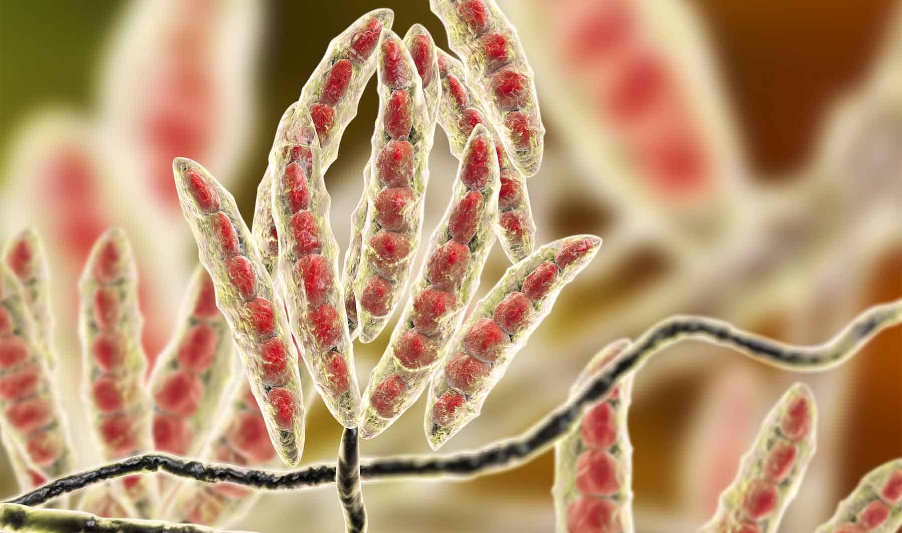
Infectious agents that potentially play a role in dysbacteriosis include mycotoxins, Eimeria spp., Clostridium perfringens, and other bacteria producing toxic metabolites.

Factors affecting gut health

The factors affecting broiler gut health can be summarized as follows:

  1. Feed and water quality: The form, type, and quality of feed provided to broilers can significantly impact their gut health. Consistent availability of cool and hygienic drinking water is crucial for optimum production performance.
  2. Stress: Stressful conditions, such as high environmental temperatures or poor ventilation, can lead to an imbalance in the gut microbiome and an increased risk of disease.
  3. Microbial exposure: Exposure to pathogens or other harmful bacteria can disrupt the gut microbiome and lead to gut health issues.
  4. Immune system: A robust immune system is important for maintaining gut health, as it helps to prevent the overgrowth of harmful bacteria and promote the growth of beneficial bacteria.
  5. Sanitation: Keeping the broiler environment clean and free of pathogens is crucial for maintaining gut health, as bacteria and other pathogens can easily spread and disrupt the gut microbiome.
  6. Management practices: Proper management practices, such as proper feeding and watering, and litter management can help to maintain gut health and prevent gut-related issues.

Chat GutFig. 2. Key factors affecting broilers’ gut health

Key approaches for managing gut health without antibiotics

Two key approaches for managing gut health in poultry without the use of antibiotics are outstandingly successful.

Proper nutrition and management practices

Ensuring the birds have access to clean water, high-quality feed, and a stress-free environment is crucial for ABF poultry production. A balanced diet in terms of protein, energy, and essential vitamins and minerals is essential for maintaining gut health.

The environment in which birds have kept plays a major role in maintaining gut health. Proper sanitation and ventilation, as well as the right temperature and humidity, are crucial to prevent the spread of disease and infection. There is no alternative to the strict implementation of stringent biosecurity measures to prevent the spread of disease.

Early detection and treatment of diseases can help to prevent them from becoming more serious problems affecting the profitability of ABF production. It is important to keep a close eye on birds for signs of disease, such as diarrhea, reduced water, and feed consumption.

Gut health-promoting feed additives

Another approach to maintaining gut health in antibiotics-free poultry production is using gut health-supporting feed additives. A variety of gut health-supporting feed additives including phytochemicals/essential oils, organic acids, probiotics, prebiotics, exogenous enzymes, etc. in combination or alone are used in animal production. Particularly, phytogenic feed additives (PFAs) have gained interest as cost-effective feed additives with already well-established effects on improving broiler chickens’ intestinal health.

Plant secondary metabolites and essential oils (generically called phytogenics, phytochemicals, or phytomolecules) are biologically active compounds that have recently garnered interest as feed additives in poultry production, due to their capacity to improve feed efficiency by enhancing the production of digestive secretions and nutrient absorption. This helps reduce the pathogenic load in the gut, exert antioxidant properties and decrease the microbial burden on the animal’s immune status (Abdelli et al. 2021).

Plant extracts – Essential oils (EOs) /Phytomolecules

Phytochemicals are naturally occurring compounds found in plants. Many phytomolecules have been found to have antimicrobial properties, meaning they can inhibit the growth or kill microorganisms such as bacteria, viruses, and fungi. Examples of phytomolecules with antimicrobial properties include compounds found in garlic, thyme, and tea tree oil. Essential oils (EOs) are raw plant extracts (flowers, leaves, roots, fruit, etc.) whereas phytomolecules are active ingredients of essential oils or other plant materials. A phytomolecule is clearly defined as one active compound. Essential oils (EOs) are important aromatic components of herbs and spices and are used as natural alternatives for replacing antibiotic growth promoters (AGPs) in poultry feed. The beneficial effects of EOs include appetite stimulation, improvement of enzyme secretion related to food digestion, and immune response activation (Krishan and Narang, 2014).

A wide variety of herbs and spices (thyme, oregano, cinnamon, rosemary, marjoram, yarrow, garlic, ginger, green tea, black cumin, and coriander, among others), as well as EOs (from thyme, oregano, cinnamon, garlic, anise, rosemary, citruses, clove, ginger), have been used in poultry, individually or mixed, for their potential application as AGP alternatives (Gadde et al., 2017).

Table DataFig. 3: Phytomolecule-based feed additive outperforms AGPs with improved broiler performance (42 Days field study)

One of the primary modes of action of EOs is related to their antimicrobial effects which allow for controlling potential pathogens (Mohammadi and Kim, 2018).

Phytomolecule blend  Clostridium perfringens Enterococcus caecorum Enterococcus hirae Escherichia coli Salmonella typhimurium  Staphylococcus aureus
Ventar D 1250 2500 5000 2500 5000 2500

Fig. 4: Effectivity of phytomolecule-based feed additive (Ventar D) against enteropathogenic bacteria (MIC value in PPM)

Phytomolecules have been shown to have anti-inflammatory properties. These compounds include flavonoids, polyphenols, carotenoids, and terpenes, among others. One of the ways in which phytomolecules exhibit anti-inflammatory effects is through their ability to inhibit the activity of pro-inflammatory enzymes and molecules. For example, polyphenols have been shown to inhibit the activity of nuclear factor-kappa B (NF-kB), a transcription factor that plays a key role in regulating inflammation.

Phytomolecules also have antioxidant properties, which can help to protect cells from damage caused by reactive oxygen species (ROS) and other reactive molecules that can contribute to inflammation. Plant extracts are also proposed to be used as antioxidants in animal feed, protecting animals from oxidative damage caused by free radicals. The presence of phenolic OH groups in thymol, carvacrol, and other plant extracts act as hydrogen donors to the peroxy radicals produced during the first step in lipid oxidation, thus retarding the hydroxyl peroxide formation (Farag et al., 1989, Djeridane et al., 2006). Thymol and carvacrol are reported to inhibit lipid peroxidation (Hashemipour et.al. 2013) and have strong antioxidant activity (Yanishlieva et al., 1999).

Overall, the anti-inflammatory effects of phytomolecules are thought to be due to a combination of their ability to inhibit the activity of pro-inflammatory enzymes and molecules, their antioxidant properties, and their ability to modulate the immune system. Plant extracts (i.e. carvacrol, cinnamaldehyde, eugenol. etc.) inhibit the production of pro-inflammatory cytokines and chemokines from endotoxin-stimulated immune cells and epithelial cells (Lang et al., 2004, Lee et al., 2005, Liu et al., 2020). It has been indicated that anti-inflammatory activities may be partially mediated by blocking the NF-κB activation pathway (Lee et al., 2005).

Table DataFig. 5: Anti-inflammatory effect of phytomolecule-based feed additive (Ventar D) – the reduced activity of inflammatory cytokines

Proper protection of EOs/Phytomolecules is key to optimum results

Several phytogenic compounds have also been shown to be largely absorbed in the upper GIT, meaning that without proper protection, the majority would not reach the lower gut where they would exert their major functions (Abdelli et al. 2021). The benefits of supplementing the broiler diet with a mixture of encapsulated EOs were higher than the tested PFA in powdered, non-protected form (Hafeez et al. 2016). Novel delivery technologies have been developed to protect PFAs from the degradation and oxidation process during feed processing and storage, ease the handling, allow a slower release, and target the lower GIT (Starčević et al. 2014). The specific protection techniques used during the commercial production of an EO/phytomolecule blend are crucial in delivering on all the objectives with remarkable consistency.

Table Data

Fig. 6: Pelleting stability of phytomolecule – based feed additive (Ventar D) at high temperature and longer conditioning time

Phytomolecule blend optimizes production performance

Removal of antibiotics in poultry production can be challenging for controlling mortality and maintaining the production performance of the birds. Phytogenic feed additives have been shown to improve production performance of chicken due to their antimicrobial, anti-inflammatory, antioxidant, and digestive properties. Possible mechanisms behind improved nutrient digestibility by phytogenic feed additives (PFAs) supplementation could be attributed to the ability of these feed additives to stimulate appetite, saliva secretion, intestinal mucus production, bile acid secretion, and activity of digestive enzymes such as trypsin and amylase as well as to positively affect the intestinal morphology (Oso et al. 2019). EOs are perceived as growth promoters in poultry diets, with strong antimicrobial and anticoccidial activities (Zahi et al., 2018). PFAs have positive effects on body weight gain and FCR in chickens (Khattak et al. 2014, Zhang et el. 2009).

Table Data
Fig. 7: Phytomolecule-based feed additive improved broiler FCR and mortality in field trial

Conclusion

In conclusion, managing gut health is a significant challenge in ABF broiler production that must be addressed to achieve optimal performance and welfare of the birds. The use of antibiotics as a preventative measure in broiler production has been widely used, but with the increasing demand for antibiotic-free products, alternative methods to maintain gut health must be implemented. These include using gut health-supporting feed additives, and proper management practices such as implementing biosecurity measures, maintaining optimal environmental conditions, providing adequate space and ventilation, and reducing stress. However, it is essential to note that there is no one-size-fits-all solution for gut health management in ABF broiler production. It is important to continuously monitor and assess their flock’s gut health and make adjustments as necessary. Additionally, research and development in this field should be encouraged to identify new and innovative ways to maintain gut health in ABF broiler production.

Overall, managing gut health is a critical challenge that requires a multi-faceted approach and ongoing monitoring and management. By implementing the appropriate strategies and utilizing new technologies, poultry operators can ensure the health and well-being of their flocks while meeting the growing demand for antibiotic-free products sustainably.

References:

Abdelli N, Solà-Oriol D, Pérez JF. Phytogenic Feed Additives in Poultry: Achievements, Prospective and Challenges. Animals (Basel). 2021 Dec 6;11(12):3471.

Bailey R. A. 2010. Intestinal microbiota and the pathogenesis of dysbacteriosis in broiler chickens. PhD thesis submitted to the University of East Anglia. Institute of Food Research, United Kingdom

Choct M. Managing gut health through nutrition. British Poultry Science Volume 50, Number 1 (January 2009), pp. 9—15.

De Gussem M, “Coccidiosis in Poultry: Review on Diagnosis, Control, Prevention and Interaction with Overall Gut Health,” Proceedings of the 16th European Symposium on Poultry Nutrition, Strasbourg, 26-30 August, 2007, pp. 253-261.H.J. Dorman, S.G. Deans. Antimicrobial agents from plants: antibacterial activity of plant volatile oils. J Appl Microbiol, 88 (2000), pp. 308-316

Djeridane A., M. Yousfi M, Nadjemi B, Boutassouna D., Stocker P., Vidal N. Antioxidant activity of some Algerian medicinal plants extracts containing phenolic compounds. Food Chem, 97 (2006), pp. 654-660

Farag R. S., Daw Z.Y., Hewedi F.M., El-Baroty G.S.A. Antimicrobial activity of some Egyptian spice essential oils. J Food Prot, 52 (1989), pp. 665-667

Gadde U., Kim W.H., Oh S.T., Lillehoj H.S. Alternatives to antibiotics for maximizing growth performance and feed efficiency in poultry: A review. Anim. Health Res. Rev. 2017;18:26–45.

Guo, F.C., Kwakkel, R.P., Williams, B.A., Li, W.K., Li, H.S., Luo, J.Y., Li, X.P., Wei, Y.X., Yan, Z.T. and Verstegen, M.W.A., 2004. Effects of mushroom and herb polysaccharides, as alternatives for an antibiotic, on growth performance of broilers. British Poultry Science45(5), pp.684-694.

Hafeez A., Männer K., Schieder C., Zentek J. Effect of supplementation of phytogenic feed additives (powdered vs. encapsulated) on performance and nutrient digestibility in broiler chickens. Poult. Sci. 2016;95:622–629.

Hammer K.A., Carson C.F., Riley T.V. Antimicrobial activity of essential oils and other plant extracts. J Appl Microbiol, 86 (1999), pp. 985-990

Hashemipour H, Kermanshahi H, Golian A, Veldkamp T. Effect of thymol and carvacrol feed supplementation on performance, antioxidant enzyme activities, fatty acid composition, digestive enzyme activities, and immune response in broiler chickens. Poultry Science. Volume 92. Issue 8. 2013, Pp 2059-2069,

Khattak F., Ronchi A., Castelli P., Sparks N. Effects of natural blend of essential oil on growth performance, blood biochemistry, cecal morphology, and carcass quality of broiler chickens. Poult. Sci. 2014;93:132–137

Kraehenbuhl, J.P. & Neutra, M.R. (1992) Molecular and cellular basis of immune protection of mucosal surfaces. Physiology Reviews, 72: 853–879.Krishan and Narang J. Adv. Vet. Anim. Res., 1(4): 156-162, December 2014

Lang A., Lahav M., Sakhnini E, Barshack I., Fidder H. H., Avidan B. Allicin inhibits spontaneous and TNF-alpha induced secretion of proinflammatory cytokines and chemokines from intestinal epithelial cells. Clin Nutr, 23 (2004), pp. 1199-1208

Lee S.H., Lee S.Y., Son D.J., Lee H., Yoo H.S., Song S. Inhibitory effect of 2′-hydroxycinnamaldehyde on nitric oxide production through inhibition of NF-kappa B activation in RAW 264.7 cells Biochem Pharmacol, 69 (2005), pp. 791-799

Liu, S., Song, M., Yun, W., Lee, J., Kim, H. and Cho, J., 2020. Effect of carvacrol essential oils on growth performance and intestinal barrier function in broilers with lipopolysaccharide challenge. Animal Production Science, 60(4), pp.545-552.

Mitsch, P., Zitterl-Eglseer, K., Köhler, B., Gabler, C., Losa, R. and Zimpernik, I., 2004. The effect of two different blends of essential oil components on the proliferation of Clostridium perfringens in the intestines of broiler chickens. Poultry science, 83(4), pp.669-675.

Mohammadi Gheisar M., Kim I.H. Phytobiotics in poultry and swine nutrition—A review. Ital. J. Anim. Sci. 2018;17:92–99.

Oso A.O., Suganthi R.U., Reddy G.B.M., Malik P.K., Thirumalaisamy G., Awachat V.B., Selvaraju S., Arangasamy A., Bhatta R. Effect of dietary supplementation with phytogenic blend on growth performance, apparent ileal digestibility of nutrients, intestinal morphology, and cecal microflora of broiler chickens. Poult. Sci. 2019;98:4755–4766

Oviedo-Rondón, Edgar O., et al. “Ileal and caecal microbial populations in broilers given specific essential oil blends and probiotics in two consecutive grow-outs.” Avian Biology Research 3.4 (2010): 157-169.

Petersen C. and June L. Round. Defining dysbiosis and its influence on host immunity and disease. Cellular Microbiology (2014)16(7), 1024–1033

Starčević K., Krstulović L., Brozić D., Maurić M., Stojević Z., Mikulec Ž., Bajić M., Mašek T. Production performance, meat composition and oxidative susceptibility in broiler chicken fed with different phenolic compounds. J. Sci. Food Agric. 2014;95:1172–1178.

Yanishlieva, N.V., Marinova, E.M., Gordon, M.H. and Raneva, V.G., 1999. Antioxidant activity and mechanism of action of thymol and carvacrol in two lipid systems. Food Chemistry64(1), pp.59-66.

Yegani, M. and Korver, D.R., 2008. Factors affecting intestinal health in poultry. Poultry science, 87(10), pp.2052-2063.

Zhai, H., H. Liu, Shikui Wang, Jinlong Wu and Anna-Maria Kluenter. “Potential of essential oils for poultry and pigs.” Animal Nutrition 4 (2018): 179 – 186.

Zhang G.F., Yang Z.B., Wang Y., Yang W.R., Jiang S.Z., Gai G.S. Effects of ginger root (Zingiber officinale) processed to different particle sizes on growth performance, antioxidant status, and serum metabolites of broiler chickens. Poult. Sci. 2009;88:2159–2166.


Cryptosporidia in calves – chickens can help

Header Calf Standing Fotolia L

By Lea Poppe, Regional Technical Manager, EW Nutrition

Diarrhea due to infestation with cryptosporidia is one of the most pressing problems in calf rearing. These protozoa, along with rotaviruses, are now considered the most common pathogens in infectious calf diarrhea. Due to their high resistance and thus limited possible control and prevention measures, they have now overtaken other pathogens such as coronaviruses, salmonellae, and E. coli.

Cryptosporidia show complex development

Cryptosporidia are single-celled intestinal parasites. In calves, Cryptosporidium parvum and Cryptosporidium bovis are most commonly found. C. bovis is normally considered nonpathogenic. Accordingly, the disease known as cryptosporidiosis is caused by C. parvum. The rapid tests for determining the diarrheal pathogens, which are increasingly widespread, are usually unsuitable for distinguishing between the individual strains, which can lead to false positive results.

Resistant in the environment, active in the animal

In the environment, cryptosporidia are distributed as oocysts. The oocysts are only about 5 µm in size and have a very resistant shell. They can remain infectious for up to 6 months in high humidity and moderate temperatures. Drought and extreme temperatures (below -18°C and above 65°C) cause the oocysts to die.

After oral ingestion, the oocysts are reactivated by conditions in the gastrointestinal tract (low pH and body temperature): As sporozoites, the parasites attach to the posterior small intestine, causing diarrhea symptomatology. There, they surround themselves with a special protective membrane, and the complex life cycle continues. Only a few days after infection, reproductive forms are detectable in the calf’s intestine, and excretion of infectious oocysts in the feces begins.

Header Calf En
Figure 1 (Olias et al., 2018): Life cycle of cryptosporidia: ingested oocysts release four sporozoites that invade host enterocytes (intestinal epithelial cells). There, they develop into trophozoites before asexual and sexual reproduction ensues, and thin- and thick-walled oocysts are formed. Thick-walled oocysts are excreted through the intestine. Thin-walled oocysts may break apart, and the sporozoites may infect other enterocytes, resulting in relapse or prolonged diarrhea. Infestation of the cells leads to their destruction, resulting in villi atrophy or fusion.

Oocysts bring the disease to the animal

Cryptosporidiosis is transmitted either by direct contact of calves with feces from infected animals or indirectly by ingesting contaminated feed, bedding, or water. Each gram of feces excreted by calves showing symptoms may contain up to 100 million oocysts. According to experimental studies, as few as 17 orally ingested oocysts are sufficient to trigger infection. In addition, some multiplication forms can infect other intestinal cells directly within the intestine and thus further advance the disease by autoinfection.

Cryptosporidiosis caused by cryptosporidia often presents with typical diarrhea symptoms and occurs primarily in calves up to 3 weeks of age. Older calves may also be infected with cryptosporidia but usually show no symptoms. Pathogen excretion and, thus, the spread of disease within the herd is nevertheless likely due to the minimal infectious dose.

Damage to the intestinal wall leads to retardation of growth

Attachment of cryptosporidia to the intestinal wall is associated with an inflammatory reaction, regression and fusion of the intestinal villi, and damage to the microvilli. As a result, nutrient absorption in the small intestine is impaired, and more undigested nutrients enter the colon. The microflora starts a fermentation process with lactose and starch, leading to increased lactate levels in the blood and, thus, hyperacidity in the calf. Faintness, unwillingness to drink, recumbency, and growth disorders are the consequences.

Diarrhea often occurs late or not at all and, accordingly, is not considered the main symptom of cryptosporidiosis. When diarrhea occurs, it lasts about 1-2 weeks. The feces are typically watery, greenish-yellow, and are often described as foul-smelling. Due to diarrhea, there is a loss of electrolytes and dehydration.

Studies show: Cryptosporidia are the most prevalent diarrheal pathogens

Several studies in different regions, which examined calf diarrhea and its triggers in more detail, came to a similar conclusion: Cryptosporidia are one of the most common causes of calf diarrhea. In addition, mixed infections often occur.


Country or region Number Age/Health status % Crypto-sporidia % Rota viruses Combined infections with crypto-sporidia Others (%) Source
Switzerland 2 – 21 DL

Ill and healthy

43 46 1 case of E. coli Luginbühl et al., 2012
Switzerland 63 1 – 4 DL

Ill and healthy

————–

7 – 20 DL

————–

26 – 49 DL

34.4

—————

54.0

—————

33.3

3.1

—————-

28.6

—————-

13.3

2 EP – 1.6

4 EP – 3.2

—————-

2 EP – 19

3 EP – 3.2

4 EP – 0

—————-

2 EP – 30

3 EP – 11.7

4 EP – 6.7

Corona 4.7

E. coli 4.7

Giardia 1.6

—————

Corona 0

E. coli 3.2

Giardia 6.3

—————

Corona 0

E. coli 15

Giardia 35


Weber et al., 2016


Weber et al., 2016 EN

Switzerland 147 Up to 3rd WL;

Diarrhea

55 58.7 5.5 % Rota

7.8 % BCV

Lanz Uhde et al., 2014
Sweden 782 1 – 7 DL

Diarrhea

25.3 Detected with Giardia, E. coli, Rota, Eimeria Silverlås et al., 2012
USA (East coast) 503 Pre-weaning 50.3 Santin et al., 2004
USA 30 2 weeks old

1-8 weeks old

3-12 months

12-24 months

96.7

45.8

18.5

2.2

Santin et al., 2008
Germany 521 32 9 Losand et al., 2021
Ethiopia 360 18.6 Ayele et al., 2018
Argentina 1073 n.m. / Ill and healthy 25.5 Lombardelli et al., 2019
UK n.m. Ill ?? 37 25 20 Coccidia 8

E. coli 4

Corona 3

Co infections not including Crypto-sporidia 3

APHA, SRUC, Veterinary investigation diagnosis analysis (VIDA) report (2014)

DL = days of life WL = weeks of life n.m. = not mentioned  EP = enteropathogen

Cryptosporidia reduces profit

Infection with cryptosporidia and sometimes subsequent diarrhea entails treatment of the animals and generates costs (veterinarian, medication, electrolyte drinks). In addition, poorer feed conversion, lower growth, and animal losses result in lower production efficiency.

A Scottish study shows 34 kg less gain in the first six months of life compared to healthy calves in beef calves that experienced severe cryptosporidiosis in the first three weeks of life. Similar results are described in lambs, also a susceptible species to cryptosporidia. These studies suggest a long-term negative effect of cryptosporidia on growth performance and production efficiency.

Here’s how you can support your calves against cryptosporidia

High resistance of the pathogens to environmental influences, a very low necessary infection dose combined with an elevated excretion of infectious oocysts, and the possibility of autoinfection make cryptosporidia tough opponents. This is also reflected in their worldwide distribution.

What is the treatment?

Suitable drugs for the treatment of cryptosporidiosis are currently unavailable on the market. The only medicine that can be used in case of cryptosporidiosis infestation may only be administered to calves that have had diarrhea symptoms for 24 hours or less. Accordingly, this agent is usually used only for prevention. Scientific studies on its effectiveness are contradictory; some suggest that it merely delays the onset of the disease. In addition, it is not always easy to use due to the exact dosage that must be followed. Doubling the dose (sometimes happening already due to incorrectly observed intervals between doses) can lead to a toxic overdose.

Accordingly, only the symptoms of the disease – diarrhea with its accompanying symptoms – can be treated. Electrolyte and water losses must be continuously compensated with the help of a high-quality electrolyte drink. The buffer substances contained also reduce the hyperacidity of the blood caused by faulty fermentation in the intestines. For successful treatment, the electrolyte drink should be given in addition to the milk drink. Under no circumstances should the feeding of milk or milk replacer be discontinued because the sick calf urgently needs energy and nutrients. Opinions to the contrary are outdated.

As always: prevention is better than treatment

To make it more difficult for cryptosporidiosis to spread from the outset, it is worth looking at the risk factors. These include direct contact with other calves and general herd size. Furthermore, organic farms seem to have more problems with cryptosporidia. Weather also influences calves born during warmer and, at the same time, wetter weather periods (temperature-humidity index) often get sick.

Due to the limited possibilities for treatment, prevention is of greater importance. For other diarrheal pathogens such as rotavirus, coronavirus, and E. coli, it has become established practice to vaccinate dams to achieve better passive immunization of the calf. However, commercial vaccination against cryptosporidia is not currently available, making dam vaccination as unavailable as calf vaccination.

Accordingly, optimal colostrum management is the first way to protect the calf from cryptosporidia infection. This also confirms the general discussion on the Failure of Passive Transfer: various studies suggest that calves with poor immunoglobulin supply suffer from diarrhea more frequently than calves with good supply, although a concrete link to cryptosporidia itself cannot always be established with certainty.

Furthermore, it is essential to break the chain of infection within farms. In addition to the separate housing of the calves, it is necessary to ensure consistent hygiene. One should take advantage of the pathogen’s weakness as well as its sensitivity to high temperatures and ensure that the water temperature is sufficiently high when cleaning the calf pens and calving area. When disinfecting afterward, it is crucial to consider the spectrum of activity of the agent used, as not all are effective against cryptosporidia.

Egg immunoglobulins support animals against cryptosporidia

Egg immunoglobulins were initially designed to help chicks get started. In this process, hens form antibodies against pathogens they are confronted with. As studies have shown, this also works with cryptosporidia. Cama and Sterling (1991) tested their produced antibodies in the neonatal mouse model and achieved a significant (P≤0.001) reduction in parasites there. Kobayashi et al. (2004) registered decreased binding of sporozoites to the intestinal cell model and their decreased viability in addition to oocyst reduction.

In the IRIG Research Institute (2009, unpublished), feeding egg powder with immunoglobulins against cryptosporidia (10 g/day) to 15 calves reduced oocyst excretion. Before administration, calves excreted an average of 106.42 oocysts/g of feces. After administration of egg powder, only two calves still showed 103.21 oocysts/g feces, and the other 13 of the 15 calves showed no oocyst excretion. All these results are confirmed by positive customer feedback on IgY-based feed supplements.

Egg immunoglobulins and optimal colostrum management as a key solution

Since there are no effective drugs against cryptosporidia, animals must be prophylactically protected against this disease as much as possible. In addition to optimal colostrum management, which means feeding high-quality colostrum (IgG≥50g/L) to the calf as soon as possible after birth, we have products with egg immunoglobulins available to support the calf as a prophylactic against cryptosporidia infestation and thus prevent significant performance losses, especially during rearing.

References

Brainard, J., Hooper, L., McFariane, S., Hammer, C. C., Hunter, P. R., & Tyler, K. (2020). Systemic review of modifiable risk factors shows little evidential support for most current practices in Cryptosporidium management in bovine calves. Parasitology research 119, 3572-3584.

Cama, V. A., and C. R. Sterling. “Hyperimmune Hens as a Novel Source of Anti-Cryptosporidium Antibodies Suitable for Passive Immune Transfer.” University of Arizona. Wiley-Blackwell, January 1, 1991. https://experts.arizona.edu/en/publications/hyperimmune-hens-as-a-novel-source-of-anti-cryptosporidium-antibo.

Kobayashi, C, H Yokoyama, S Nguyen, Y Kodama, T Kimata, and M Izeki. “Effect of Egg Yolk Antibody on Experimental Infection in Mice.” Vaccine 23, no. 2 (2004): 232–35. https://doi.org/10.1016/j.vaccine.2004.05.034.

Lamp, D. O. (25. Januar 2020). Rinder aktuell: Kälberdurchfall durch Kryptosporidien – Hartnäckig und weitverbreitet. BAUERNBLATT, S. 52-53.

Losand, B., Falkenberg, U., Krömker, V., Konow, M., & Flor, J. (2. März 2021). Kälberaufzucht in MV – Alles im grünen Bereich? 30. Milchrindtag Mecklemburg-Vorpommern.

Luginbühl, A., K. Reitt, A. Metzler, M. Kollbrunner, L. Corboz, and P. Deplazes. “Feldstudie Zu Prävalenz Und Diagnostik Von Durchfallerregern Beim Neonaten Kalb Im Einzugsgebiet Einer Schweizerischen Nutztierpraxis.” Schweizer Archiv für Tierheilkunde 147, no. 6 (2005): 245–52. https://doi.org/10.1024/0036-7281.147.6.245.

Olias, P., Dettwiler, I., Hemphill, A., Deplazes, P., Steiner, A., & Meylan, M. (2018). Die Bedeutung der Cryptosporidiose für die Kälbergesundheit in der Schweiz. Schweiz Arch Tierheilkd, Band 160, Heft 6, Juni 2018, 363-374.

Santín, M., Trout, J. M., Xiao, L., Zhou, L., Greiner, E., & Fayer, R. (2004). Prevalence and age-related variation of Cryptosporidium species and genotypes in dairy calves. Veterinary Parasitology 122, 103-117.

Shaw, H. J., Innes, E. A., Marrison, L. J., Katzer, F., & Wells, B. (2020). Long-term production effects of clinical cryptosporidiosis in neonatal calves. International Journal for Parasitology 50, 371-376.

Silverlås, C., H. Bosaeus-Reineck, K. Näslund, and C. Björkman. “Is There a Need for Improved Cryptosporidium Diagnostics in Swedish Calves?” International Journal for Parasitology 43, no. 2 (2013): 155–61. https://doi.org/10.1016/j.ijpara.2012.10.009.

Thomson, Sarah, Carly A. Hamilton, Jayne C. Hope, Frank Katzer, Neil A. Mabbott, Liam J. Morrison, and Elisabeth A. Innes. “Bovine Cryptosporidiosis: Impact, Host-Parasite Interaction, and Control Strategies.” Veterinary Research 48, no. 1 (2017). https://doi.org/10.1186/s13567-017-0447-0.

Uhde, F., Kaufmann, T., Sager, H., Albini, S., Zanoni, R., & Schelling, E. (2008). Prevalence of four enteropathogens in the feces of young diarrhoeic dairy calves in Switzerland. Veterinary Record (163), 362-366.


Xylanase solutions for broiler feed: Enzyme innovation finally hits the market

Group of broiler chicks

By Dr. Ajay Awati, Global Category Manager for Gut Health and Nutrition, EW Nutrition, and Dr. Howard Simmins, InSci Associates

After 30 years of stagnating solutions, in-feed xylanase innovation has finally arrived – with a complete focus on the needs of the broiler feed industry.

Group of broilers

It has been over 30 years since xylanase was first introduced in broiler diets in Europe. In the meantime, it has been widely used worldwide with few, if any, major improvements. While the animal feed industry evolved in terms of production landscape, feed processing technologies and use of various by-products, xylanase enzyme technology did not keep pace. In fact, it did not evolve to meet customers’ changing needs and provide that much-needed flexibility of diet formulation for a commercial nutritionist. The wait is over: new in-feed xylanase technology is about to revolutionize broiler nutrition.

Why we need innovative xylanase enzymes for broiler production

Initially, in the 1980s, xylanase was leveraged from industries unrelated to animal production into the feed business. Gut viscosity had been a continuing problem in broiler chickens fed wheat-based diets. It led to an increased risk of enteric disease, generally reducing performance. Xylanase was shown to reduce gut viscosity in wheat-based feed by breaking down soluble arabinoxylans.

As a result, the birds grew as well as if they were fed a low-viscosity corn/soya diet. An additional benefit was lower disease risks from the reduced level of anti-nutritional factors (ANFs) and the multiple negative effects of viscosity in the intestine.

In addition to reducing viscosity, xylanase augments the release in the small intestine of nutrients from previously undigested feedstuffs. The outcome has been the use of an energy matrix value for xylanase, which essentially helps diets through least-cost formulation.

These effects account for the growth of xylanase use in the monogastric feed market. Today, the penetration is above 50%.

Limitations of existing xylanase solutions

Leveraging xylanases from other industries for viscosity reduction in poultry comes with a couple of distinct limitations:

  1. Most broiler diets around the globe are based on a corn-soybean formulation, which contains far higher levels of insoluble arabinoxylans than soluble arabinoxylans. In such cases, viscosity is a relatively minor issue compared to the anti-nutritional effect of insoluble arabinoxylans.
  2. The reduction of gut viscosity is less relevant in other poultry sectors, such as laying hens and turkeys.

Commercial xylanases would be required to break down insoluble NSPs in which substrate activity may be limited and difficult to predict. Fiber constituents of different cereal grains used in feed are highly variable. By- and co-products derived from cereals contain even more complex fiber components, altered further by the manner of processing that the raw material has undergone.

Additionally, poultry response is highly variable:  For an individual bird, the effectiveness of xylanase depends on the enzyme’s interaction with feed in the gastrointestinal tract (GIT) of the animal, which varies depending on the species and the animal’s age. This may explain why xylanase penetration on the feed market is not as high as that of phytase.

GH10: the next-level xylanase for feed application

A xylanase for feed is required to provide multiple functionalities, of which four are essential:

  1. Capacity to break down soluble and insoluble arabinoxylan across a range of typical feedstuffs
  2. Rapid activity at optimal pH in the preferred section of the GIT
  3. No inhibition in the presence of xylanase inhibitors
  4. Comprehensive feed processing thermostability

The GH11 family of xylanases commonly used in feed does not offer these aggregated benefits. They successfully reduce soluble NSPs in wheat-based diets, hence lowering the viscosity level in the broiler GIT. However, they are less effective in the presence of insoluble NSPs in which the arabinoxylan backbone is more complex.

Why GH10 instead of GH11?

The explanation for this can be found in the 3-dimensional structure of the GH11 xylanase. The activity of GH11 xylanases requires 3 or 4 consecutive unsubstituted xylan monomers on the backbone to find an active site. That is why they are hindered by the presence of branches, or side chains, on arabinose backbones. Consequently, they are highly specific, favoring the particularly low-branching wheat backbone.

Xylanases from the GH10 family are entirely different. Although well known, they have not been used in feed yet. The GH10 xylanases require two or fewer consecutive unsubstituted xylan monomers on the backbone to find an active site. Therefore, they can act on xylose residues near branches. This results in both more and shorter xylo-oligomers than found with GH11 xylanases. In simple terms, the GH10 xylanases have a less deep cleft than the GH11 xylanases, providing greater catalytic versatility (Pollet 2010).

Significantly, this potentially allows a broader range of feedstuffs to be incorporated into the complete diet, including co- and by-products, while maintaining performance. Therefore, with GH10, higher levels of cheaper ingredients may be included, with a significant value proposition of further reducing feed costs.

Axxess Xy Is Effective Against Soluble And Insoluble Arabinoxylans

GH10 xylanases generate a range of important prebiotics

As early as 1995, it was proposed that xylanase may affect microbial activity in the gastrointestinal tract through the provision of fermentable oligosaccharides and low molecular weight polysaccharides. These are produced from the hydrolysis of soluble and insoluble arabinoxylans in cereals.

A development of particular interest is that the GH10 xylanases break down the backbone of different fibre components into small xylooligosaccharides (XOS) and arabino-xylanoligosaccharides (AXOS). This action, research shows, has value in supporting the selective growth of fibre-degrading bacteria in the large intestine, conferring positive effects on the host’s health.

The most well-known probiotic strains belong to bifidobacteria and lactobacilli, which have quite different XOS and AXOS utilization systems. Bifidobacterium adolescentis has been shown to consume AXOS and XOS, whereas Lactobacillus brevis utilises only XOS. The outcome is that AXOS releases butyrate, the short-chain fatty acid, which can improve the host’s gut barrier function, as well as reduce Salmonella colonization in broilers. Alongside these health benefits, their presence may improve performance also by reducing FCR. (Courtin et al. 2008; Ribeiro et al. 2018)

As mentioned earlier, the GH10 xylanase requires only two consecutive unsubstituted xylan monomers to cleave the xylan main chain, whereas a GH11 xylanase requires 3 or 4 consecutive unsubstituted xylan monomers. Therefore, the number of potential AXOS and XOS oligomers is higher from the action of the GH10 xylanase. This results in a wider size range of oligomers. The range is valuable as the effect is spread across the large intestine, each oligomer having a different fermentation rate. Consequently, the large intestine’s microbial activity becomes saccharolytic, which potentially reduces the undesirable products of proteolytic degradation, such as phenols and cresols.

Prebiotic combinations will vary depending on the substrate available. However, there is more flexibility in breaking down insoluble NSPs across different feedstuffs using GH10 xylanase compared to GH-11 xylanase.Data showing Axxess XY Efficacy In Both Corn Soy And Wheat Soy Diets

The future of xylanase: Reducing feed costs through flexible formulation

EW Nutrition’s GH10-based AXXESS XY xylanase, specifically developed for animal feed, has a wide-ranging activity across typical substrates, both in corn-soy and wheat-soy diets. It also allows for a greater proportion of cheaper ingredients, enabling increased flexibility in feedstuff choices and resulting in more stable feed pricing. The activity of the GH10 xylanase in producing oligomers from the breakdown of the arabinoxylan backbone also indicates that it can produce a greater number and diversity of valuable prebiotics that sustain the growth of fiber-degrading microbiota. Consequently, the metabolism of the large intestine is shifted from proteolytic to saccharolytic, which supports the animal’s general health.

The combination of these benefits from using this xylanase results in a bird with a balanced digestive system that is more robust in the face of environmental and health challenges, supporting better performance. Furthermore, this novel enzyme solution gives nutritionists a reliable tool to reduce feed costs by being flexible in diet formulation and opportunistic in using raw materials while maintaining consistency in animal performance. Especially in these times of supply problems and raw material price hikes, such advantages are invaluable.

The naturally thermostable AXXESS XY 1000G is the most advanced xylanase yet. It is a GH10 xylanase that delivers what the industry has been asking for: a fiber-degrading enzyme suited for all poultry feed.


References

Courtin, Christophe M, Katrien Swennen, Willem F Broekaert, Quirine Swennen, Johan Buyse, Eddy Decuypere, Christiaan W Michiels, Bart De Ketelaere, and Jan A Delcour. “Effects of Dietary Inclusion of Xylooligo- Saccharides, Arabinoxylooligosaccha- Rides and Soluble Arabinoxylan on the Microbial Composition of Caecal Contents of Chickens.” Journal of the Science of Food and Agriculture 88, no. 14 (2008): 2517–22. https://doi.org/10.1002/jsfa.3373.

Ribeiro, T., V. Cardoso, L.M.A. Ferreira, M.M.S. Lordelo, E. Coelho, A.S.P. Moreira, M.R.M. Domingues, M.A. Coimbra, M.R. Bedford, and C M Fontes. “Xylo-Oligosaccharides Display a Prebiotic Activity When Used to Supplement Wheat or Corn-Based Diets for Broilers.” Poultry Science 97, no. 12 (2018): 4330–41. https://doi.org/10.3382/ps/pey336.

Pollet, Annick. “Functional and Structural Analysis of Glycoside Hydrolase Family 8, 10 and 11 Xylanases with Focus on Bacillus Subtilis Xylanase A,” 2010. https://www.biw.kuleuven.be/m2s/clmt/lmcb/publications/docs/apollet


The risk of using by-products as feed alternatives – and the solutions

feed milling byproduct

By Marisabel Caballero, Global Technical Manager Poultry, and Dr. Inge Heinzl, Editor, EW Nutrition

Consistently rising feed prices compel feed producers to resort to alternative feed ingredients. By-products of milling and ethanol distillation would be good options. The following article shows what should be paid attention to when using these feeds.

Factory for high-quality animal protein

Keeping high-quality animal protein affordable requires cost-efficient alternatives

For a high percentage of consumers, the price of food products is one of the most decisive purchase factors; however, quality and sustainable use of resources are also of high importance. So, to comply with market requirements, meat producers must find cost-efficient and sustainable sources of feed ingredients. Feed prices already increased during the COVID-19 pandemic. Shortage of workforce and high shipping costs led to discontinuity in the supply chain, long delivery times, and increased costs for certain raw materials. Due to the Ukrainian crisis, there is no improvement to be seen. Alternatives must be considered more vigorously to compensate for this limited feed supply.

Grain by-products are an option

The use of grain by-products occurring at milling or ethanol production can cover a part of animal nutritional demands. Additionally, it contributes to sustainable usage of the available sources, as the remains of the production of human consumables are put back into the food chain.

However, increasing levels of by-products in the feed also have their sticking points. The raw materials grains or corn are often contaminated with mycotoxins, impacting the quality of this kind of feed.

Milling processes reduce mycotoxins in food

Before the whole process of milling, the grains are sorted and cleaned. Kernels with extensive mold growth, broken kernels, fine materials, and dust are removed.

When it comes to reducing mycotoxins by sorting and cleaning, the results vary a lot. They are influenced by several factors, including the initial condition of the grains, the type and level of contamination, and the type and efficiency of the cleaning process (Pinotti et al., 2016). The cleaning process has been shown to remove from 5 to 80 % of DON and NIV, 5 to 40 % of ZON (Schaarschmidt & Fauhl-Hassek, 2018), and 50 to 60 % of T2/HT2 contamination in wheat (Pascale et al., 2011). Debranning, the mechanical process by which the outer layers of wheat grains are removed, further reduces mycotoxin content in wheat grain from 15 to 80% of the initial contamination (Aureli et al., 2007; Rios et al., 2009). However, neither the cleaning and debranning nor the milling process include a step that destroys mycotoxins.

In white flour for human consumption, mycotoxin levels typically range from 50 to 70% of the wheat grain (Cheli et al., 2013).

The milling of maize shows a reduction factor of about 4 for aflatoxins and about 10 for zearalenone from the grain to the final human products. Contrarily, concentration triplicates for both aflatoxins and zearalenone in the case of the by-products such as germs, bran, and animal flour.

Milling processes concentrate mycotoxins in animal-feed fractions

The milling and pre-milling processes reduce the content of mycotoxins in products for human consumption, but what about the parts removed and normally used in animal feeds? Several studies (Tibola et al., 2015; Hoffmans et al., 2022) indicate that the concentration of mycotoxins is higher in the wheat fractions intended for animal feeds such as bran, flour shorts screenings, and middlings. However, their level in feedstuffs is variable and affected by several factors such as the type of mycotoxins, the level and extent of fungal contamination, and the complexity of the cereal processing technology.

Compared to the concentration in wheat grain, these concentrations in by-products may be up to 800 % but more typically range from 150 % to 340 % (Cheli et al., 2013). EW Nutrition’s worldwide mycotoxin survey shows a similar trend (Figures 1 and 2), in which DON levels are nine times higher in wheat midds than wheat grains, and fumonisin is eight times higher in wheat bran.

Mycotoxins levels in grain and by-products bar graphs EW Nutrition's worldwide mycotoxin survey bar graphs

Figure 1 + 2: Mycotoxins levels in grain and by-products

Highest concentrations in germ and bran fraction

After corn milling, animal feed fractions such as germ and bran have a low yield ranging from 5 to 7 % and are mostly composed of the outer parts of the kernels; as a consequence, an important concentration of mycotoxins occurs in these fractions (Schollenberger et al., 2008). When taking corn grains as the base, the contamination of aflatoxins goes up to three times in corn germ and up to nine times in bran (Brera et al., 2006; Pietri et al., 2009). For fumonisins, a double concentration can be expected (Brera et al., 2004), and for zearalenone, up to four times (Brera et al., 2006). Recently, Park and collaborators (2018) evaluated the distribution of 12 mycotoxins during wet milling of corn and found higher concentrations in corn gluten feed and corn bran.

Milling is a crucial step in the post-production of rice, in which the husk and the bran layers are removed. Rice bran is a common ingredient for animal feeds, in which aflatoxin is a common contaminant. It is believed that most of the aflatoxin contamination in rice bran occurs due to non-optimal storage conditions (Takahashi et al., 1989); however, a concentration of the toxin during milling of stored paddy rice also occurs, and the levels can triplicate compared with the grains (Trucksess et al., 2011).

The concentration of mycotoxins in DDGS during the ethanol production

Fresh MaizeDestillers’ dried grains with solubles (DDGS), a by-product of ethanol production, is a valuable feed ingredient, particularly as a source of protein for ruminants and monogastric animals at a competitive price.

Also here, mycotoxin contamination raises concerns with regard to their use in animal feeds. Mycotoxins are not destroyed during the ethanol fermentation process or during the production of DDGS. Moreover, a concentration of DON, ZEA, and fumonisin from corn to DDGS of 2–3.5 times has been reported for industrial ethanol production (Bennett et al., 1981; Schaafsma et al., 2009; Bowers & Munkvold, 2014).

In summary, studies on the fate of mycotoxins during food processing have shown that mycotoxins are concentrated in the fractions commonly used as animal feed. Moreover, high variability in mycotoxin contamination of cereal by-products has been evidenced, representing barriers to an increased acceptance of several food by-products as feed ingredients.

Feed formulation: Consider the mycotoxin contamination in by-products

Higher inclusions of cereals have an impact on their safe use in feeds. To evaluate this impact, we can simulate two different scenarios with different inclusions of by-products:

Table 1: Different levels of by-products’ inclusion rates

*Risk Tool (masterrisktool.com)

In the first lower inclusion scenario, the risk for broilers in the starting phase considers the low inclusion of raw materials; the losses related to the contamination (without management) are mild. When increasing the levels of by-products, the risk category also increases. The losses are more important for the operation, ranging from gut barrier alterations with impaired production parameters to alterations in the immune response and increased susceptibility to disease.

Mycotoxins in by-products effective toxin risk management can help!

Given the pros of including cereal by-products in animal feeds, such as their saving potential and their link with sustainability of resources, their utilization is advisable; however, understanding how mycotoxin distribution and concentration change during grain processing is critical. Today’s knowledge is limited to a few mycotoxins in cereal milling.

Therefore, when considering using these by-products in the animal feed, we must bear in mind that:

  • modified mycotoxins and mycotoxin co-contamination can be present, contributing to additive/synergistic effects on animal health.
  • toxin risk management strategies, including analysis, risk evaluation, and risk mitigation must be pursued to prevent those undesired effects.

References:

Aureli, G., and M.G. D’Egidio. “Efficacy of Debranning on Lowering of Deoxynivalenol (DON) Level in Manufacturing Processes of Durum Wheat.” Tecnica Molit. 58 (2007): 729–33.

Bennett, G. A., A. A. Lagoda, O. L. Shotwell, and C. W. Hesseltine. “Utilization of Zearalenone- Contaminated Corn for Ethanol Production.” Journal of the American Oil Chemists’ Society 58, no. 11 (1981): 974–76. https://doi.org/10.1007/bf02659774.

Bowers, Erin, and Gary Munkvold. “Fumonisins in Conventional and Transgenic, Insect-Resistant Maize Intended for Fuel Ethanol Production: Implications for Fermentation Efficiency and DDGS Co-Product Quality.” Toxins 6, no. 9 (2014): 2804–25. https://doi.org/10.3390/toxins6092804.

Brera, Carlo, Carla Catano, Barbara de Santis, Francesca Debegnach, Marzia de Giacomo, Elena Pannunzi, and Marina Miraglia. “Effect of Industrial Processing on the Distribution of Aflatoxins and Zearalenone in Corn-Milling Fractions.” Journal of Agricultural and Food Chemistry 54, no. 14 (2006): 5014–19. https://doi.org/10.1021/jf060370s.

Brera,Carlo, Francesca, Debegnach, Silvana Grossi, and Marina Miraglia. “Effect of Industrial Processing on the Distribution of Fumonisin B1 in Dry Milling Corn Fractions.” Journal of Food Protection 67, no. 6 (2004): 1261–66. https://doi.org/10.4315/0362-028x-67.6.1261.

Cheli, Federica, Luciano Pinotti, Luciana Rossi, and Vittorio Dell’Orto. “Effect of Milling Procedures on Mycotoxin Distribution in Wheat Fractions: A Review.” LWT – Food Science and Technology 54, no. 2 (2013): 307–14. https://doi.org/10.1016/j.lwt.2013.05.040.

Park, Juhee, Dong-Ho Kim, Ji-Young Moon, Jin-Ah An, Young-Woo Kim, Soo-Hyun Chung, and Chan Lee. “Distribution Analysis of Twelve Mycotoxins in Corn and Corn-Derived Products by LC-MS/MS to Evaluate the Carry-over Ratio during Wet-Milling.” Toxins 10, no. 8 (2018): 319. https://doi.org/10.3390/toxins10080319.

Pascale, Michelangelo, Miriam Haidukowski, Veronica Maria Lattanzio, Marco Silvestri, Roberto Ranieri, and Angelo Visconti. “Distribution of T-2 and HT-2 Toxins in Milling Fractions of Durum Wheat.” Journal of Food Protection 74, no. 10 (2011): 1700–1707. https://doi.org/10.4315/0362-028x.jfp-11-149.

Pietri, A., M. Zanetti, and T. Bertuzzi. “Distribution of Aflatoxins and Fumonisins in Dry-Milled Maize Fractions.” Food Additives & Contaminants: Part A 26, no. 3 (2009): 372–80. https://doi.org/10.1080/02652030802441513.

Pinotti, Luciano, Matteo Ottoboni, Carlotta Giromini, Vittorio Dell’Orto, and Federica Cheli. “Mycotoxin Contamination in the EU Feed Supply Chain: A Focus on Cereal Byproducts.” Toxins 8, no. 2 (2016): 45. https://doi.org/10.3390/toxins8020045.

Ríos, G., L. Pinson-Gadais, J. Abecassis, N. Zakhia-Rozis, and V. Lullien-Pellerin. “Assessment of Dehulling Efficiency to Reduce Deoxynivalenol and Fusarium Level in Durum Wheat Grains.” Journal of Cereal Science 49, no. 3 (2009): 387–92. https://doi.org/10.1016/j.jcs.2009.01.003.

Schaafsma, Arthur W, Victor Limay-Rios, Diane E Paul, and J David Miller. “Mycotoxins in Fuel Ethanol Co-Products Derived from Maize: A Mass Balance for Deoxynivalenol.” Journal of the Science of Food and Agriculture 89, no. 9 (2009): 1574–80. https://doi.org/10.1002/jsfa.3626.

Schaarschmidt, Sara, and Carsten Fauhl-Hassek. “The Fate of Mycotoxins during the Processing of Wheat for Human Consumption.” Comprehensive Reviews in Food Science and Food Safety 17, no. 3 (2018): 556–93. https://doi.org/10.1111/1541-4337.12338.

Schollenberger, M., H.-M. Müller, M. Rüfle, S. Suchy, and W. Drochner. “Redistribution of 16FusariumToxins during Commercial Dry Milling of Maize.” Cereal Chemistry Journal 85, no. 4 (2008): 557–60. https://doi.org/10.1094/cchem-85-4-0557.

Takahashi, H., H. Yazaki, M. Manabe, S. Matsuura, and S. Kimura. “Distribution of Citrinin and Aflatoxins in Steamed Milled Rice Kernels Inoculated with Penicillium Citrinum and Aspergillus Flavus.” Mycotoxins 1990, no. 31 (1989): 49–53. https://doi.org/10.2520/myco1975.1990.49.

Trucksess, M.W., H.K. Abbas, C.M. Weaver, and W.T. Shier. “Distribution of Aflatoxins in Shelling and Milling Fractions of Naturally Contaminated Rice.” Food Additives & Contaminants: Part A 28, no. 8 (2011): 1076–82. https://doi.org/10.1080/19440049.2011.576441.