The hidden danger of endotoxins in animal production

e coli photo 1

by Technical Team, EW Nutrition

Find out why LPS can cause endotoxemia and how intelligent toxin mitigation solutions can support endotoxin management.

Each E. coli bacterium contains about 100 lipopolysaccharides molecules in its outer membrane

Lipopolysaccharides (LPS) are the major building blocks of the outer walls of Gram-negative bacteria. Throughout its life cycle, a bacterium releases these molecules, which are also known as endotoxins, upon cell death and lysis. The quantity of LPS present in Gram-negative bacteria varies between species and serotypes; Escherichia coli, for example, contain about 100 LPS/bacterial cell. When these are released into the intestinal lumen of chickens or swine, or in the rumen of polygastric animals, they can cause serious damage to the animal’s health and performance by over-stimulating their immune system.

How lipopolysaccharides cause disease

LPS are rather large and structured chemical molecules with a weight of over 100,000 D. They are highly thermostable; boiling in water at 100°C for 30 minutes does not destabilize their structure. LPS consist of three chemically distinct sections: a) the innermost part, lipid A, consisting mostly of fatty acids; b) the core, which contains an oligosaccharide; and c) the outer section, a chain of polysaccharides called O-antigen (Figure 1).

Figure 1: Structure of an LPS

The toxicity of LPS is mainly caused by lipid A; however, both lipid A and O-antigen stimulate the immune system. This happens when the LPS pass the mucosa and enter the bloodstream or when they attack the leukocytes.

The intestinal mucosa is the physical immune barrier that protects the microvilli from external agents (bacteria, free LPS viruses, etc.). Despite its strength (the thickness, for example, amounts to ≈830 µm in the colon and ≈123 µm in the jejunum), vulnerable points exist (cf. Zachary 2017).

LPS can easily come into contact with the cells of the lamina propria (a layer of connective tissue underneath the epithelium) through the microfold (M) cells of the Peyer’s patches (which consist of gut-associated lymphoid tissue). The M cells are not covered by mucus and thus exposed.

Secondly, LPS can also pass through the mucosa, where they become entangled in this gelatinous structure. There, they come into contact with the lymphocytes or can reach the regional lymph nodes through the afferent lymphatic vessels.

Thirdly, LPS might affect the tight junctions, the multiprotein complexes that keep the enterocytes (cells that form the intestinal villi) cohesive. By destabilizing the protein structures and triggering enzymatic reactions that chemically degrade them, LPS can break the tight junctions, reaching the first capillaries and, consequently, the bloodstream.

The presence of endotoxins in the blood, endotoxemia, can trigger problematic immune responses in animals. An innate immune stimulation leads to an increase in the concentration of pro-inflammatory cytokines in the blood and, consequently, to an induced febrile response in the animal: heat production increases, while the available metabolic energy decreases.  As a result, performance suffers, and in the worst-case scenario, septic shock sets in. Furthermore, when LPS compromise intestinal integrity, the risk of secondary infections increases, and production performance may decline.

LPS’ modes of action

How does all of this happen? The physiological consequences of endotoxemia are quite complex. Simplified, the immune system response to LPS in the blood takes three forms:

  • The stimulation of TLR4 (toll-like receptor 4) induces monocytes and macrophages to secrete critical pro-inflammatory cytokines, primarily interleukin (IL) IL-1β, IL-6, IL-8, and tumor necrotic factor (TNF) α and β. TLR4 is a structure on the cell membrane of mainly macrophages and leukocytes, which is activated by the LPS-binding protein (LBP).
  • The complement cascade constitutes about 10% of plasma proteins and determines the chemotaxis and activation of leukocytes. It can form a membrane attack complex (MAC), which perforates the membranes of pathogenic cells, enabling lysis.
  • The Hagemann factor, also known as coagulation factor XII: once stimulated by LPS, it initiates the formation of fibrin (through the intrinsic coagulation pathway), which might lead to thrombosis. The Hagemann factor directly stimulates the transformation of prekallikrein to kallikrein (enzymes involved in regulating blood pressure).

Figure 2: How LPS leads to endotoxemia – 3 modes of action

These three modes of action of inflammatory stimulation lead to important physiological reactions:

  • Pro-inflammatory cytokines (see above) modulate the functional expression of other immune cell types during the inflammatory response;
  • Metabolites of arachidonic acid (prostaglandins, leukotrienes, and lipoxins), intra- and extracellular messengers that influence the coagulation cascade;
  • Synthesis in the blood of bradykinin, a peptide responsible for the typical symptoms of inflammation, such as swelling, redness, heat and pain;
  • PAF (platelet-activating factor), which creates inflammatory effects through narrowing of the blood vessels and constriction of the airways, but also through the degranulation of leukocytes.

The symptoms of endotoxemia are:  hypotension, metabolic acidosis, hemoconcentration, intestinal hemorrhage, fever, activations of neutrophils and endothelial cells, and predisposition to thrombosis.

In case of a progression to septic shock, the following sequence takes place:

1) Reduction in blood pressure and increased heart rate (hemodynamic alterations)

2) Abnormalities in body temperature

3) Progressive hypoperfusion at the level of the microvascular system

4) Hypoxic damage to susceptible cells

Up to here, symptoms follow a (severe) endotoxemia pathogenesis. A septic shock furthermore entails:

5) Quantitative changes in blood levels of leukocytes and platelets

6) Disseminated intravascular coagulation (see Hageman factor)

7) Multi-organ failure

8) Death of animal

If an animal is continously challenged with endotoxins, experiences septic shock, or comes close to it, it risks developing LPS tolerance, also known as CARS (compensatory anti-inflammatory response syndrome). This syndrome essentially depresses the immune system to control its activity. The anti-inflammatory prerogative of CARS is not to interfere directly with the elimination of pathogens but to regulate the “excessive” inflammatory reaction in a hemostatic way. However, this regulation can be extremely dangerous as the syndrome involves a lack of homeostasis control, and an excessive depression of the immune system leaves the organism exposed to the actual pathogens.

Farm animal research on endotoxemia pathogenesis

Lipopolysaccharides are difficult to quantify in the intestine of a live animal. One way to evaluate a possible endotoxemia is to analyze biomarkers present in the bloodstream. The most important one is the LPS themselves, which can be detected in a blood sample taken from the animal via ELISA. Other biomarkers include pro-inflammatory interleukins, such as TNF α and β, IL-6 or IL-8, and fibrin and fibrinogen (though they are not specific to endotoxemia). It is vital to carry out a blood sample analysis to deduce a possible endotoxemia from symptoms and performance losses in the animal.

How the metabolic effects of endotoxemia depress performance

One of the biggest issues caused by endotoxemia is that animals reduce their feed intake and show a poor feed conversion rate (FCR). Why does this happen? The productive performance of farm animals (producing milk, eggs, or meat) requires energy. An animal also requires a certain baseline amount of energy for maintenance, that is, for all activities related to its survival. As a result of inflammation and all those physiological reactions mentioned above, endotoxemia leads to a feverish state. Maintenance needs to continue; hence, the energy required for producing heat will be diverted from the energy usually spent on producing milk, egg, meat, etc., and performance suffers.

The inflammation response can result in mitochondrial injury to the intestinal cells, which alter the cellular energy metabolism. This is reflected in changes to the levels in adenosine triphosphate (ATP), the energy “currency” of living cells. A study by Li et al. (2015) observed a respective reduction of 15% and 55% in the ATP levels of the jejunum and ileum of LPS-challenged broilers, compared to the unchallenged control group. This illustrates the extent to which animals lose energy while they experience (more or less severe) endotoxemia.

Figure 3: Reduction in ATP level in Jejunum and Ileum in broilers (adapted from Li et al., 2015)

A piglet study by Huntley, Nyachoti, and Patience (2017) took this idea further (Figure 4):  3 groups of 10 Yorkshire x Landrace pigs, weighing between 11 and 25 kg, were studied in metabolic cages and in respiratory chambers. This methodology allows for simultaneous measurement of oxygen consumption, CO2 production, energy expenditure, physical activity, and feed/water intake. The study found that LPS-challenged pigs retained 15% less of the available metabolizable energy and showed 25% less nutrient deposition. These results show concrete metabolic consequences caused by the febrile response to endotoxemia we discussed above.

Figure 4: Retained Energy as % of ME intake and nutrient deposition of pigs in metabolic cages (adapted from Huntley, Nyachoti, and Patience, 2017)

Control treatment (CON) = Pigs fed by a basal diet
Immune system stimulation treatment (ISS) = Pigs given LPS (E. coli serotype 055:B5) injection

A loss of energy retained due to a reduction in available metabolizable energy leads to losses in performance as the amount of energy available for muscle production and fat storage will be lower. Furthermore, the decrease in feed intake creates a further energy deficit concerning production needs.

A trial carried out at the University of Illinois examined the effects of repeated injections of 400 μg E. coli LPS on chick performance from 11 to 22 days after hatching. The chicks were fed casein-based diets with graded levels of arginine. LPS administration reduced weight gain (P<0.05) and feed intake, and these effects tended to be worse at higher levels of arginine supplementation (Figure 5). The researchers hypothesize that, in response to endotoxin and elevated cytokine levels, macrophages use more arginine to produce nitric oxide, diverting it from protein production for muscle development.

Figure 5: Effects of LPS on feed intake and body weight gain in chicks fed graded level of arginine (based on Webel, Johnson, and Baker, 1998)

NC = negative control

This data on poultry complements the results for swine, again showing that endotoxin-induced energy losses quantifiably depress animal performance even in milder disease cases.

The way forward: Endotoxin mitigation

Animals suffering from endotoxemia are subject to severe metabolic dysfunctions. If they do not perish from septic shock, they are still likely to show performance losses. Moreover, they at great risk of immunosuppression caused by the immune system “overdrive.” Effective endotoxin mitigating agents can help to prevent these scenarios.

EW Nutrition’s Mastersorb Gold is not only a leading anti-mycotoxin agent; thanks to its specific components, it effectively binds bacterial toxins. An in vitro study conducted at the Hogeschool Utrecht laboratory (part of Utrecht University) evaluated the binding capacity of Mastersorb Gold on LPS compared to three different competitor products. All products were tested at two different inclusion rates. At an inclusion rate of 0.25%, only Mastersorb Gold reduced the toxin load on the solution by 37%. At 1% inclusion, Mastersorb Gold bound 75% of the toxin, while only one competitor product demonstrated any binding (10%).

Figure 6: LPS adsorption capacity (%) – Mastersorb Gold clearly outperforms other anti-endotoxin products

Lipopolysaccharides are a constant challenge for animal production. The quantity of Gram-negative bacteria in an animal intestine is considerable; therefore, the danger of immune system over-stimulation through endotoxins cannot be taken lightly. Producers need to prioritize the maintenance of intestinal eubiosis in production animals proactively; for instance, through targeted gut health-enhancing additives based on phytomolecules and, possibly, organic acids.

Most importantly, the detrimental impact of LPS can be mitigated by using a high-performance agent such as Mastersorb Gold. To limit losses from an energy point of view yields positive results in terms of production levels and the prevention of secondary infections, preserving animal health and farms’ economic viability.

References

Adib-Conquy, Minou, and Jean-Marc Cavaillon. “Compensatory Anti-Inflammatory Response Syndrome.” Thrombosis and Haemostasis 101, no. 01 (2009): 36–47. https://doi.org/10.1160/th08-07-0421.

Huntley, Nichole F., C. Martin Nyachoti, and John F. Patience. “Immune System Stimulation Increases Nursery Pig Maintenance Energy Requirements.” Iowa State University Animal Industry Report 14, no. 1 (2017). https://doi.org/10.31274/ans_air-180814-344.

Li, Jiaolong, Yongqing Hou, Dan Yi, Jun Zhang, Lei Wang, Hongyi Qiu, Binying Ding, and Joshua Gong. “Effects of Tributyrin on Intestinal Energy Status, Antioxidative Capacity and Immune Response to Lipopolysaccharide Challenge in Broilers.” Asian-Australasian Journal of Animal Sciences 28, no. 12 (2015): 1784–93. https://doi.org/10.5713/ajas.15.0286.

Mani, Venkatesh, James H Hollis, and Nicholas K Gabler. “Dietary Oil Composition Differentially Modulates Intestinal Endotoxin Transport and Postprandial Endotoxemia.” Nutrition & Metabolism 10, no. 1 (2013): 6. https://doi.org/10.1186/1743-7075-10-6.

Webel, D.M., R.W. Johnson, and D.H. Baker. “Lipopolysaccharide-Induced Reductions in Body Weight Gain and Feed Intake Do Not Reduce the Efficiency of Arginine Utilization for Whole-Body Protein Accretion in the Chick.” Poultry Science 77, no. 12 (1998): 1893–98. https://doi.org/10.1093/ps/77.12.1893.

Zachary, James F. “Chapter 4 – Mechanisms of Microbial Infections.” Essay. In Pathologic Basis of Veterinary Disease, 132–241. St Louis, MO: Mosby, 2017. https://doi.org/10.1016/B978-0-323-35775-3.00004-7.


Understanding and managing Strep suis in swine: The essentials

shutterstock 281988731

Strep suis causes vast losses in pig production and threatens human health, too. We still rely on antibiotics to control it – but we will have to change tactics to contain antimicrobial resistance.


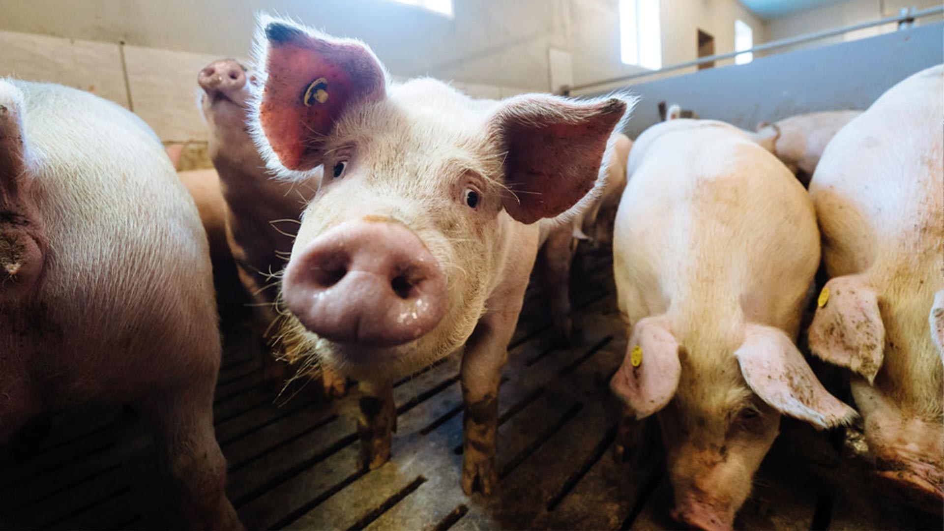
Streptococcus suis is one of the most economically harmful pathogens for the global swine industry. When I started working in pig production 25 years ago, S. suis was already a problem on practically all the farms that I visited. Back then, our understanding of the pathogen and hence our control strategies were rudimentary: in farrowing rooms, we cut piglets’ teeth, used gentian violet spray on their navels, and sometimes applied penicillin lyophilized with iron. For the nursery phase, we only had penicillin or phenoxymethylpenicillin at our disposal – until the first amoxicillin-based premixes arrived, which turned out to be highly effective.

To this day, we control S. suis mainly through oral beta-lactam antibiotics (in feed or water) or injectable solutions, administered to piglets at an early age. However, pig production has evolved dramatically over the past decades, and so has the scientific research on this complex pathogen. Crucially, we now know that the excessive use of antibiotics contributes to the development of antimicrobial resistance.

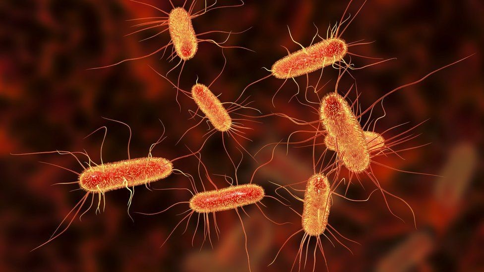
Recent Australian research has discovered S. suis strains (both in humans and pigs) with a high degree of resistance to macrolides or tetracyclines, strains with intermediate sensitivity to Florfenicol, and others that are developing resistance to penicillin G. Additionally, we now know that S. suis is a zoonotic bacteria that affects not only at-risk farm or slaughterhouse personnel: S. suis is among the leading causes of death from meningitis in countries such as Thailand, China or Vietnam. In light of these threats to human health, we in the swine industry more than ever have a duty to help control this pathogen.

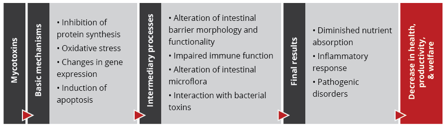
This article first reviews our current state of knowledge about the epidemiology and pathogenesis of Strep suis; it then lays out virulence factors and the role of coinfections. The second part considers the dimensions of a holistic approach to S. suis prevention and control and highlights the central role of intestinal health management.

What we know about S. suis epidemiology and pathogenesis

Practically all farms worldwide have carrier animals, but the percentage of animals colonized “intra-farm” varies between 40 and 80%, depending on several factors such as environmental conditions, hygiene measures, and the virulence of the S. suis strains involved.

How S. suis strains are classified

S. suis strains were once classified into 35 serotypes, according to their different capsular polysaccharides(CPS), theoutermost layer of the bacterial cell. Due to phylogenic and genomic sequencing, some of the old serotypes (20, 22, 26, 32, 33, and 34) are now reclassified, either in other bacterial genera or in other Streptococcus species. This has reduced the total to 29 S. suis serotypes.

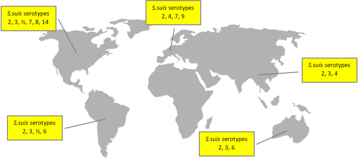
Globally, the prevalence of the disease varies between 3% and 30%. The main serotypes affecting pig population are type 2 (28%), 9 (20%), and 3 (16%); differences in the geographical distribution are shown in Figure 1.

Figure 1: Global distribution of S. suis serotypes
Based on different sources, incl. Goyette-Desjardins et al. (2014), Zimmermann et al. (2019), and Gebhart (2019)

In addition to the serotype classification based on CPS antigens, S. suis has also been genetically differentiated into “sequence types” using the MLST (Multi Locus Sequence Typing) technique. The distribution of both porcine and human sequence types is detailed in Figure 2.

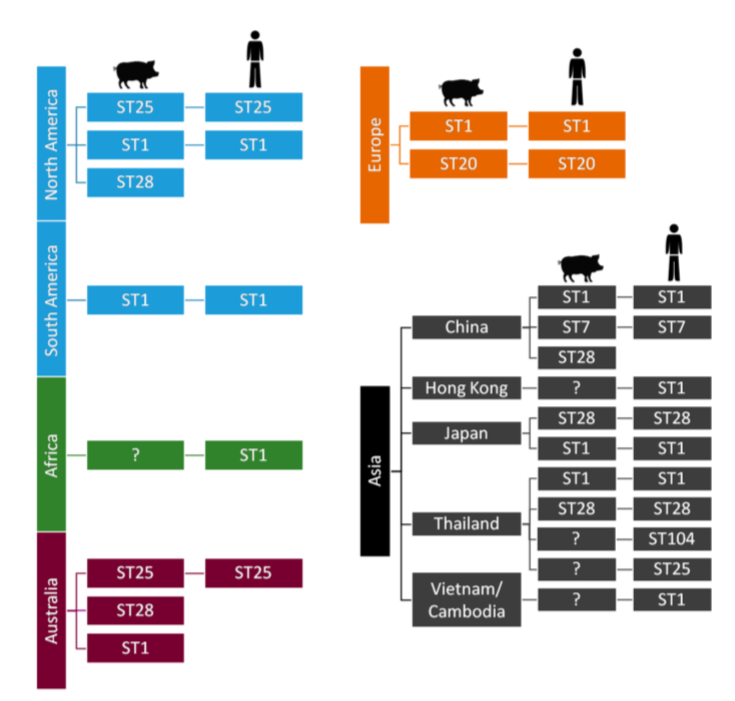
Figure 2: S. suis sequence types and their worldwide distribution

How S. suis is transmitted in swine

The main transmission routes are, firstly, the vertical sow-piglet route; the mucosa of the vagina is the first point of contamination. In the farrowing room, respiratory transmission from the sow to the piglets takes place. Horizontal transmission between piglets has also been proven to occur, especially during outbreaks in the post-weaning phase. This form of transmission happens through aerosols, feces, and saliva.

While in humans, the possibility of infection via the digestive tract has been confirmed, there are discussions about this route for swine. De Greeff et al. (2020) argue, based on in vitro and in vivo data, that infection through the digestive tract is associated with specific serotypes. Serotype 9, for example, would have a greater capacity for colonizing the gastrointestinal tract, and from there, the bacteria’s translocation takes place. The same authors point out that, in Western Europe, S. suis serotype 9 has become one of the most prevalent serotypes in recent years.

How S. suis colonization occurs

Although there are still unknown mechanisms in the pathogenesis of the disease, it can be schematically summarized how colonization occurs (Figure 3). From the different infection routes, the pathogen always passes through the mucosa. When S. suis enter the bloodstream, it can lead to a systemic infection, ending in septicemia, meningitis, endocarditis, or pneumonia, or a local infection at the joints level, causing arthritis.

According to Haas and Grenier (2018), different pathogenicity factors intervene in each of the processes. The CPS, for example, are relevant during colonization and the initial progression (indicated by black arrows). Microvesicles released by S. suis cell membranes are more involved in the passage to the bloodstream or, for example, the progression towards local or systemic infection (indicated by white arrows).


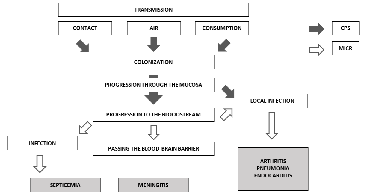
Figure 3: Pathogenesis of S. suis infection
Source: based on Haas and Grenier (2018)


Depending on the host and the immune response, the well-known clinical signs of the disease will occur. Although they may begin in the lactation phase, the highest prevalence of meningitis (the main clinical symptom) usually occurs between the 5th and the 10th week of life, that is, between two and three weeks after weaning.

How to diagnose S. suis infection

Diagnosing S. suis is relatively simple at a clinical level; however, we need to know how to differentiate it from G. parasuis in the case of animals with nervous symptoms. We also need to distinguish S. suis from other pathogens responsible for producing arthritis, such as M. hyosynoviae or the fibrin-producing agent M. hyorhinis.

Laboratory techniques are developing on two fronts. Among molecular techniques, multilocus sequence typing (MLST) is considered the gold standard for serotyping. It is still costly and not yet practicable for large samples at the farm level. In contrast, several types of polymerase chain reaction (PCR) show greater practical applicability. Quantitative PCRs (qPCR) are used for the evaluation of bacterial load, and some PCRs are based on the identification of specific virulence genes.

Due to the relevance of S. suis for human health, more complex techniques are also available, such as the complete sequencing of the bacterial genome. This type of method aims to develop epidemiological analyzes together with the differentiation between virulent and non-virulent S. suis strains. Research is also underway in serology, particularly on evaluating maternal immunity and its interference with the piglet, as well as autogenous vaccines monitoring.

Why S. suis sometimes causes disease: Virulence factors and coinfections

Streptococcus suis is a pathobiont, i.e., a microorganism that belongs to the commensal flora of animals but generates disease under certain conditions. In their daily work on farms, clinical veterinarians, for instance, find that S. suis often colonizes the upper respiratory tract, nasal cavity, and tonsils without causing disease. S. suis pathogenicity is associated with an astounding range of different circumstances or triggering factors; some sources list more than 100 virulence factors. Several factors are considered essential in the development of pathogenesis; others, however, are the subject of ongoing research (cf. Xia et al., 2019, and Segura et al., 2017).

Critical virulence factors

  • One of the most important proteins is the CPS that establishes serotypes. The CPS largely determines the bacteria’s adhesion and colonization behavior. It can modify its thickness depending on the stage: it becomes thinner when adhering to the mucociliary apparatus and thicker when circulating through the bloodstream, protecting the bacteria against possible attacks by immune system cells.
  • Likewise, suis has an adhesin known as Protection Factor H (FHB) that protects it from phagocytosis by macrophages and can also interfere with the complement activation pathways of the immune system.
  • Suilysin is one of the most critical suis‘ protein toxins. This toxin plays a fundamental role in the interaction with host cells (modulating them to facilitate invasion and replication within the host cells) as well as in the inflammatory response.
  • S. suis is a mucosal pathogen and, hence, triggers a mucosal immunity response, mainly by immunoglobulins A (IgA). S. suis has developed proteases capable of destroying both IgA and IgG.
  • Research is still in progress, but both suis serotype 2 and 9 encode the development of adhesion proteins that facilitate mucociliary colonization when salivary glycoproteins are present (these are called antigens 1 and 2).
  • Other than Suilysin, two of the bacteria’s protein components that have been studied in-depth to develop subunit vaccines are the MRP (Muramidase Release Protein) and EF (Extracellular Factor) protein. Whether the expression of these proteins is associated with virulence depends on the serotype.
  • Recent research indicates that greater biofilm production capacity is associated with the more virulent suis strains. The production of biofilm is closely related to the production of fibrinogen, which allows the bacteria to develop resistance to the action of antimicrobials, to colonize tissues, to evade the immune system, etc.

Concomitant factors for S. suis infection

Even though S. suis is a primary pathogen that can cause disease by itself, many factors can exert a direct or indirect influence on whether or not and to which extent disease develops.

Veterinarians and producers are well aware of the influence of environmental and management factors such as temperature variations, poor ventilation together with poor air quality, irritants for the respiratory tract, as well as correct densities for animals’ welfare. Occasionally, depending on the geographical location, S. suis can be considered as a seasonal pathogen, showing a higher prevalence during the coldest months of the year when ventilation is lower or not well-controlled.

At the level of the individual animal, concomitant pathogens, environmental changes, diet changes, previous pathologies, piglet handling problems, etc., all come into play. Younger piglets tend to be more susceptible because of the decrease in maternal immunity or insufficient colostrum intake; diarrhea during the lactation phase also increases disease vulnerability.

Recently, researchers have started to explore the hypothesis that a change in the digestive tract microbiome balance may favor a pathogenic trajectory. Some results indicate that changes in the microbiota around the moment of weaning could indeed trigger disease. I will return to the vital topic of the digestive tract in S. suis pathogenesis below.

The role of coinfections

The virulence of S. suis can increase in the presence of other pathogens, both viral and bacterial. Among the main viruses, key interactants are the PRRS virus, the influenza virus (SIV), as well as Porcine Circovirus (PCV) and Porcine Respiratory Coronavirus (PRCV). At the bacterial level, Bordetella bronchiseptica and Glaesserella parasuis have the most direct interaction with S. suis (Brockmeier, 2020).

There are several mechanisms by which coinfections might increase S. suis virulence: some of them (i.e., B. bronchiseptica and SIV) alter the epithelial barrier, facilitating the translocation of S. suis. Moreover, viruses such as PRRS either cause an alteration in the response of the immune system or destroy relevant immune system cells.

Valentin-Weigand et al. (2020) posit that the influenza virus increases the pathogenic capacity of S. suis so that, for specific strains, the disease can develop even in the absence of the key virulence factor suilysin. This highlights the importance of controlling coinfections for successful S. suis management.

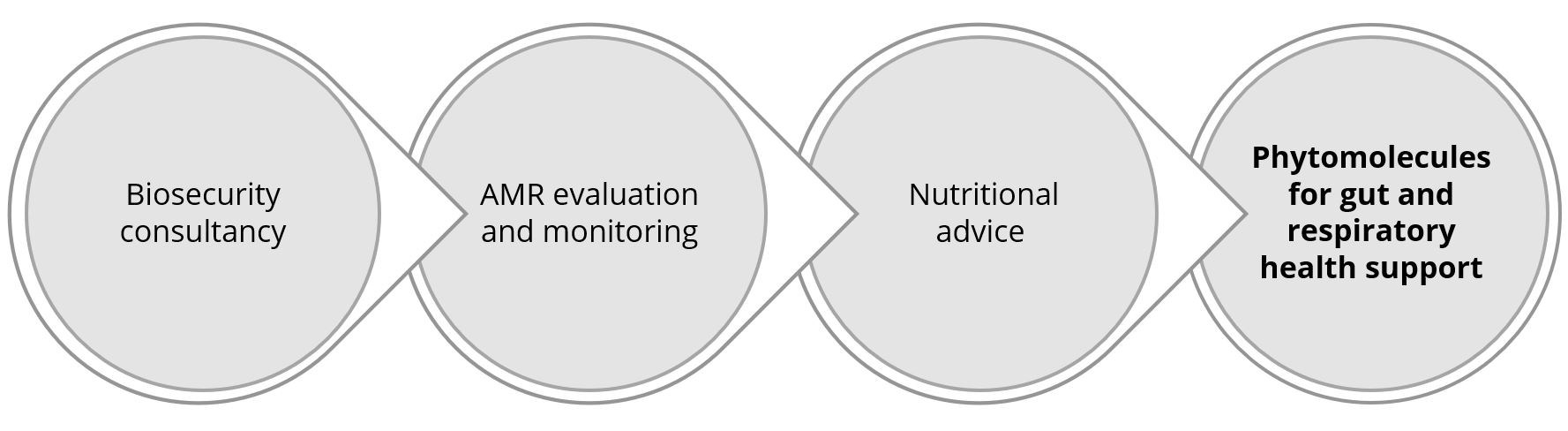
The five pillars of holistic S. suis management in swine

The challenge of managing this problematic pathogen with limited use of antibiotics prompts a review of all strategies within our reach. From birth to slaughterhouse, interventions must be coordinated and cannot work independently.

1. Biosecurity

The principles of biosecurity are easily understood. Yet, across different locations and production systems, farms struggle with consistently executing biosecurity protocols. For the moment, it appears unrealistic to avoid the introduction of new S. suis strains altogether. Also, complete eradication is not feasible with the currently available tools.

Genetic companies and research centers will likely continue to explore how to reduce bacterial colonization in animals, to produce piglets that have no or only minimal S. suis populations. Again, this option is not available for now.

At the farm level, the most promising and feasible approach is to reduce the risk of bacterial transmission, i.e., to optimize internal biosecurity. This extends to controlling both viral and bacterial coinfections. The two major viruses affecting the nursery stage are the PRRS virus and Swine Influenza virus. Bacteria that can contribute to the disintegration of the mucosa, both at the respiratory level and the digestive level, are Atrophic Rhinitis (progressive or not) and digestive pathogens such as E. coli, Rotavirus and Eimeria suis. All possible measures to reduce the prevalence and spread of these co-infectants must be executed to help control S. suis.

2. The pre-weaning period

We need to consider several elements in the first hours after birth that influence the spread of the bacteria in the farrowing rooms:

  • How is the colostrum distribution between the litters and the subsequent distribution of the piglets carried out?
  • How is the “processing” of the piglets carried out after farrowing: iron administration, wound management, and tail docking?
  • Are we taking any measure to prevent iatrogenic transmission of pathogens through needle exchange?

Until today, it is common practice to administer systemic (in-feed) or local (vaginally applied) antibiotics during the pre-weaning phase, albeit with partial or inconsistent successes in terms of reducing infection pressure. Notably, during the pre-weaning phase, the development of the piglet’s microbiota begins to take shape, and the systematic and prophylactic application of antibiotics in young animals can reduce bacterial diversity of the microbiome (Correa-Fiz et al., 2019). This, in turn, leads to a proliferation of bacteria with a pathogenic profile that could detrimentally influence subsequent pathology.

piglets suckling

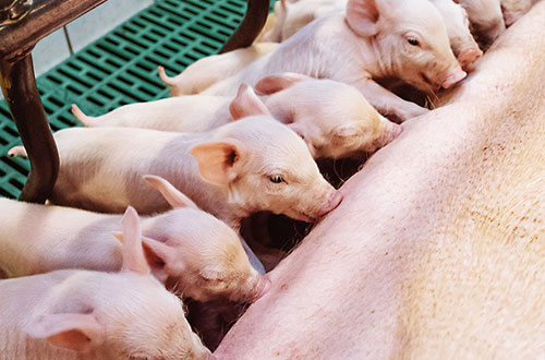
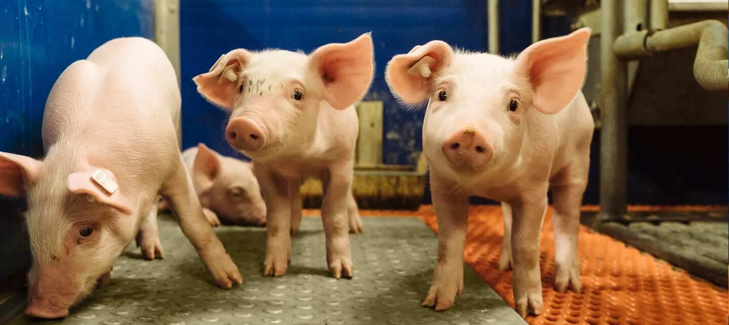
S. suis is an ultra-early colonizer; piglets can get infected already at birth

3. The post-weaning period

The post-weaning period undoubtedly constitutes the most critical stage of the piglets’ first weeks of life. In addition to social and nutritional stress, piglets are exposed to new pathogens. While maternal immunity is decreasing, piglets have not developed innate immunity yet; they are now most susceptible to the horizontal transmission of diseases. Hence, S. suis prevention during this phase center on measures that improve piglet quality. Key parameters include:

  • Do we have a correct and homogeneous weight/age ratio at weaning?
  • What is the level of anorexia in piglets? Do we practice suitable corrective measures to encourage the consumption of post-weaning feed?
  • How are we feeding them? What medications do they routinely receive?
  • How are housing facilities set up concerning density, environment, and hygiene?

Again, gut health is critical: Ferrando and Schultsz (2016) suggest that the status of the piglet’s weaning gastrointestinal tract centrally influences the subsequent development of the disease. Their research supports the idea that some specific S. suis serotypes can develop their pathogenesis from the digestive tract, just as in human medicine. While in humans, this digestive route is associated with the consumption of raw or insufficiently processed pork, in swine, the most susceptible moments are sudden changes in diet. The transition from milk to solid feed, in particular, leads to an increase in alpha-glucans that favor bacteria proliferation. Likewise, an increase in susceptibility occurs when the integrity of the intestinal wall is lost, for example, due to viral and bacterial coinfections.

4. Treatments and vaccination

Since weaning is such a difficult phase for the life of the piglet, it is a common practice on farms across the world to include one or several antibiotics in the post-weaning phase. Sometimes, when the legal framework allows, producers use a systematic antibiotic (i.e., beta-lactams or tetracyclines) and another one with a digestive profile (e.g., pharmacological doses of ZnO, trimethoprim, sulfa drugs and derivatives).

While antibiotics, for the most part, effectively prevent infection in the post-weaning phase, they can have adverse effects on the digestive tract. According to Zeineldin, Aldrige, and Lowe (2019), continued antibiotics use:

  • might increase the susceptibility to other infections because of the imbalance of the microbiome,
  • the immune system might be weakened, together with an alteration in metabolism,
  • and it fosters a greater accumulation of bacteria that are resistant to antibiotics.

The effectiveness of curative antibiotics treatments varies considerably. In any case, early detection is critical; affected animals need to be isolated and provided with a comfortable environment. Therapeutic parenteral antibiotics are best combined with high-dose corticosteroids. Some sick animals are unable to stand or walk. As a complementary measure, it is recommended, where possible, to help them ingest some feed and water.

Much research attention is focused on finding suitable vaccines to control the disease. This is a challenging task: S. suis shows high genetic diversity, making the identification of common proteins difficult, and is protected against antibody binding by a sugar-based envelope. The research group around Mariela Segura and Marcelo Gottschalk, for example, is working on a subunit vaccine strategy that addresses both dimensions. Recently, Arenas et al. (2019) identified infection-site specific patterns of S. suis gene expression, which could serve as a target for future vaccines.

The arrival of a universal, affordable S. suis vaccine is still a distant hope, though. Inactivated vaccines generally offer low levels of antibodies at the mucosal level and would need some adjuvant to increase them. A multiple injection protocol will not work from a commercial and practical point of view. On the other hand, live attenuated vaccines risk re-developing virulence with potentially drastic effects on human health. To complicate the topic of vaccination further, there is a controversy regarding the time of application and what animals we should vaccinate – sows, piglets, both?

Today, though with variable results, the alternative to scarce commercial vaccines is autogenous vaccines. These are based on the suspected serotype(s) present on a particular farm. This strategy hinges on the difficult procedure of isolating the strain from the meninges, spleen, or joints of the animals. If this step is successful, a laboratory can then develop the autogenous vaccine. Immunization occurs mainly in piglets, but occasionally some sows are vaccinated during the lactation period.

5. Hygiene

Just as for any other pathogen, hygiene management is critical. The infection pressure can be lowered through simple steps, such as washing the breeders before they enter the farrowing room. It is, or it should be, standard practice to maximize hygiene in the processing of piglets, avoiding injuries or pinching of the gums during teeth cutting, as well as disinfecting the umbilical area.

We know that S. suis is usually very sensitive to most disinfectants, but that is can form a biofilm that allows it to withstand hostile conditions. Physical or chemical methods to eliminate biofilm-formation are thus vital for combatting S. suis effectively.


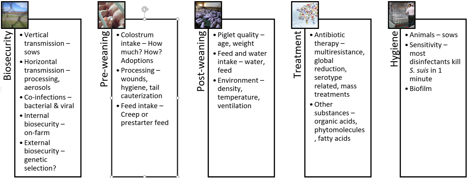
Figure 4: The 5 pillars of S. suis control and prevention

S. suis control and prevention: The future lies in the gut

There is no ideal solution for totally controlling S. suis yet: autogenous vaccines are only partially effective, and since we cannot continue to administer antibiotics systematically, it is necessary to look for alternatives. Pending the arrival of a universal vaccine, the most promising efforts focus on the gastrointestinal tract.

Microbiome balance to keep S. suis in check

The gastrointestinal tract is not only the site where nutrient absorption takes place.  The gut is the largest immune system organ in the body and most exposed to different antigens; therefore, what happens at the digestive level has a considerable influence on the immune system, locally and systemically.

The microbiome can be defined as the set of autochthonous bacteria that reside in the digestive system of animals. This group of bacteria is continually evolving and changes at critical moments in the life of animals. Simply put, a healthy microbiome is one that has a high bacterial diversity in the digestive tract (alpha diversity). The diversity between animals, on the other hand, should be low (beta diversity). A healthy microbiota implies the absence of dysbiosis and pathogens. Finally, one wants to promote the presence of bacteria that can produce substances with a bactericidal effect, such as short-chain fatty acids or bacteriocins.

Can we influence the microbiome to have fewer S. suis problems? Research by Wells, Aragon, and Bessems (2019) compared microbiota samples of the palatine tonsils from healthy and infected animals. They found that animals that would later develop the disease showed less diversity and, in particular, a diminished presence of the genus Moxarella. Importantly, they found that these differences in the microbiome’s composition of animals that later developed the disease were noticeable before weaning and at least two weeks before the outbreak occurred.

The same authors investigated in more depth, which bacteria in the microbiome were able to maintain homeostasis at the digestive level, finding that this was mostly the case for the genera Actinobacillus, Streptocuccus, and Moraxella. Moreover, they found that Prevotellacea and Rhotia produce antibacterial substances against S. suis.

Nutrition can impact the microbiome through targeted ingredients

We have to think about the microbiome of locations other than the digestive system as well. As we previously saw, the bacteria are transmitted through the mucosal route in the vagina, through the respiratory route, and there are recent studies that consider saliva as a leading source of infection in oral transmission.

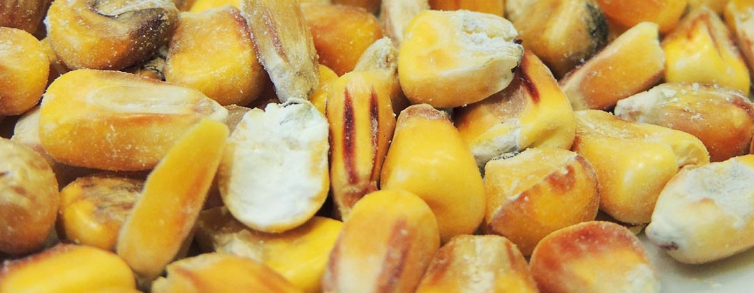
This research contributes insights into how we might approach S. suis management through nutritional strategies. The question for nutritionists is, can you formulate feed that reduces the availability of S. suis’ favorite nutrients? S. suis appears to develop best when the feed contains large quantities of carbohydrates or starches. Other nutritional factors include the feed’s buffering capacity and the stomach pH of the piglets.


In times of antimicrobial resistance, additives are crucial for S. suis control and prevention

Gut health and nutrition approaches come together in the area of additives: targeted gut health-enhancing additives to feed or water will become a cornerstone of S. suis control. What we want to see in such products are molecules or substances that are capable of limiting, inhibiting, or slowing down the growth of S. suis by altering the membrane or interfering with the energy mechanisms of the bacteria.

There are already several products on the market with different active ingredients, such as phytomolecules, medium-chain fatty acids, organic acids, prebiotics, probiotics, etc. Soon, those products or combinations of them will be a part of our strategy for controlling this pathogen of such importance to our industry.

By Technical Team, EW Nutrition

References

Arenas, Jesús, Ruth Bossers-De Vries, José Harders-Westerveen, Herma Buys, Lisette M. F. Ruuls-Van Stalle, Norbert Stockhofe-Zurwieden, Edoardo Zaccaria, et al. “In Vivo Transcriptomes of Streptococcus Suis Reveal Genes Required for Niche-Specific Adaptation and Pathogenesis.” Virulence 10, no. 1 (2019): 334–51. https://doi.org/10.1080/21505594.2019.1599669.

Brockmeier, Susan L. “Appendix F – The role of concurrent infections in predisposing to Streptococcus suis and other swine diseases: Proceeding from the 4th International Workshop on S. suis.” Pathogens 9, no. 5 (2020): 374. https://doi.org/10.3390/pathogens9050374.

Correa-Fiz, Florencia, José Maurício Gonçalves Dos Santos, Francesc Illas, and Virginia Aragon. “Antimicrobial Removal on Piglets Promotes Health and Higher Bacterial Diversity in the Nasal Microbiota.” Scientific Reports 9, no. 1 (2019): Article number: 6545. https://doi.org/10.1038/s41598-019-43022-y.

De Greeff, Astrid, Xiaonan Guan, Francesc Molist, Manon Houben, Erik van Engelen, Ton Jacobs, Constance Schultsz et al. “Appendix A – Streptococcus suis serotype 9 infection: Novel animal models and diagnostic tools: Proceeding from the 4th International Workshop on S. suis.” Pathogens 9, no. 5 (2020): 374. https://doi.org/10.3390/pathogens9050374.

Ferrando, M. Laura, Peter Van Baarlen, Germano Orrù, Rosaria Piga, Roger S. Bongers, Michiel Wels, Astrid De Greeff, Hilde E. Smith, and Jerry M. Wells. “Carbohydrate Availability Regulates Virulence Gene Expression in Streptococcus Suis.” PLoS ONE 9, no. 3 (2014). https://doi.org/10.1371/journal.pone.0089334.

Ferrando, Maria Laura, and Constance Schultsz. “A Hypothetical Model of Host-Pathogen Interaction OfStreptococcus Suisin the Gastro-Intestinal Tract.” Gut Microbes 7, no. 2 (2016): 154–62. https://doi.org/10.1080/19490976.2016.1144008.

Gebhart, Connie. “Cracking the Streptococcus Suis Code.” Pijoan Lecture. Lecture presented at the University of Minnesota Allen D. Leman Swine Conference, 2019. https://drive.google.com/file/d/1-E5tgFbteuPcDnMquOj_YhSKHYlaCqwO/view.

Goyette-Desjardins, Guillaume, Jean-Philippe Auger, Jianguo Xu, Mariela Segura, and Marcelo Gottschalk. “Streptococcus Suis, an Important Pig Pathogen and Emerging Zoonotic Agent—an Update on the Worldwide Distribution Based on Serotyping and Sequence Typing.” Emerging Microbes & Infections 3, no. 1 (2014): 1–20. https://doi.org/10.1038/emi.2014.45.

Haas, B., and D. Grenier. “Understanding the Virulence of Streptococcus Suis : A Veterinary, Medical, and Economic Challenge.” Médecine et Maladies Infectieuses 48, no. 3 (2018): 159–66. https://doi.org/10.1016/j.medmal.2017.10.001.

Murase, Kazunori, Takayasu Watanabe, Sakura Arai, Hyunjung Kim, Mari Tohya, Kasumi Ishida-Kuroki, Tấn Hùng Võ, et al. “Characterization of Pig Saliva as the Major Natural Habitat of Streptococcus Suis by Analyzing Oral, Fecal, Vaginal, and Environmental Microbiota.” Plos One 14, no. 4 (2019). https://doi.org/10.1371/journal.pone.0215983.

O’Dea, Mark A., Tanya Laird, Rebecca Abraham, David Jordan, Kittitat Lugsomya, Laura Fitt, Marcelo Gottschalk, Alec Truswell, and Sam Abraham. “Examination of Australian Streptococcus Suis Isolates from Clinically Affected Pigs in a Global Context and the Genomic Characterisation of ST1 as a Predictor of Virulence.” Veterinary Microbiology 226 (2018): 31–40. https://doi.org/10.1016/j.vetmic.2018.10.010.

Segura, Mariela, Nahuel Fittipaldi, Cynthia Calzas, and Marcelo Gottschalk. “Critical Streptococcus Suis Virulence Factors: Are They All Really Critical?” Trends in Microbiology 25, no. 7 (2017): 585–99. https://doi.org/10.1016/j.tim.2017.02.005.

Segura, Mariela, Virginia Aragon, Susan Brockmeier, Connie Gebhart, Astrid Greeff, Anusak Kerdsin, Mark O’Dea, et al. “Update on Streptococcus Suis Research and Prevention in the Era of Antimicrobial Restriction: 4th International Workshop on S. Suis.” Pathogens 9, no. 5 (2020): 374. https://doi.org/10.3390/pathogens9050374.

Tenenbaum, Tobias, Tauseef M Asmat, Maren Seitz, Horst Schroten, and Christian Schwerk. “Biological Activities of Suilysin: Role InStreptococcus Suispathogenesis.” Future Microbiology 11, no. 7 (2016): 941–54. https://doi.org/10.2217/fmb-2016-0028.

Valentin-Weigand, Peter, Fandan Meng, Jie Tong, Désirée Vötsch, Ju-Yi Peng, Xuehui Cai, Maren Willenborg et al. “Appendix G – Viral coinfection replaces effects of suilysin on adherence and invasion of Streptococcus suis into respiratory epithelial cells grown under air–liquid interface conditions: Proceeding from the 4th International Workshop on S. suis.Pathogens 9, no. 5 (2020): 374. https://doi.org/10.3390/pathogens9050374.

Wells, Jerry, Virginia Aragon, and Paul Bessems. “Report on the deep analysis of the microbiota composition in healthy and S. suis-diseased piglets.” European Commission Program for Innovative Global Prevention of Streptococcus suis. Ref. Ares(2019)6305977, 2019. https://cordis.europa.eu/project/id/727966/results

Xia, Xiaojing, Wanhai Qin, Huili Zhu, Xin Wang, Jinqing Jiang, and Jianhe Hu. “How Streptococcus Suis Serotype 2 Attempts to Avoid Attack by Host Immune Defenses.” Journal of Microbiology, Immunology and Infection 52, no. 4 (2019): 516–25. https://doi.org/10.1016/j.jmii.2019.03.003.

Zeineldin, Mohamed, Brian Aldridge, and James Lowe. “Antimicrobial Effects on Swine Gastrointestinal Microbiota and Their Accompanying Antibiotic Resistome.” Frontiers in Microbiology 10 (2019). https://doi.org/10.3389/fmicb.2019.01035.

Zimmerman, Jeffrey J., Locke A. Karriker, Alejandro Ramirez, Kent J. Schwartz, Gregory W. Stevenson, and Jianqiang Zhang. Diseases of Swine. 11th ed. Hoboken, NJ: Wiley-Blackwell, 2019.


Do we have the tools to reduce antibiotics in swine production?

SOW iStock 000000850516 web horiz. 1

The global swine industry is going through unprecedented challenges. On the one hand, the threat of the African Swine Fever virus is global, despite the fact it hasn’t arrived in all markets. The virus is today alive among the wild boars in the Polish and Belgian forests. Every day it keeps gaining a few more meters to the border, threatening the German swine industry, one of the largest in the European Union.

If this happens, we might be seeing important changes to the pork supply chain on the meat market worldwide – in Europe in addition to current issues in the USA meat plants. The profitability of swine businesses depends in many ways on the export capacity of large corporations based in Germany, Spain, Denmark, etc.

On the other hand, the presence of COVID-19 in most countries is changing human behavior, meat consumption at home, and the way we look at the future. Perhaps a virus overload via the news, some “fake news” conveying wrong messages on what’s coming, and suddenly we feel the future will never be the same.

The future of the swine industry

At least for the swine industry, the future will indeed never be exactly the same. We will be facing different challenges. Some of these will be structural, such as the issue of decreased manpower and how to substitute manpower by machines, through the implementation of Precision Livestock Farming, for instance.

We are also facing important health challenges to our animals: not just ASF, but also new and more aggressive PRRS strains, among other pathogens. Our sows´ production capacity is increasing annually, yet in some cases 25% of the new-born piglets are lost from birth to market. Increasingly, we may start to see increased levels of mortality not only in the nursery but in fattening pigs and sows as well.

It is becoming clearer all the time: the future of the global swine industry lies in producing more pigs with reduced antibiotics. To stay the course, we need to take further action and implement corrective measures.

Why we should remove antibiotics in pig production

Pressure from stakeholders and regulators

There is, and there will be, increasing pressure from many stakeholders worldwide to work toward pig production with reduced or no antibiotics. Meat suppliers, slaughterhouses and processors, governments at different levels, and, of course, the European Union – all are demanding reductions in the level of antibiotics in livestock production.

There is also an increasing awareness at the global societal level regarding antimicrobial resistance related to antibiotic usage in farming production. Consumer pressure will grow exponentially as the terrible COVID-19 experience will be “digested” by the global population.

Pressure to accede to the pork market

There is yet another important reason to start working in that direction: the global swine meat market. Today, China’s pork meat shortage is opening the market. Now any producer could potentially sell meat, either to China or to any other country. We are starting to see moves from companies in the USA or Brazil banning the use of Ractopamine in their operations because they want to get access to the ractopamine-free market (Europe & Asia, over 70% of the global population).

According to M. Pierdon (AASV 2020 Proceedings), there will be two types of markets: the “Niche ABFree” and the “Commodity ABFree”. Companies will have to analyse what their future is on the meat market. Not all the producers may be willing to enter this new phase, but for sure many will try.


Strategies for antibiotic reduction

In Europe, the time has arrived. Zinc oxide will be banned in June 2021 and there is now more than a trend in production with less or no antibiotic use. In some cases, there is a need; in others, this is simply profitable.

Challenges to antibiotic reduction

Producing pigs completely without antibiotics is not easy, and not affordable for all. Initially we may have to give up some performance parameters in order to achieve the balance between what we want and what we can achieve in animal performance. But the time will arrive when these two objectives will converge; there is no alternative.

To that end, we will have to include in our pig production strategy all the available tools and technologies: genetic selection, immunization against some key pathogens, environmental control (mandatory but often forgotten), early detection of diseases, etc.

In this new era we are entering, nutrition and feed additives will play a key role. It will be crucial to find solutions targeting the microbiome’s stabilization and diversification, creating and maintaining healthy farms and achieving all the performance parameters.

Do we have the tools for antibiotic reduction?

Even today there are companies able to produce completely antibiotic-free pigs – proof that yes, the tools are already in place.

Nevertheless, for most producers, the answer to – Can we produce without antibiotics? is most likely “probably not”. This will require a holistic approach, given the specific case of piglets.

The microbiome of the piglet is strongly influenced by birth and the subsequent weeks. What, then, are the elements that will be part of this new game that comprises a new approach?

    • The colostrum intake & the management of the piglets
    • Antibiotic usage and its influence on the gut
    • The piglets’ microbiome and its evolution during the periweaning period
    • The weaning process, appetite, and water intake
    • Zinc oxide removal and its influence on the microbiome
    • The immune system and the relationship with the GIT status
    • Inflammation and its modulation at the gut level
    • The health status and the effect on the concomitant infections: which ones are key and which ones are secondary pathogens
    • The all-important biosecurity, management, and hygiene

To summarize: there is no one tool, but rather a holistic approach to face this new challenge that the swine industry is facing nowadays. The answer is not a silver bullet, but a journey that we all must undertake.


Available in Spanish here.


A complex battlefield: mycotoxins in the gastrointestinal tract

shutterstock 494829349 fusarium mycotoxins 1 scaled

Most grains used as feed raw materials are susceptible to mycotoxin contamination. These toxic secondary metabolites are produced by fungi before or after harvest and cause severe economic losses all along agricultural value chains. For livestock, negative consequences include acute effects such as impaired liver and kidney function, vomiting, or anorexia, as well as chronic effects such as immunosuppression, growth retardation, and reproductive problems. Mycotoxin management is, therefore, of the utmost priority for animal producers worldwide.

But how is it that mycotoxins cause such damage in the first place? This article delves into the complex processes that take place when mycotoxins come into contact with the gastrointestinal tract (GIT). The intestinal epithelium is the first tissue to be exposed to mycotoxins, and often at higher concentrations than other tissues. A deeper understanding of how mycotoxins affect the GIT allows us to appreciate the cascading effects on animal health and performance, why such damage already occurs at contamination levels well below official safety thresholds – and what we can do about it.

The intestinal epithelium: the busy triage site for nutrients and harmful substances

When mycotoxins are ingested, they encounter the GIT’s intestinal epithelium (Figure 1). This single layer of cells lining the intestinal lumen serves two conflicting functions: firstly, it must be permeable enough to allow the absorption of nutrients. On the other hand, it constitutes the primary physiological barrier against harmful agents such as viruses, microorganisms, and toxins.

Within the intestinal epithelium, several types of highly specialized cells are involved in epithelial regeneration, nutrient absorption, innate defense, transport of immunoglobulins, and immune surveillance. The selective barrier function is maintained due to the formation of complex networks of proteins that link adjacent cells and seal the intercellular space. Besides, the intestinal epithelium is covered with mucus produced by goblet cells, which isolates its surface, preventing the adhesion of pathogens to the enterocytes (intestinal absorptive cells).

Due to its dual involvement in digestive and immune processes, the intestinal epithelium plays a pivotal role in the animal’s overall health. Importantly, the epithelium is directly exposed to the entire load of ingested mycotoxins. Hence their effects can be problematic even at low concentrations.

Figure 1: The intestinal epithelium

The intestinal epithelium


Problematic effects of mycotoxins on the intestinal epithelium

Most mycotoxins are absorbed in the proximal part of the gastrointestinal tract (Table 1). This absorption can be high, as in the case of aflatoxins (~90%), but also very limited, as in the case of fumonisins (<1%); moreover, it depends on the species. Importantly, a significant portion of unabsorbed toxins remains within the lumen of the gastrointestinal tract.

Some of the mycotoxins that enter the intestinal lumen can be bio-transformed into less toxic compounds by the action of certain bacteria. This action, however, predominantly happens in the large intestine – therefore, no detoxification takes place before absorption in the upper parts of the GIT. Part of the absorbed mycotoxins can also re-enter the intestine, reaching the cells from the basolateral side via the bloodstream. Furthermore, they re-enter through enterohepatic circulation (the circulation of substances between the liver and small intestine). Both actions increase the gastrointestinal tract’s overall exposure to the toxins.

Table 1: Rate and absorption sites of different mycotoxins

Rate and absorption sites of different mycotoxins

Adapted from: Biehl et al., 1993; Bouhet & Oswald, 2007; Devreese et al., 2015; Ringot et al., 2006

The damaging impact of mycotoxins on the intestinal epithelium initially occurs through:

  • A decrease in protein synthesis, which reduces barrier and immune function (Van de Walle et al., 2010)
  • Increased oxidative stress at the cellular level, which leads to lipid peroxidation, affecting cell membranes (Da Silva et al., 2018)
  • Changes in gene expression and the production of chemical messengers (cytokines), with effects on the immune system and cellular growth and differentiation (Ghareeb et al., 2015)
  • The induction of programmed cell death (apoptosis), affecting the reposition of immune and absorptive cells (Obremski & Poniatowska-Broniek, 2015)

Importantly, studies based on realistic mycotoxin challenges (e.g., Burel et al., 2013) show that the mycotoxin levels necessary to trigger these processes are lower than the levels reported as safe by EFSA, the Food Safety Agency of the European Union. The ultimate consequences range from diminished nutrient absorption to inflammatory responses and pathogenic disorders in the animal (Figure 2).

Figure 2: Mycotoxins’ impact on the GIT and consequences for monogastric animals

Mycotoxins’ impact on the GIT and consequences for monogastric animals

1. Alteration of the intestinal barrier‘s morphology and functionality

The mycotoxins DON, fumonisin, and T2 induce a reduction in the rate of epithelial cell proliferation and differentiation. This causes a decrease in the height and the surface of the intestinal villi, which in turn leads to a reduction in nutrient absorption. Additionally, some nutrient transporters are inhibited by the action of mycotoxins such as DON and T2, for example, negatively affecting the transport of glucose.

Several studies indicate that mycotoxins such as aflatoxin B1, DON, fumonisin B1, ochratoxin A, and T2, can increase the permeability of the intestinal epithelium of poultry and swine (e.g. Pinton & Oswald, 2014). This is mostly a consequence of the inhibition of protein synthesis. As a result, there is an increase in the passage of antigens into the bloodstream (e.g., bacteria, viruses, and toxins). This increases the animal’s susceptibility to infectious enteric diseases. Moreover, the damage that mycotoxins cause to the intestinal barrier entails that they are also being absorbed at a higher rate.

2. Impaired immune function in the intestine

The intestine is a very active immune site, where several immuno-regulatory mechanisms simultaneously defend the body from harmful agents. Immune cells are affected by mycotoxins through the initiation of apoptosis, the inhibition or stimulation of cytokines, and the induction of oxidative stress. Studies demonstrate that aflatoxin, DON, fumonisin, T2, and zearalenone interact with the intestinal immune system in such a manner that the animal’s susceptibility to viral and bacterial infections increases (e.g., Burel et al., 2013). Moreover, by increasing their fecal elimination, the horizontal transmission of pathogens is extended.

For poultry production, one of the most severe enteric problems of bacterial origin is necrotic enteritis, which is caused by Clostridium perfringens toxins. Any agent capable of disrupting the gastrointestinal epithelium – e.g. mycotoxins such as DON, T2, and ochratoxin – promotes the development of necrotic enteritis. The inhibition of the intestinal immune system caused by mycotoxins such as aflatoxin, DON, and T2 also collaborates with the development of this disease.

3. Alteration of the intestinal microflora

The gastrointestinal tract is home to a diverse community of bacteria, fungi, protozoa, and viruses, which lines the walls of the distal part of the intestine. This microbiota prevents the growth of pathogenic bacteria through competitive exclusion and the secretion of natural antimicrobial compounds, volatile fatty acids, and organic acids.

Recent studies on the effect of various mycotoxins on the intestinal microbiota show that DON and other trichothecenes favor the colonization of coliform bacteria in pigs. DON and ochratoxin A also induce a greater invasion of Salmonella and their translocation to the bloodstream and vital organs in birds and pigs – even at non-cytotoxic concentrations. It is known that fumonisin B1 may induce changes in the balance of sphingolipids at the cellular level, including for gastrointestinal cells. This facilitates the adhesion of pathogenic bacteria, increases in their populations, and prolongs infections, as has been shown for the case of E. coli.

From the perspective of human health, the colonization of the intestine of food-producing animals by pathogenic strains of E. coli and Salmonella is of particular concern. Mycotoxin exposure may well increase the transmission of these pathogens, posing a risk for human health.

4. Interaction with bacterial toxins

When mycotoxins induce changes in the intestinal microbiota, this can lead to an increase in the endotoxin concentration in the intestinal lumen. Endotoxins or lipopolysaccharides (LPS) are fragments of Gram-negative bacteria’s cell walls. They are released during bacterial cell death, growth, and division. Hence endotoxins are always present in the intestine, even in healthy animals. Endotoxins promote the release of several cytokines that induce an enhanced immune response, causing inflammation, thus reducing feed consumption and animal performance, damage to vital organs, sepsis, and death of the animals in some cases.

The synergy between mycotoxins and endotoxins can result in an overstimulation of the immune system. The interaction between endotoxins and estrogenic agents such as zearalenone, for example, generates chronic inflammation and autoimmune disorders because immune cells have estrogen receptors, which are stimulated by the mycotoxin. The combination of DON at low concentrations and endotoxins in the intestine, on the other hand, has been shown to engender a decrease in transepithelial resistance and to alter the balance of the microbiota.

What to do? Proactive toxin risk management

To prevent the detrimental consequences of mycotoxins on animal health and performance, proactive solutions are needed that support the intestinal epithelium’s digestive and immune functionality and help maintain a balanced microbiome in the GIT. Moreover, it is crucial for any anti-mycotoxin product to feature both anti-mycotoxin and anti-bacterial toxin properties and that it supports the organs most targeted by mycotoxins, e.g., the liver. EW Nutrition’s Mastersorb Gold premix is based on the synergistic combination of natural clay minerals, yeast cell walls, and phytomolecules. Its efficacy has been extensively tested, including as a means for dealing with E. coli endotoxins.

Mastersorb Gold: anti-mycotoxin activity stabilizes performance and strengthens liver health

A field trial conducted in Germany on male Ross 308 broilers showed that for broilers receiving a diet contaminated with DON and zearalenone, adding 1kg of Mastersorb Gold per ton of feed to their diet led to significant performance enhancements. Not only did they recuperate the mycotoxin-induced weight loss (6% increase relative to the group receiving only the challenge), but they gained weight relative to the control group (which received neither the challenge nor Mastersorb Gold). Feed conversion also improved by 3% relative to the group challenged with mycotoxins.

A scientific study of crossbred female pigs showed that Mastersorb Gold significantly reduced the deleterious effects of fumonisin contamination in the feed. The decrease in weight gain and the decline of feed conversion could be mitigated by 6.7% and 13 FCR points, respectively (Figure 3). Also, the sphinganine/sphingosine (Sa/So) ratio, a biomarker for fumonisin presence in the blood serum, could be decreased by 22.5%.

Figure 3: Mastersorb Gold boosts performance for pigs fed a fumonisin-contaminated diet

Mastersorb® Gold boosts performance for pigs fed a fumonisin-contaminated diet

Another study of crossbred female piglets, carried out at a German university, investigated whether Mastersorb Gold could support performance as well as liver health under a naturally occurring challenge of ZEA (~ 370ppb) and DON (~ 5000ppb).  Mastersorb Gold significantly improved weight gain and feed conversion in piglets receiving the mycotoxin-contaminated diet: daily body weight gain was 75g higher than that of a group receiving only the challenge, and the FCR improved by 24% (1.7 vs. 2.25 for the group without Mastersorb Gold). Moreover, Mastersorb Gold significantly improved the liver weight (total and relative) and the piglets’ AST levels (aspartate aminotransferase, an enzyme indicating liver damage). A tendency to improve spleen weight and GGT levels (gamma-glutamyl transferase, another enzyme indicative of liver issues) was also evident, all of which indicate that Mastersorb Gold effectively counteracts the harmful impact of mycotoxin contamination on liver functionality.

In-vitro studies demonstrate Mastersorb Gold’s effectiveness against mycotoxins as well as bacterial toxins

Animal feed is often contaminated with two or more mycotoxins, making it important for an anti-mycotoxin agent to be effective against a wide range of different mycotoxins. Besides, to prevent mycotoxins damaging the GIT, an effective product should ideally adsorb most mycotoxins in the first part of the animal’s intestine (under acidic conditions). In-vitro experiments at an independent research facility in Brazil showed that an application of 0.2% Mastersorb Gold binds all tested mycotoxins at rates from 95 to 97% at a pH level of 3, using realistic challenges of 1000ppb (Aflatoxin B1 and ZEA) and 2500ppb (Fumonisin B1 and DON). The binding results achieved for Fumonisin and DON, which are often considered outright “nonbinding,” under challenging close to neutral conditions (pH 6), are particularly encouraging.

Figure 4: Mastersorb Gold binding capacity against different mycotoxins (%)

Concerning its efficacy against endotoxins, an in vitro study conducted at Utrecht University, among other studies, has shown Mastersorb Gold to be a strong tool against the LPS released by E. coli. For the test, four premium mycotoxin binders were suspended in a phosphate buffer solution to concentrations of 0.25% and 1%. E. coli LPS were suspended to a final concentration in each sample of 50ng/ml. Against this particularly high challenge, Mastersorb Gold achieved a binding rate of 75% at an inclusion rate of 1%: clearly outperforming competing products, which at best showed a binding rate of 10%.

Conclusion

A healthy gastrointestinal tract is crucial to animals’ overall health: it ensures that nutrients are optimally absorbed, it provides effective protection against pathogens through its immune function, and it is key to maintaining a well-balanced microflora. Even at levels considered safe by the European Union, mycotoxins can compromise different intestinal functions such as absorption, permeability, immunity, and microbiota balance, resulting in lower productivity and susceptibility to disease.

To safeguard animal performance, it is important to continually strive for low levels of contamination in feed raw materials –  and to stop the unavoidable mycotoxin loads from damaging the intestinal epithelium through the use of an effective anti-mycotoxin agent, which also supports animals against endotoxins and boosts liver function. Research shows that Mastersorb Gold is a powerful tool for proactive producers seeking stronger animal health, welfare, and productivity.

By Technical Team, EW Nutrition


5 key facts pig producers need to know about the EU’s ZnO ban

shutterstock 126738341 piglets drinking water

We all know the headlines, “European Commission adopts ZnO ban” or “Zinc oxide to be phased out at EU level by 2022”. Clearly, EU legislation has far-reaching consequences for European pig producers – but in the jungle of acronyms and legalistic jargon, it’s not always clear which institution gets to decide what and why. Here are five key facts that help pig producers make sense of the EU’s zinc oxide ban.

1. Zinc oxide can only be used as a feed additive (low dosage)

Pigs require zinc to maintain various metabolic functions, hence it is included in their diet as a feed additive. This use will not be banned: ZnO is included as a source of zinc in the so-called register of feed additives, which applies to the whole EU. The European Commission decides which products are included in the register based on the opinions of the European Food Safety Authority (EFSA), which also advises the Commission on topics like animal welfare and African swine fever. The EFSA currently suggests that a total level of 150ppm meets the animals’ physiological needs for zinc. The European Commission has turned this recommendation into law, hence 150ppm is the legal limit for zinc supplementation for piglets.

2. The EU sets common rules for veterinary medicinal products

ZnO-based products to treat post-weaning diarrhea in piglets, on the other hand, contain pharmacological doses of zinc oxide. A commonly administered dosage is 100mg per kg body weight per day for 14 consecutive days, amounting to 2500ppm zinc in the feed. These products are classified as veterinary medicinal products (VMPs) and are thus covered by Directive 2001/82/EC on medicinal products for veterinary use and by Regulation (EC) No 726/2004. These pieces of legislation set out the EU’s rules for the production, distribution, and authorizations of VMPs, and they establish the European Medicines Agency (EMA). Just as the EFSA advises the European Commission on feed additives, they turn to the EMA regarding VMPs.

Zinc oxide – two different uses, two different situations

zinc oxide feed additive medicinal product

3. ZnO products licenses are a national topic – but subject to EU scrutiny

One of EMA’s key topics are marketing authorizations: VMPs can only be sold and traded in the EU if they have received a marketing authorization, which is basically a license. Depending on the type of VMP and on when it was first released, the marketing authorization is either issued by the EMA or by national authorities. Veterinary medicines containing zinc oxide are (or rather were) within the remit of national authorization procedures. However, national authorities are supposed to turn to the EMA’s Committee for Medicinal Products for Veterinary Use (CVMP) if they have any issues with an application that is submitted to them. This is what happened in the case of zinc oxide.

4. France and the Netherlands initiated the review of zinc oxide

A European company in the feed industry had applied for marketing authorization for its ZnO-based medicated feeding stuff for piglets in the United Kingdom, hoping for a so-called decentralized authorization procedure to take place. This procedure would mean that the marketing authorization issued in the UK would also be valid in other EU countries. However, France and the Netherlands objected to this on the grounds of environmental concerns. Initially, the CVMP ruled that the marketing authorization could be granted, but France and the Netherlands persisted. In a second round, they raised doubts about the efficacy of risk mitigation measures and the added issue of antimicrobial resistance. This time, they were successful.

5. Bottom line: ZnO products will no longer get a marketing authorization

In March 2017, the CVMP concluded that zinc oxide’s benefits of preventing diarrhea do not outweigh the risks to the environment. Therefore the panel recommended that national authorities withdraw existing marketing authorizations for zinc oxide-based VMPs and that they no longer grant new authorizations. Shortly after that, on 26 June 2017, the European Commission adopted the CVMP’s recommendation, which means that all EU countries have to implement it. This decision also says that countries may defer withdrawing the marketing authorizations if they think that the lack of available alternatives and necessary changes in farming practices put too much pressure on their pig sectors. They can only defer for five years though; hence, the decision must be implemented no later than 26 June 2022.

Today we stand at the half-way point before the ban of VMP ZnO as a veterinary medicinal product kicks in across the EU. Hence the search is on for effective strategies to control post-weaning diarrhea: without zinc but through continuous improvements in management and feed practices, as well as the support of targeted, functional feed additives.


By Technical Team, EW Nutrition
Article available in german, dutch and spanish.


Why we need to replace zinc oxide in tackling post-weaning diarrhea

SOW Schmidtkord DSC2695 website

Piglets experience significant stress when they are weaned from the sow and change diet, making them susceptible to gastrointestinal disorders. Primarily during the first two weeks after weaning, they are likely to suffer from post-weaning diarrhea (PWD). PWD is a significant problem for pig producers worldwide: it leads to severe dehydration, stunted growth and mortality rates of up to 20-30%. Treatment and additional labor costs further squeeze farm profitability and necessitate unwanted antibiotic interventions.

Zinc oxide: an effective but highly problematic tool

Since the early 1990s zinc oxide (ZnO) has been used to control post-weaning diarrhea and promote growth in piglets, mainly at pharmacological dosages of 2500 to 3000ppm. Its mode of action is still not entirely understood; effects on immune or metabolic processes, altered microbiota, or post-absorptive metabolism are likely to play a role. What is clear is that the use of ZnO in European pig production has strongly increased since the EU banned the use of antibiotic growth promoters such as colistin in 2006 to curb the development of antimicrobial resistance.

Pigs depend on a continuous supply of zinc. Among other roles, this trace element constitutes a functional component of around 300 biochemical enzymes, making it pivotal to most metabolic processes, and by extension to optimal health, production and reproduction.  Modern pig diets thus include zinc supplementation to meet the animals’ requirements. The European Food Safety Authority (EFSA) currently suggests that a total level of 150ppm of zinc in feed matches the animals’ physiological need for zinc. The EFSAs concerns are solely connected to the environmental concerns arising from pharmacological high dosages of ZnO.

These concerns are grave indeed: zinc is a heavy metal after all. Too much zinc is toxic for the animal, hence its physiology ensures that excessive zinc intake is excreted. The bioavailability and absorption of zinc from zinc oxide is particularly low. Therefore most of the zinc given to piglets in this way accumulates in their manure – which is widely used as an organic fertilizer for agricultural soils.

The continual application of manure gradually increases topsoil zinc concentrations; leaching and run-off then lead to contamination of groundwater, surface waters, and sediment. As zinc is non-volatile and non-degradable, it is only a matter of time before concentrations lead to ecotoxic effects, including food crops, aquatic life, and drinking water. Classic mitigation measures such as diluting the manure or keeping certain minimum distances between application areas and surface waters can only slow down the environmental accumulation of zinc, not prevent it.

EU ban: ZnO to be phased out by 2022

In 2017, the European Medicines Agency (EMA) – the EU agency responsible for the scientific evaluation, supervision and safety monitoring of medicines, including veterinary medicinal products – conducted an overall risk-benefit analysis for ZnO. It concluded that the benefits of preventing diarrhea in pigs did not outweigh the significant environmental risks caused by zinc pollution. By June 2022 all EU member states will thus have to withdraw marketing authorizations for veterinary medicinal products containing zinc oxide that are administered orally to food-producing species.

In its decision, the EMA’s Committee for Medicinal Products for Veterinary Use also points out the risk that, due to co-resistance, the use of zinc oxide might promote the development of antimicrobial resistance. High doses of zinc supplementation have been shown to increase the proportion of multidrug-resistant E. coli and Salmonella, two of the most important pathogens in pig production.

What is more, studies show that excessive zinc can accumulate in the liver, the pancreas, and blood serum, and that it permanently reduces the lactobacilli population of the gut flora. With what consequences for performance in the fattening phase? Hence, there are plenty of reasons why getting rid of zinc oxide is a good thing and will ultimately result in even better, more sustainable pig production – but, of course, only if effective replacement strategies to control PWD and boost piglet performance are in place.

Towards zero ZnO: smart feed additives optimize gut health

The search for ZnO alternatives takes us right back to the start, to the piglets’ challenged gastrointestinal tract. During their first three months of life, pigs’ gastrointestinal system undergoes a complex maturation process of its epithelial, immune, and enteric nervous systems. Only once all of these systems are fully developed is the gut capable of delivering its normal functions (digestion, nutrient absorption, immunity, etc.), while also providing an effective barrier against the pathogens, antigens, and toxins in the lumen.

Unlike in nature, where weaning occurs around the time when GIT functions have matured, weaning in commercial pig production takes place during this vulnerable developmental period. Post-weaning diarrhea is ultimately a consequence of intestinal dysbiosis, a state of imbalance in the intestinal microbiome which in turn is induced by the dietary, behavioral, and environmental stressors of the weaning phase (such as separation from the sow, vaccinations, transport, introduction of solid feed).

PWD control thus starts with managing these stressors, which includes ensuring sufficient colostrum intake, gradual feed changes, and meticulous nursery hygiene. Critically, the weaning diet needs to optimally support gut health. Intelligent feed additive solutions are able to

  • reduce the pathogenic load in the piglet’s GIT,
  • strengthen the piglet’s maturing gut barrier functionality, and
  • selectively induce the development of beneficial microorganisms within the microbiome.

A synergistic combination of phytomolecules, medium-chain fatty acids, glycerides of butyric acid, and prebiotics achieves these objectives in a reliable and cost-effective manner. Thanks to their antimicrobial, anti-inflammatory, and digestive properties these selected ingredients effectively support piglets during this critical phase of their postnatal gut development, while also boosting their feed intake.

In the past decade, the European pig sector has successfully adapted to the 2006 ban on antibiotic growth promoters through significant improvements in management and feed practices. Cutting out zinc oxide is an ambitious challenge – but with the support of targeted, functional feed additives, producers will be able to set their piglets up for a strong, sustainable, zero ZnO health and growth performance.

*You can find this article in polish and italian.

References

Amezcua, Rocio, Robert M. Friendship, Catherine E. Dewey, Carlton Gyles, and John M. Fairbrother. “Presentation of postweaning Escherichia coli diarrhea in southern Ontario, prevalence of hemolytic E. coli serogroups involved, and their antimicrobial resistance patterns.” Canadian Journal of Veterinary Research 66, no. 2 (April 2002): 73-8. https://www.ncbi.nlm.nih.gov/pmc/articles/PMC226986/.

Bednorz, Carmen, Kathrin Oelgeschläger, Bianca Kinnemann, Susanne Hartmann, Konrad Neumann, Robert Pieper, Astrid Bethe, et al. “The Broader Context of Antibiotic Resistance: Zinc Feed Supplementation of Piglets Increases the Proportion of Multi-Resistant Escherichia Coli in Vivo.” International Journal of Medical Microbiology 303, no. 6-7 (2013): 396–403. https://doi.org/10.1016/j.ijmm.2013.06.004.

Brugger, Daniel, and Wilhelm M. Windisch. “Strategies and Challenges to Increase the Precision in Feeding Zinc to Monogastric Livestock.” Animal Nutrition 3, no. 2 (March 24, 2017): 103–8. https://doi.org/10.1016/j.aninu.2017.03.002.

Burrough, Eric R., Carson De Mille, and Nicholas K. Gabler. “Zinc Overload in Weaned Pigs: Tissue Accumulation, Pathology, and Growth Impacts.” Journal of Veterinary Diagnostic Investigation 31, no. 4 (June 6, 2019): 537–45. https://doi.org/10.1177/1040638719852144.

De Mille, Carson, Emma T. Helm, Eric R. Burrough, and Nicholas K. Gabler. “Zinc oxide does not alter ex vivo intestinal integrity or active nutrient transport in nursery pigs.” Paper presented at the Zero Zinc Summit, Copenhagen, Denmark, June 17-18, 2019. https://svineproduktion.dk/Services/-/media/3E0A1D2A4CAC409FAA6212B91DFEA537.ashx.

Moeser, Adam J., Calvin S. Pohl, and Mrigendra Rajput. “Weaning Stress and Gastrointestinal Barrier Development: Implications for Lifelong Gut Health in Pigs.” Animal Nutrition 3, no. 4 (December 2017): 313–21. https://doi.org/10.1016/j.aninu.2017.06.003.

Rhouma, Mohamed, Francis Beaudry, William Thériault, and Ann Letellier. “Colistin in Pig Production: Chemistry, Mechanism of Antibacterial Action, Microbial Resistance Emergence, and One Health Perspectives.” Frontiers in Microbiology 7 (November 11, 2016): Article 1789. https://doi.org/10.3389/fmicb.2016.01789.

Starke, Ingo C., Robert Pieper, Konrad Neumann, Jürgen Zentek, and Wilfried Vahjen. “The Impact of High Dietary Zinc Oxide on the Development of the Intestinal Microbiota in Weaned Piglets.” FEMS Microbiology Ecology 87, no. 2 (February 1, 2014): 416–27. https://doi.org/10.1111/1574-6941.12233.


Mind the immunity gap: egg immunoglobulins bolster piglets’ immune system

EW Nutrition article piglets 6

egg immunoglobulins bolster piglets’ immune system

In contrast to humans, piglets do not receive any maternal immunoglobulins via the placenta. It is therefore of vital importance for these young animals to receive maternal antibodies via the colostrum as soon as possible after birth. Otherwise, they are more vulnerable to illnesses in their early stages of life.

In this article, we look in-depth at how the immune system works and which role antibodies play in it.  We then consider why immunoglobulins from the egg (IgY) might potentially be a powerful tool for supporting young animals immunologically, allowing producers to maintain young animals’ health and to promote their performance.

How the immune system defends the body: three barriers

The immune system aims to prevent pathogens such as viruses, bacteria, and fungi from entering the body or to eliminate them when they have already entered. Furthermore, it seeks to prepare the body for quicker reactions, in case of subsequent infections, by building an immunological memory. Generally, in case of an attack by pathogens, there are three barriers against the “enemy” (Figure 1).

Figure 1: The three barriers of the immune response

First barrier: the immediate, physical defense upon contact with pathogens

The animal body has several anatomical features that prevent pathogens from entering in the first place, such as cilia and mucus. Skin, intestines and nose lining are colonized by a community of beneficial micro-organisms that form a physical barrier against pathogens. Other barriers include the urinary system, the acid pH of the stomach, as well as tears and saliva, which contain antibacterial lysozymes.

Second barrier: the unspecific, native defense that does most of the work

If the mechanical mechanisms of defense were not successful, the unspecific, innate immune defense enters into play (Murphy and Weaver, 2018, 47ff.). At this stage, the body needs to differentiate between “known” and “alien” agents, and between “potentially harmful” and “harmless” ones.

To identify alien, potentially harmful agents, the unspecific defense looks for so-called PAMPs (pathogen associated molecular patterns). These are general characteristics often displayed by pathogens, such as lipopolysaccharides in the bacterial membrane or double-stranded RNA in viruses. Everything that shows PAMPs is heavily targeted.

The unspecific defense can be further divided into the humoral and the cellular defense. The humoral defense consists of substances dissolved in the body fluids, such as enzymes, reactive oxygen compounds, signal molecules and a whole cascade of proteins. Some of these substances kill pathogens directly; others “mark” the pathogens and “call for” the help of leucocytes.

The cellular defense consists of different leucocytes, also known as white blood cells (because they do not contain any red hemoglobin). The main task of leucocytes is the defense of the body against pathogens, hence many leucocytes are capable of phagocytosis (the ingestion of other cells). To prevent phagocytes from accidentally ingesting the body’s own cells, these own cells are marked with the so-called major histocompatibility complex (MHC). This acts as a red flag, saying “I belong to the body!”.

The cellular defense consists of:

  • Neutrophil granulocytes (60-70% of the leucocytes), which mainly act against bacteria
  • Eosinophil and basophil granulocytes (1.5% of the leucocytes), which mainly act against parasites
  • Natural killer cells, which mainly act against viruses
  • Monocytes (3-8% of the leucocytes; they differentiate into macrophages and dendritic cells)

Figure 1: The three barriers of the immune response


The monocytes, as well as their macrophage and dendritic cell “offspring”, are the bridge to the next step, the specific defense. When these phagocytes digest pathogens, minuscule protein structures (antigens) of the pathogens remain. These antigens are unique to each pathogen. During a process called antigen presentation, the antigens are tied to the cell’s MHC and transported to the cell surface. This triggers the production of specific antibodies, the immune system’s third barrier.

Third barrier: the specific immune defense that creates antibodies and immunological memory

The specific (also called adaptive or acquired) immune response kicks in a few days after contact with specific pathogens and is mostly carried out by lymphocytes called T and B cells (Murphy and Weaver, 2018, 177ff.). They are active at the cellular and the humoral level, respectively.

T cells possess receptors on their surface through which they can recognize the antigens presented to them by phagocytes. What they do subsequently depends on the subtype of the T cell:

  • Cytotoxic T cells (CD8+) directly destroy the antigen-phagocyte-combination
  • T helper cells (CD4+) attract other cells that can destroy the pathogens (e.g. macrophages) and stimulate B cells to produce antibodies against them

B cells also possess receptors through which they can recognize antigens. Once they spot an antigen (and T helper cells “confirm” that an immune response is required), they divide and mature into so-called plasma cells. Plasma cells, in turn, secrete plenty of antibodies (or immunoglobulins) into the bloodstream and the lymphatic system. Antibodies are protein structures that lock onto and neutralize antigens through different mechanisms.

The chemical reaction between antibodies and antigens is the body’s most powerful immune response through which it can protect itself from pathogens and their toxins. Antibody production continues for several days to remove the antigens, and antibodies usually remain in circulation for a few months.

Moreover, certain T and B cells memorize the first attack of a pathogen and turn into memory cells. The T memory cells CD4+CD8+, for instance, match the antigens from certain past, latent, and particularly persistent viral infections. This immunological memory, created by acquired immunity, can be thought of as a library of antibodies that the body adds to whenever it deals with a new pathogen or receives a vaccine. In case of a subsequent contact with the pathogen, the right antibody “model” already exists and mass production can start up very quickly.

Why young animals’ immune defense is so vulnerable – and what IgY can do about that

Building one’s immunological memory takes time. A lot of new-born animals are in a vulnerable position: they have not had time yet to acquire immunity of their own, but they are also particularly fragile and susceptible to being attacked by commonly occurring pathogens such as corona and rotaviruses, E. coli and clostridia. The toxins that E. coli and clostridia, for instance, release, may cause diarrhea, edema, endotoxic shock, and even death.

To be protected during the first critical days of their lives, new-born animals thus need to receive a foundational stock of antibodies (passive immunity) from their mother. Humans receive maternal immunoglobulins via the placenta. Piglets, because a sow’s placenta is constructed differently, are dependent on receiving them through the colostrum after birth. If this is not the case – due to inadequate quantity or quality of the colostrum – they need to receive immune support in a different way.

Egg-yolk antibodies have been proposed as a powerful tool for supporting young animals during the critical period after birth. These special proteins support the colostrum supply and guarantee that every animal in the herd has some degree of protection. This protection mostly takes place in the gut. The IgY recognize and tie up pathogens and render them ineffective.

Trial: can egg immunoglobulins support piglet immunity?

In 2009, research at the National Veterinary Research Institute in Pulawy, Poland, was conducted to probe this hypothesis. The objective of the trial was to evaluate whether an oral application of egg immunoglobulins would have a quantifiable, positive influence on the immune system of the piglets. Different immunological parameters were measured, including different types of leucocytes.

Trial design

The test consists of 6 litters with 67 piglets in total divided into two groups. The control group (n=32) received the prophylaxis customary on the farm; the trial group (n=35) additionally received a product based on egg powder (EP)[1], applied at the inclusion rate recommended by the producer. Blood samples were taken on days 0 (before application of the product), 7, 14, and 28. They were analyzed with respect to the percentages of different types of lymphocytes.

Trial results

For the group receiving egg powder, the number of leucocytes in peripheral blood was significantly elevated compared to the control group on the 7th day of life (table 1). The amounts of lymphocytes and monocytes – indicators for the specific immunological defense – were also significantly increased on day 7, whereas the total amount of granulocytes – indicator for the innate, unspecific immune defense – remained constant. Hence, already during the first days, the piglets supplied with EP disposed of a higher level of adaptive (specific) immune defense, compared to the animals in the control group. In addition, there was a significant increase in the number of CD4-positive (CD4+) and CD4-CD8-double positive (CD4+CD8+) T cells in the EP group, compared to the control animals, indicating an active stimulation of the immune system.

Except for CD4+CD8+ T cells (which remained elevated in the EP group), on day 14, the differences in cellular immune response were no longer significant. This is most probably the case because by that time the immune system of the control group had activated its own protective response. The EP product therefore supported the young animals precisely when it was necessary, during the critical first days of life.

Table 1: Hematological parameters measured in piglets after prophylactic application of an egg powder based product (EP1)

1Ig-PRO P (EW Nutrition)

The improvement of immune status, as indicated by the presence of the specific immune cells, was confirmed by the results for the incidence of diarrhea and mortality (table 2). The animals of the control group showed a nearly 1.5 times higher incidence of diarrhea and a 1.6 times higher rate of mortality. Another explanation of these results could be the mode of action of egg immunoglobulins: by neutralizing the pathogens directly in the gut, they prevent them from causing diarrhea in the first place.

Table 2: Incidence of diarrhea and mortality

Frequency of diarrhoea and mortality of piglets

In conclusion, this trial demonstrates that immunoglobulins from eggs (IgY) effectively support the immune system of piglets during the critical period of the first days of life.
Thanks to the stimulation of the young animals’ specific immune defense and the direct neutralization of pathogens in the gut, the incidence of diarrhea – one of the main causes of losses during the first weeks of life – decreases. Hence, mind the immunity gap: providing piglets with a suitable egg powder based product sets them up for long-term health, growth, and performance.

By I. Heinzl, Editor, EW Nutrition


References:

Foley, J. A., and D. E. Otterby. “Availability, Storage, Treatment, Composition, and Feeding Value of Surplus Colostrum: A Review 1, 2.” Journal of Dairy Science 61, no. 8, 1033-1060. doi.org/10.3168/jds.S0022-0302(78)83686-8.
Heinzl, Inge, and Fellipe Barbosa. “Egg Antibody Technology for Nursery Pig Application.” Swineweb.com. June 24, 2019. Accessed July 17, 2019. http://www.swineweb.com/egg-antibody-technology-for-nursery-pig-application/.
Marquardt, Ronald R., L. Z. Jin, Jung-Woo Kim, Lin Fang, Andrew A. Frohlich, and Samuel K. Baidoo. “Passive Protective Effect of Egg-yolk Antibodies against Enterotoxigenic Escherichia Coli K88 Infection in Neonatal and Early-weaned Piglets.” FEMS Immunology and Medical Microbiology 23, no. 4 (1999): 283-288. https://doi.org/10.1111/j.1574-695X.1999.tb01249.x.
Murphy, Kenneth M., and Casey Weaver. 2018. Janeway Immunologie. 9th ed. Translated by Lothar Seidler. Berlin: Springer.
Nascimbeni, Michelina, Eui-Cheol Shin, Luis Chiriboga, David E. Kleiner, and Barbara Rehermann. “Peripheral CD4 CD8 T Cells Are Differentiated Effector Memory Cells with Antiviral Functions.” Blood 104, no. 2 (2004): 478-486. doi:10.1182/blood-2003-12-4395.
Yokoyama, Hideaki, Robert C. Peralta, Roger Diaz, Sadako Sendo, Yutaka Ikemori, and Yoshikatsu Kodama. “Passive Protective Effect of Chicken Egg Yolk Immunoglobulins.” Infection and Immunity 60, no. 3 (March 1992): 998-1007. https://iai.asm.org/content/iai/60/3/998.full.pdf.


Optimal conditions in the farrowing unit put piglets in pole position

EW Nutrition article sow and piglets 6

Optimal conditions in the farrowing unit put piglets in pole position

The most important parameters for a pig producer are the number of healthy pigs weaned/sow/year and their weaning weight. Due to improved genetics, it is possible today to find production systems that deliver more than 30 pigs weaned/sow/year. Strategies to increase sow productivity need to take into account the management, feeding, and health of both the piglets and the sows.

Pigs’ start in life – limited energy reserves and practically no immune protection

It is generally known that pigs are born physiologically immature. Their energy reserves are limited. They only possess 1-2 % fat, the main part of which is subcutaneous or structural fat protecting organs, joints and skin. Thus, the young pigs depend on the glucose of the glycogen deposits in the liver as main source of energy. This energy supply only meets their requirements for the first few hours.
Besides that, pigs cannot count on maternal antibodies. Unlike in humans, a sow’s placenta is not built to enable the transfer of these protective cells within the womb. At birth, the amount of protective cells in a pig’s intestine, the main site of pathogenic contamination, is therefore virtually zero. As they are born without any immune protection, new-born pigs rely on an early supply of antibodies from the maternal colostrum. During the first 24-36 hours after birth, antibodies are absorbed in the intestine and pass directly to the bloodstream. The intestinal barrier then closes. Importantly, the content of antibodies in the colostrum decreases with every hour after birth.

Prevention – the best way to protect the progeny!

Given this difficult situation in the early stages of life, it is clear that the farrowing unit should be as comfortable as possible for the young animals:

  • It should be warm, as low temperatures contribute to hypoglycemia. The search for body heat at the sow additionally increases the risk of crushing, one of the main causes of pig losses. The problem arising here is that sows and the new-born pigs have different temperature requirements. One good solution is a heat lamp, installed specifically for the piglets.
  • It should be clean, and pathogenic pressure should be as low as possible. Due to their poor immune status, young pigs are susceptible to diarrhea-causing pathogens like E. coli and Clostridium perfringens during their first days of life. In order to meet hygiene requirements, the first step is a careful cleaning and disinfection of the farrowing unit prior to placing the sows/gilts.

Sows’ manure – the first source of contamination

Cleaning both the farrowing unit and also the sows/gilts before placing them is helpful. Producers, however, have to understand that a sow is continuously shedding pathogens through her feces and that her young come into contact with them. In fact, sow manure is the first source of contamination for new-born pigs.
There are several methods to decrease pathogens within the sow’s gut. Feeding them natural substances such as probiotics or phytomolecules (also known as secondary plant compounds) in order to improve gut health is one possibility: beneficial microbes such as lactobacilli or bifidobacteria compete with pathogens such as E. coli or clostridia for nutrients and prevent their proliferation. Phytomolecules such as carvacrol and cinnamaldehyde, on the other hand, were found to have antimicrobial properties.
Could feeding them egg immunoglobulins be another possibility?

Egg immunoglobulins – the key to reduce pathogenic pressure?

Yokoyama et al. (1992 and 1997) already showed that immunoglobulins from eggs applied to piglets bind to pathogens within their intestinal tract. If they also bind pathogens in the sow’s gut – generating harmless complexes – this could be the key to reduce pathogenic pressure in the farrowing unit.

Trial

Method
To evaluate this possibility a trial was conducted in Japan. Two groups of eight sows were used. The sows of the control group received standard lactation feed. The trial group was also fed standard feed, but additionally received a supplement containing egg powder product (EPP) with immunoglobulins* at a dosage of 5g/sow twice daily during the last ten days before and the first seven days after delivery. The feces of the sows were obtained by rectal stimulation (in order to rule out contamination from the environment) on day 10 before and day 7 after delivery. The amount of colony-forming units (CFU) of total E. coli, E. coli O141 and Clostridium perfringens was determined.

Results
The results are shown in figure 1. At the beginning of the trial, before the application of the EPP, both groups showed nearly the same level of the pathogens evaluated, with a slight disadvantage for the EPP group. After 17 days of using the EPP, the sows of the EPP group showed lower levels of pathogens in their excrements than the sows of the control group. A reduction in the colony-forming units of total Escherichia coli (from 107.12 to 106.3), Escherichia coli O141 (from 106.8 to 105.6) and of Clostridium perfringens (from 105.17 to 104.24) could be seen.

*The product used in this trial was Globimax® Sow, EW Nutrition.

Egg immunoglobulins – a tool to optimize conditions in the farrowing unit

It is important for pig producers to understand how they can combat adverse influences on their animals’ performance. The results of this trial showed that supplementing the standard sows’ diets with the EPP substantially reduced the amount of pathogenic colonies in sow’s manure. Reducing pathogenic pressure in the farrowing unit is central to reducing the incidence of diarrhea and pre-weaning mortality. Giving young pigs the best possible start in life sets them up for delivering the best possible performance – and more healthy and heavy pigs weaned/sow/year means a more profitable farm.

Figure 1: Amounts of total E. coli, O 141 E. coli and Clostridium perfringens in the feces of sows 10 days before delivery (before the first application of EPP) and 7 days after delivery (after the last application of EPP)


By Inge Heinzl, Editor, EW Nutrition


References:

Yokoyama, H., Peralta, R. C., Diaz, R., Sendo, S., Ikemori, Y. and Kodama, Y. (1992): Passive protective effect of chicken egg yolk immunoglobulins against experimental enterotoxigenic Escherichia coli infection in neonatal piglets. Infection and Immunity, March edition, 998-1007.
Yokoyama, H., Hashi, T., Umeda, K., Icatlo, F. C., Kuroki, M., Ikemori, Y. and Kodama, Y. (1997): Reduced intestinal colonization with F18-positive enterotoxigenic Escherichia coli in weaned pigs fed chicken egg antibody against the fimbriae. FEMS Immunology and Medical Microbiology (18), 153-161.


Diarrhea? Egg powder to the rescue

Calf lying Stable Fotolia 84943524 XL

Another tool to reduce the use of antibiotics is the use of immunoglobulins from eggs.
Trials showed that this product is effective to support a calf’s start in life and also to offer support when challenged by various forms of diarrhoea.

The main cause for calf losses during the first two weeks of life is diarrhea. In general diarrhoea is characterised by more liquid being secreted than that being resorbed. However, diarrhoea is not a disease, but actually only a symptom. Diarrhea has a protective function for the animal, because the higher liquid volume in the gut increases motility and pathogens and toxins are excreted faster. Diarrhoea can occur for several reasons. It can be caused by incorrect nutrition, but also by pathogens such as bacteria, viruses and protozoa.

Bacteria in the gut
E. coli belong to the normal gut flora of humans and animals and can be mainly found in the colon. Only a fraction of the serotypes causes diseases. The pathogenicity of E.coli is linked to virulence factors. Decisive virulence factors are for example the fimbria used for the attachment to the gut wall and the bacteria’s ability to produce toxins.

Salmonella in general plays a secondary role in calf diarrhea, however, salmonellosis in cattle is a notifiable disease. Disease due to Clostridia is amongst the most expensive one in cattle farming globally. In herbivores, clostridia are part of the normal gastro-intestinal flora, only a few types can cause serious disease. In calves, Clostridium perfringens occurs with the different types A, C, and D. Rotaviruses are the most common viral pathogens causing diarrhoea in calves and lambs. They are mainly found at the age of 5 to 14 days. Coronaviruses normally attack calves at the age of 5 to 21 days. Cryptosporidium parvum is a protozoa and presumed to be the most common pathogen causing diarrhoea (prevalence up to more than 60 %) in calves.

Undigested feed and incorrect use of antibiotics
Plant raw materials (mainly soy products) are partly used in milk replacers as protein sources. These products contain carbohydrates, that cannot be digested by calves which can lead to diarrhea. The transition from milk to milk replacer can also be a reason.

An early application of tetracyclines and neomycin to young calves can lead to a change in the villi, malabsorption and therefore to slight diarrhoea. Longer therapies using high dosages of antibiotics can also lead to a bacterial superinfection of the gut. The problem is that in a disease situation, antibiotics are often used incorrectly. The use of antibiotics only makes sense when there is a bacterial diarrhea and not due to viruses, protozoa or poor feed management. To keep the use of antibiotics as low as possible, alternatives need to be considered.

Egg powder to add immunoglobulins
In order to achieve optimal results in calf rearing two approaches are possible. Firstly, the prophylaxis approach. This is the method of choice as diarrhoea can mostly be prevented. Therefore, it is necessary to supply the calf with the best possible equipment. As antibodies are one crucial but limiting factor in the colostrum of the “modern” cow, this gap needs to be minimised.  A study conducted in Germany in 2015 demonstrated that more than 50% of the new-born calves had a deficiency of immunoglobulins in the blood. Only 41% of the calves showed an adequate concentration of antibodies in the blood (>10 mg IgG/ml blood serum). Immunoglobulins contained in hen eggs (IgY) can partly compensate for poor colostrum quality and serve as a care package for young animals. A trial was conducted with an egg powder product* on a dairy farm (800 cows) in Brandenburg, Germany. In total 39 new-born calves were observed until weaning (65th day of life). Before birth, the calves were already divided into control and trial group according to the lactation number of their mother cow. All calves were fed the same and received four litres of colostrum with ≥ 50 mg IgG /ml on the first day of life.

Control (n=20):            no additional supplementation
Trial group (n=19):      day 1 – 5: 100 g of the egg powder product per animal per day mixed into the colostrum or milk.

It was shown that the calves in the trial group showed a significantly higher (13%) weaning weight (105.74 kg compared to 93.45 kg in the control group) and 18%  higher average daily gain (999 g compared to 848 g in the control group) (Figure 1 and Figure 2).

Support during acute diarrhea
When diarrhea occurs, the calf has to be treated. So the second approach is to find the best and quickest solution. It is not always necessary to use antibiotics, as they do not work against virus or protozoa. Egg antibodies can be an answer when combined with electrolytes as the following trial shows. On a dairy farm (550 cows) in Germany a feeding trial with a product based on egg powder and electrolytes** was conducted from December 2017 to May 2018. Two groups of calves were used. Before birth the animals were allocated into the two groups according to the calving plan and were examined from day one until weaning (77th day of life). All calves suffering from diarrhea (38 in total, 17 in the control and 21 in the trial group) were treated as follows:

Control (n=17):            Application of electrolytes
Trial group (n=21):      50 g of the egg powder and electrolytes product twice daily, stirred into the milk replacer until diarrhea stopped.

If the diarrhea did not stop or even got worse, the animals were treated with antibiotics. It was shown that in the control group the antibiotic treatment necessary was nearly twice as long as needed in the trial group (Figure 3). This means also that nearly twice the amount of antibiotics were used. This leads to the conclusion that calves in the trial group had an improved health status compared to calves in the control group. A further result from the improved health status was an increase in performance in the trial group (Figure 4).

The average daily weight gain of the trial group was 20% higher than in the control (600 vs. 500 g per day) leading to a significantly higher weaning weight (87.8 kg) than in the control (80.7 kg).

By Dr. Inge Heinzl, Editor EW Nutrition
Published in Dairy Global (Online and Printed), 10/2018


Fewer pathogens with egg immunoglobulins

1487476672 c92daf8f6d o TRATADA 1 1024x474

Piglets nursing

For newborn pigs there are often a host of different challenges – think of crushing or contamination of the farrowing pen.
For the last problem, solutions exist. A dietary approach can help to relieve pathogenic pressure through sow manure.

The main objective of a piglet producer is to maximise the number of healthy weaned piglets per animal per year. Nowadays, it is not difficult to find production systems delivering more than 30 piglets weaned/sow/year. Combining strategies on management, feeding, and health of both piglets and sows, is crucial for increasing sow’s productivity. A unique environment that can determine the success of a piglet farm is the farrowing unit. It is important to reduce as much as possible losses during this period. Pre-weaning mortality must always be monitored and targets must be set. In European conditions, it ranges between 8-10%.

One important driver in reducing pre-weaning mortality is understanding the fragility of newborn piglets. At birth, the resources of a piglet are very scarce: low energy reserves and practically no immune defence against existing pathogens in their new environment. Problems are prone to happen and will be mostly caused by pathogens present in the environment, in the feed, in the water and most important, in the faeces of the sow. The main contamination source for newborn piglets is their mother’s manure. And this first contamination can be quite severe causing diarrhoea and increasing piglet mortality.

Together with crushing, diarrhoea definitely causes a high percentage of total losses during the first days of life. In most of the cases, the disease is caused not only by one agent but by a combination of enteric infections from different pathogens or at least different strains of a pathogenic species. E. coli and clostridia are two of the most important diarrhoea causing pathogens during the first weeks after birth.

Pathogens during the first days
E. coli is well known as one of the main responsible pathogens for pre-weaning diarrhoea. And although it belongs to the normal intestinal flora of pigs, part of the different E. coli strains are pathogenic. E. coli cause about 80% of diarrhoeas in piglets and 50% of losses in piglet production. The factors making E. coli pathogenic, the so-called virulence factors include e.g. fimbria to attach to the intestinal wall and the capacity to produce toxins.

The Clostridium species are another important pathogen class. During the suckling phase, piglets are quite susceptible to Clostridium perfringens type C. This bacteria causes necrotic enteritis in piglets and the clinical symptoms appear during the first days of life. This disease provokes serious disturbances in the organism with a mortality up to 100%. It causes significant decrease in daily gain and in weaning weight.

Strategy to protect the piglets
In order to maximise the sow’s performance – measured in piglets weaned per year – it is crucial to provide the best possible conditions to the piglets. Therefore the reduction of the pathogenic pressure in the farrowing unit ranks first. Cleaning of the pen is a way to get rid of germs like E. coli and Clostridium species, the most important pathogens during the first days. This should be completed by an effective gut health management in sow and piglets. For this purpose natural ingredients can be used. Supplying natural and active immune cells, the so called antibodies, has been proven to be quite efficient in supporting gut health. Applied to piglets, immunoglobulins from the egg bind to pathogens within the intestinal tract. They show efficiency in supporting piglets’ performance, decreasing the incidence of diarrhoea, mortality and increasing daily gain.

The idea was to check if these immunoglobulins from the egg could also bind pathogens in the sow’s gut and generate harmless complexes. That way pathogenic pressure for the piglets could be reduced. Thus a trial was conducted in Japan to check this thesis.


*Globigen Sow

Trial
In the trial two groups contained eight sows each. The sows of the control group received standard lactation feed, the trial group was also fed standard feed with a supplement containing egg immunoglobulins (Globigen Sow, EW Nutrition, at a dosage of 5 g/sow twice daily) on top during the last ten days before and the first seven days after delivery. The faeces of the sows were obtained by rectal stimulation (in order to get no contamination from the environment) on day 10 before and day 7 after delivery. The amount of colony forming units (CFU) of total E. coli, E. coli O141 and Clostridium perfringens were determined.

Results are shown in Figure 1. At the beginning of the trial, before the application of the immunoglobulin supplement, both groups showed nearly the same level of the evaluated pathogens with a slight disadvantage for the supplement group. After 17 days of applying the product based on egg immunoglobulins, a reduction of the colony forming units of total E. coli, E. coli O141 and of Clostridium perfringens could be seen. The sows of the supplement-fed group showed a lower level of pathogens in their excrements than the sows of the control group.

Conclusion
It is important for swine producers to understand what adversely influences the results on the farm. One consideration is to improve farrowing unit conditions of the piglets, aiming to reduce pre-weaning mortality. The results of the trial showed that a supplement based on egg immunoglobulins supplied on top of standard sow diets substantially reduced the amount of pathogenic colonies in sow manure. The reduction on pathogenic pressure and therefore the incidence of diarrhoea may be an alternative for increasing the profitability of piglet producers by increasing the number of healthier piglets weaned/sow/year.

*References are available on request.

By Dr Inge Heinzl.
Published on PigProgress | 20th July, 2018.