Mycotoxins in poultry – External signs can give a hint

Part 4: Paleness

By Dr. Inge Heinzl, Editor and Marisabel Caballero, Global Technical Manager Poultry

We already showed bad feathering, mouth and beak lesions, bone issues, and foot pad lesions as signs of mycotoxin contamination in the feed, but there is another indicator: paleness. Paleness can signify a low count of red blood cells resulting from blood loss or inadequate production of these cells. Other possibilities are higher bilirubin levels in the blood due to an impaired liver, leading to jaundice or missing pigmentation.

Hen With Pale Comb And Wattles Large
Hen with pale comb and wattles (adapted from Bozzo et al., 2023)

The mycotoxins mainly causing anemia are Aflatoxins, Ochratoxin, DON, and T-2 toxin

Anemia can be diagnosed using parameters such as red blood cell count, hemoglobin levels, and hematocrit/packed cell volume (PCV). Numerous studies have examined the impact of mycotoxins on hematological parameters. They reveal their propensity to affect red blood cell production by impairing the function of the spleen and inducing hematological alterations. On the other hand, anemia can be caused by blood loss. Due to affecting coagulation factors, mycotoxins can lead to internal hemorrhages. The gut wall damage, probably due to secondary infections such as coccidiosis and necrotic enteritis, can entail bloody diarrhea in various animal species.

Impact on the production of blood cells

Low values of blood parameters such as red blood cells, hemoglobin, and hematocrit can result from inadequate production due to impacted production organs. The World Health Organization (WHO, 1990) and European Commission (European Commission, 2001) have identified hematopoietic tissues as targets for necrosis caused by T-2 toxin. Chu (2003) even stated that “the major lesion of T-2 toxin is its devastating effect on the hematopoietic system in many mammals, including humans”. Pande et al. (2006) suggested that reduced hemoglobin values result from decreased protein synthesis due to mycotoxin contamination, a notion supported by Pronk et al. (2002), who described trichothecenes as potent inhibitors of protein, DNA, and RNA synthesis, particularly affecting tissues with high cell division rates. Additionally, the European Commission (2001) highlighted the sensitivity of red blood cell progenitor cells (in this trial, the cells of mice, rats, and humans) to the toxic effects of T-2 and HT-toxins. DAS also seems to attack the hematopoietic system, as shown in humans (WHO, 1990). A further cause for anemia might be low feed intake or nutrient absorption, which inhibits adequate iron absorption and leads to iron deficiency. In their case report, Bozzo et al. (2023) assumed that renal failure and a resulting impaired excretion capacity caused by OTA might even increase the half-life of the toxins. This would enhance their effects on their target organs, such as the liver and bone marrow, and lead to anemia.

Several studies utilizing different animal species and mycotoxin dosages have been conducted to assess the effects of Aflatoxins, Ochratoxin, and T-2 Toxin on hematological parameters. The following table provides a summary of some of these studies.

Animal species Dosage Impact Reference
T-2 Toxin and other Trichothecenes
Broilers T-2 – 0, 1, 2, and 4 mg T-2 toxin/kg

n=30 per group

Significant reduction in hemoglobin at 1, 2, and 4 ppm; PCV significantly reduced at 4 ppm Pande et al., 2006
Broilers T-2 – 0 and 4 mg/kg diet

n=60 per group

Decrease in hemoglobin, mean corpuscular volume, and mean corpuscular hemoglobin concentration Kubena et al., 1989a
Broilers 4, 16, 50, 100, 300 ppm for seven days

n=5-20 chickens per group

Anemia; significant reduction of hematocrit (50 and 100 ppm); survivors had atrophied lymphoid organs and were anemic Hoerr et al., 1982
Yangzhou goslings 0, 0.2, 0.4, 0.6, 0.8, 1.0, 2.0 mg/kg; n=6 per group Red blood cell count decreased in the 2.0 mg/kg group along with an increase in mean corpuscular hemoglobin (p<0.05) and reduced mean platelet volume (P<0.05) Gu et al., 2023
Broilers 2 ppm; 32 birds per group Anemia, as indicated by significantly (P<0.05) lower total erythrocyte count (TEC) values, lower hemoglobin levels, and packed cell volume; additional thrombocytopenia could be the cause of bleeding Yohannes et al., 2013
DON
Broilers 5 and 15 mg/kg of feed for 42 days Decrease in erythrocytes, mean corpuscular volume (MCV), and mean corpuscular hemoglobin concentration (MCHC) at 15 mg/kg; decrease in hematocrit and hemoglobin at both levels of DON.


Riahi, 2021
Piglets 0.6 mg/kg and 2.0 mg/kg Significant decrease in mean corpuscular volume Modrá et al., 2013
Broilers 16 mg/kg diet

n=60 per group

Significant decrease in mean corpuscular volume Kubena et al., 1989c
Ochratoxin
Broilers 2 mg/kg diet singly or combined with

DAS 6 mg/kg

Reduced mean corpuscular hemoglobin values Kubena et al., 1994
Broilers 2 mg/kg diet Significant decrease in hemoglobin, hematocrit, mean corpuscular volume and mean corpuscular hemoglobin concentration Kubena et al., 1989b
Aflatoxins
Broilers 2.5 µg/g Decrease in red blood cell count Huff et al., 1988
Broilers ≥1.25 µg/g Significant decrease in hemoglobin and erythrocyte count Tung et al., 1975
AFB1 + OTA
Laying hens Natural feed contamination OTA – 31 ± 3.08 µg/kg and

AFB1 – 5.6 ± 0.33 µg/kg dry weight

Anemia signs (pale appearance of combs and wattles), evidenced by the discoloration of the content of the femoral medullary cavity.


Bozzo et al., 2023


Table 1: The effects of different mycotoxins on hematological parameters – hematopoiesis

In their meta-analysis, Andretta et al. (2012) reported that the presence of mycotoxins in broiler diets decreased the hematocrit and the hemoglobin concentration by 5% and 15%, and aflatoxin alone decreased the parameters by 6% and 20%.

It should be evident that a simultaneous occurrence of several mycotoxins even aggravates the situation. In an experiment involving Sprague Dawley rats, administering T-2, DON, NIV, ZEA, NEO, and OTB decreased hematocrit and red blood cell counts across all mycotoxins. However, for DON, NIV, ZEN, and OTB, red blood cell values showed partial recovery after 24 hours (Chattopadhyay, 2013). Perhaps the organism learns to cope with the mycotoxins.

The examples show that Trichothecenes, such as T-2 toxin, DON, and others, as well as Ochratoxins and Aflatoxins, impact blood parameters such as hematocrit, hemoglobin, red blood cell count, and mean corpuscular volume. All these changes might lead to paleness of the skin and birds’ feet and combs.

Blood loss caused by bleeding or destruction of erythrocytes

The second possibility for anemia is blood loss due to injuries or lesions. In addition to directly causing hemorrhages, mycotoxins can promote secondary infections such as coccidiosis, which damages the gut and may produce bloody feces.

Parent-Massin (2004) e.g. reports on rapidly progressing coagulation problems after the ingestion of trichothecenes leading to septicemia and massive hemorrhages. Table 2 shows more examples of mycotoxins causing paleness due to blood loss.

Animal species Dosage Impact Reference
T-2 Toxin and other Trichothecenes
Cats T-2 toxin – 0.06-0.1 mg/kg body weight/day Bloody feces, hemorrhages Lutsky et al., 1978
Cats T-2 toxin – 0.08 mg/kg BW every 48 h until death Bloody feces Lutzky and Mor, 1981
Pigeon DAS in oat, sifting Emesis and bloody stools Szathmary (1983)
Calves 0.08, 0.16, 0.32, or 0.6 mg/kg BW per day for 30 days; 1 calf per treatment Bloody feces at doses ≥0.32 mg/kg BW per day Pier et al., 1976
Ochratoxin
Rats Single dosages of 0, 17, or 22 mg/kg BW in 0.1 Mol/L NaHCO3, gavage Multifocal hemorrhages in many organs Albassam et al., 1987
 
DON
Broilers 0, 35, 70, 140, 280, 560, and 1120 mg/kg body weight Ecchymotic hemorrhages throughout the intestinal tract, liver, and musculature; relationship to hemorrhagic anemia syndrome seems warranted Huff et al., 1981
Sterigmatocystin (ST)
10-12-day old chicks (93-101 g) 10 and 14 mg/kg BW intraperitoneal Hemorrhages and foci of necrosis in the liver Sreemannarayana et al., 1987
Aflatoxins
Broiler chickens 100 µg/kg feed Hemorrhages in the liver Abdel-Sattar, 2019
Turkeys 500 and 1000 ppb in the diet Bloody diarrhea, spleens with hemorrhages, petechial hemorrhages in the small intestine Giambrone et al., 1984
Broilers 0, 0.625, 1.25, 2.5, 5.0, and 10.0 mg/kg of diet combined with Infectious Bursal Disease Slight hemorrhages in the skeletal muscles; decreased hematocrit and hemoglobin due to hemolytic anemia. Chang and Hamilton, 1981
Broilers 0, 1, and 2 mg AFB1/kg of diet Downregulation of the genes involved in blood coagulation (coagulation factor IX and X) and upregulation of anticoagulant protein C precursor, an inactivator of coagulation factors Va and VIIIa, and antithrombin-III precursor with 2 mg/kg Yarru, 2009
Pigs 1-4 mg/kg, 4 weeks

0.4-0.8 mg/kg, 10 weeks

Hemorrhages Henry et al., 2001

Table 2: The effects of different mycotoxins on hematological parameters – blood loss

Poor pigmentation

The fourth reason for paleness can be inadequate pigmentation. According to Hy Line (2021), the so-called pale bird syndrome is characterized by poor skin and egg yolk pigmentation and is caused by reduced absorption of fat and carotenoid pigments in compromised birds. This is also the case when the diets contain pigment supplements. Tyczkowski and Hamilton (1986) observed in their experiment with chickens exposed to doses of 1-8 µg of Aflatoxins/g of diet for three weeks that aflatoxins can cause poor pigmentation in chickens, probably by impairing carotenoids absorption but also transport and deposition. Osborne et al. (1982) asserted that carotenoids were significantly (P<0.05) depressed by 2 ppm ochratoxin as well as by 2.5 ppm aflatoxin in the diet.

Another possibility is oxidative stress due to the mycotoxin challenge. As pigments also serve as antioxidants, they may be expended for this purpose and are no longer available for pigmentation.

Paleness in poultry – a reason to think about mycotoxins

Paleness can have different causes, some of which are influenced by mycotoxins. If your chickens or hens are pale, checking the feed concerning mycotoxins is always recommended. A feed analysis can give information about possible contamination (see our tool MasterRisk).

In the case of contamination, effective products binding the mycotoxins and mitigating the adverse effects of these harmful substances can help protect your birds. As paleness is usually not the only effect of mycotoxins but also a decrease in growth, toxin binders can help maintain the performance of your animals.

References:

Abdel-Sattar, Ward Masoud, Kadry Mohamed Sadek, Ahmed Ragab Elbestawy, and Disouky Mohamed Mourad. “The Protective Role of Date Palm (Phoenix Dactylifera Seeds) against Aflatoxicosis in Broiler Chickens Regarding Carcass Characterstics, Hepatic and Renal Biochemical Function Tests and Histopathology.” Journal of World’s Poultry Research 9, no. 2 (June 25, 2019): 59–69. https://doi.org/10.36380/scil.2019.wvj9.

Albassam, M. A., S. I. Yong, R. Bhatnagar, A. K. Sharma, and M. G. Prior. “Histopathologic and Electron Microscopic Studies on the Acute Toxicity of Ochratoxin a in Rats.” Veterinary Pathology 24, no. 5 (September 1987): 427–35. https://doi.org/10.1177/030098588702400510.

Andretta, I., M. Kipper, C.R. Lehnen, and P.A. Lovatto. “Meta-Analysis of the Relationship of Mycotoxins with Biochemical and Hematological Parameters in Broilers.” Poultry Science 91, no. 2 (February 2012): 376–82. https://doi.org/10.3382/ps.2011-01813.

Bhat, RameshV, Y Ramakrishna, SashidharR Beedu, and K.L Munshi. “Outbreak of Trichothecene Mycotoxicosis Associated with Consumption of Mould-Damaged Wheat Products in Kashmir Valley, India.” The Lancet 333, no. 8628 (January 1989): 35–37. https://doi.org/10.1016/s0140-6736(89)91684-x.

Bozzo, Giancarlo, Nicola Pugliese, Rossella Samarelli, Antonella Schiavone, Michela Maria Dimuccio, Elena Circella, Elisabetta Bonerba, Edmondo Ceci, and Antonio Camarda. “Ochratoxin A and Aflatoxin B1 Detection in Laying Hens for Omega 3-Enriched Eggs Production.” Agriculture 13, no. 1 (January 5, 2023): 138. https://doi.org/10.3390/agriculture13010138.

Chang, Chao-Fu, and Pat B. Hamilton. “Increased Severity and New Symptoms of Infectious Bursal Disease during Aflatoxicosis in Broiler Chickens.” Poultry Science 61, no. 6 (June 1982): 1061–68. https://doi.org/10.3382/ps.0611061.

Chattopadhyay, Pronobesh, Amit Agnihotri, Danswerang Ghoyary, Aadesh Upadhyay, Sanjeev Karmakar, and Vijay Veer. “Comparative Hematoxicity of Fusarium Mycotoxin in Experimental Sprague-Dawley Rats.” Toxicology International 20, no. 1 (2013): 25. https://doi.org/10.4103/0971-6580.111552.

European Commission. “Opinion of the Scientific Committee on Food on Fusarium Toxins Part 5: T-2 Toxin and HT-2 Toxin.” Food.ec.europa. Accessed May 30, 2001. https://food.ec.europa.eu/document/download/a859c348-a38e-404c-a2af-c3e29a3a8777_en?filename=sci-com_scf_out88_en.pdf.

Giambrone, J.J., U.L. Diener, N.D. Davis, V.S. Panangala, and F.J. Hoerr. “Effect of Purified Aflatoxin on Turkeys.” Poultry Science 64, no. 5 (May 1985): 859–65. https://doi.org/10.3382/ps.0640859.

Gu, Wang, Qiang Bao, Kaiqi Weng, Jinlu Liu, Shuwen Luo, Jianzhou Chen, Zheng Li, et al. “Effects of T-2 Toxin on Growth Performance, Feather Quality, Tibia Development and Blood Parameters in Yangzhou Goslings.” Poultry Science 102, no. 2 (February 2023): 102382. https://doi.org/10.1016/j.psj.2022.102382.

Henry, H., T. Whitaker, I. Rabban, J. Bowers, D. Park, W. Price, F.X. Bosch, et al. “Aflatoxin M1.” Aflatoxin M1 (JECFA 47, 2001). Accessed July 29, 2024. https://inchem.org/documents/jecfa/jecmono/v47je02.htm.

Hoerr, F., W. Carlton, B. Yagen, and A. Joffe. “Mycotoxicosis Caused by Either T-2 Toxin or Diacetoxyscirpenol in the Diet of Broiler Chickens.” Fundamental and Applied Toxicology 2, no. 3 (May 1982): 121–24. https://doi.org/10.1016/s0272-0590(82)80092-4.

Huff, W.E., J.A. Doerr, P.B. Hamilton, and R.F. Vesonder. “Acute Toxicity of Vomitoxin (Deoxynivalenol) in Broiler Chickens,” Poultry Science 60, no. 7 (July 1981): 1412–14. https://doi.org/10.3382/ps.0601412.

Huff, W.E., R.B. Harvey, L.F. Kubena, and G.E. Rottinghaus. “Toxic Synergism between Aflatoxin and T-2 Toxin in Broiler Chickens.” Poultry Science 67, no. 10 (October 1988): 1418–23. https://doi.org/10.3382/ps.0671418.

Hy-Line. “Mycotoxins: How to deal with the threat of mycotoxicosis.” Hy-Line International. Accessed July 29, 2024. https://www.hyline.com/.

Klein, P. J., T. R. Vleet, J. O. Hall, and R. A. Coulombe. “Dietary Butylated Hydroxytoluene Protects against Aflatoxicosis in Turkey.” Poisonous plants and related toxins, November 24, 2003, 478–83. https://doi.org/10.1079/9780851996141.0478.

Kubena, L.F., R.B. Harvey, T.S. Edrington, and G.E. Rottinghaus. “Influence of Ochratoxin A and Diacetoxyscirpenol Singly and in Combination on Broiler Chickens.” Poultry Science 73, no. 3 (March 1994): 408–15. https://doi.org/10.3382/ps.0730408.

Kubena, L.F., R.B. Harvey, W.E. Huff, D.E. Corrier, T.D. Philipps, and G.E. Rottinghaus. “Influence of Ochratoxin A and T-2 Toxin Singly and in Combination on Broiler Chickens.” Poultry Science 68, no. 7 (July 1989): 867–72. https://doi.org/10.3382/ps.0680867.

Kubena, L.F., R.B. Harvey, W.E. Huff, D.E. Corrier, T.D. Phillips, and G.E. Rottinghaus. “Influence of Ochratoxin A and T-2 Toxin Singly and in Combination on Broiler Chickens.” Poultry Science 68, no. 7 (July 1989): 867–72. https://doi.org/10.3382/ps.0680867.

Kubena, L.F., W.E. Huff, R.B. Harvey, T.D. Phillips, and G.E. Rottinghaus. “Individual and Combined Toxicity of Deoxynivalenol and T-2 Toxin in Broiler Chicks.” Poultry Science 68, no. 5 (May 1989): 622–26. https://doi.org/10.3382/ps.0680622.

Lutsky, I.I., and N. Mor. “Alimentary Toxic Aleukia (Septic Angina, Endemic Panmyelotoxicosis, Alimentary Hemorrhagic Aleukia): T-2 Toxin-Induced Intoxication of Cats.” The American journal of pathology, 1980. https://pubmed.ncbi.nlm.nih.gov/6973281/.

Lutsky, Irving, Natan Mor, Boris Yagen, and Avraham Z. Joffe. “The Role of T-2 Toxin in Experimental Alimentary Toxic Aleukia: A Toxicity Study in Cats.” Toxicology and Applied Pharmacology 43, no. 1 (January 1978): 111–24. https://doi.org/10.1016/s0041-008x(78)80036-2.

MEJ, Pronk, Schothorst RC, and H.P. van Egmond. “Toxicology and Occurrence of Nivalenol, Fusarenon X, Diacetoxyscirpenol, Neosolaniol and 3- and 15- Acetyldeoxynivalenol; a Review of Six Trichothecenes.” Home – Web-based Archive of RIVM Publications, November 7, 2002. https://rivm.openrepository.com/handle/10029/9184.

Modra, Helena, Jana Blahova, Petr Marsalek, Tomas Banoch, Petr Fictum, and Martin Svoboda. “The Effects of Mycotoxin Deoxynivalenol (DON) on Haematological and Biochemical Parameters and Selected Parameters of Oxidative Stress in Piglets.” Neuro Endocrinol Lett. 34, no. Suppl 2 (2013): 84–89.

Osborne, D.J., W.E. Huff, P.B. Hamilton, and H.R. Burmeister. “Comparison of Ochratoxin, Aflatoxin, and T-2 Toxin for Their Effects on Selected Parameters Related to Digestion and Evidence for Specific Metabolism of Carotenoids in Chickens,” Poultry Science 61, no. 8 (August 1982): 1646–52. https://doi.org/10.3382/ps.0611646.

Pande, Vivek, Nitin Kurkure, and A.G. Bhandarkar. “Effect of T-2 Toxin on Growth, Performance and Haematobiochemical Alterations in Broilers .” Indian Journal of Experimental Biology 44, no. 1 (February 2006): 86–88.

Pier , A.C., S.J. Cysewski, J.L. Richard , A.L. Baetz, and L. Mitchell. “Experimental Mycotoxicoses in Calves with Aflatoxin, Ochratoxin, Rubratoxin, and T-2 Toxin.” Proceedings, annual meeting of the United States Animal Health Association, 1976. https://pubmed.ncbi.nlm.nih.gov/1078072/.

Resanovic, Radmila, Ksenija Nesic, Vladimir Nesic, Todor Palic, and Vesna Jacevic. “Mycotoxins in Poultry Production.” Zbornik Matice srpske za prirodne nauke, no. 116 (2009): 7–14. https://doi.org/10.2298/zmspn0916007r.

Riahi, Insaf, Virginie Marquis, Anna Maria Pérez-Vendrell, Joaquim Brufau, Enric Esteve-Garcia, and Antonio J. Ramos. “Effects of Deoxynivalenol-Contaminated Diets on Metabolic and Immunological Parameters in Broiler Chickens.” Animals 11, no. 1 (January 11, 2021): 147. https://doi.org/10.3390/ani11010147.

Sreemannarayana, O., A. A. Frohlich, and R. R. Marquardt. “Acute Toxicity of Sterigmatocystin to Chicks.” Mycopathologia 97, no. 1 (January 1987): 51–59. https://doi.org/10.1007/bf00437331.

Stack, Jim, and Mike Carlson. “Fumonisins in Corn.” DigitalCommons@University of Nebraska – Lincoln, 2003. https://core.ac.uk/download/pdf/188054556.pdf.

Szathmary, C.I. “Trichothecene Toxicoses and Natural Occurrence in Hungary.” Essay. In Ueno, Y: Developments in Food Science IV. Trichothecenes, 229–50. New York: Elsevier, 1983.

Tung, Hsi-Tang, F.W. Cook, R.D. Wyatt, and P.B. Hamilton. “The Anemia Caused by Aflatoxin.” Poultry Science 54, no. 6 (November 1975): 1962–69. https://doi.org/10.3382/ps.0541962.

Tyczkowski, Juliusz K., and Pat B. Hamilton. “Altered Metabolism of Carotenoids during Aflatoxicosis in Young Chickens,” Poultry Science 66, no. 7 (July 1987): 1184–88. https://doi.org/10.3382/ps.0661184.

WHO. “Selected Mycotoxins : Ochratoxins, Trichothecenes, Ergot / Published under the Joint Sponsorship of the United Nations Environment Programme, the International Labour Organisation and the World Health Organization.” World Health Organization, January 1, 1990. https://apps.who.int/iris/handle/10665/39552.

Yohannes, T., A. K. Sharma, S. D. Singh, and V. Sumi. “Experimental Haematobiochemical Alterations in Broiler Chickens Fed with T-2 Toxin and Co-Infected with IBV.” Open Journal of Veterinary Medicine 03, no. 05 (2013): 252–58. https://doi.org/10.4236/ojvm.2013.35040.


Mycotoxins in poultry – External signs can give a hint

Part 3: Bone disorders and foot pad lesions

By Dr. Inge Heinzl, Editor, and Marisabel Caballero, Global Technical Manager Poultry


Bone health is essential for animals and humans. Besides giving structural support, allowing movement, and protecting vital organs, the bones release hormones that are crucial for mineral homeostasis and acid balance and serve as reservoirs of energy and minerals (Guntur & Rosen, 2012; Rath, N.C. & Durairaj, 2022; Suchacki et al., 2017).

Bone disorders and foot pad lesions are considerable challenges in poultry production, especially for fast-growing birds with high final weights. Due to pain, the animals do not move, and dominant, healthy birds may restrict lame birds’ access to feed and water. In consequence, these birds are often culled. Moreover, processing these birds is problematic, and often, they must be discarded or downgraded.

Foot pad lesions, another common issue in poultry production, can also have significant economic implications. On the one hand, pain restricts birds from eating and drinking and reduces weight gain. On the other hand, for many producers, chicken feet constitute a substantial part of the economic value of the bird; therefore, discarding them represents a significant financial loss. Additionally, to push poultry production in the right direction concerning animal health and welfare, a foot pad scoring system at the processing plant is in place in European countries.

Mycotoxins affect bones in different ways

Mycotoxins, depending on their target organs, can have diverse effects on the skeleton of birds. For example, mycotoxins that target the liver can disrupt calcium metabolism, which in turn affects the mineralization of the bones (rickets) and the impairment of chondrocytes can slow down bone growth (e.g., tibial dyschondroplasia). When the kidneys are impacted, urate clearance decreases, plasma uric acid consequently increases, and urate crystals form in the synovial fluid and tendon sheaths of various joints, particularly the hock joints. These examples highlight the complex and varied ways mycotoxins can impact poultry bone health.

Inadequate bone mineralization and strength – Rickets and layer cage fatigue

Sufficient bone mineralization is essential for the stability of the skeleton. Calcium (Ca), Vitamin D, and Phosphorous (P) deficiency leads to inadequate mineralization, weakens the bone, and can cause soft and bent bones or, in the case of layers, cage fatigue – a collapse of the spinal bone- and paralysis. Inadequate bone mineralization can be caused in different ways, among them:

  1. Decrease in the availability of the nutrients necessary for mineralization. This can occur if the digestibility of these nutrients deteriorates
  2. Impact on the Ca/P ratio—A ratio of 1 – 2:1 is vital for adequate bone development (Loughrill et al., 2016). Mycotoxins can alter absorption and transporters for one or both elements, altering their ratio.
  3. Impact on the Vitamin D receptor, affecting its expression or the transporters for Ca and P.

Aflatoxins can impair bone mineralization by different modes of action. An important one is the impairment of the digestibility of Ca and P: Kermanshahi et al. (2007) fed broilers diets with high levels of aflatoxins (0.8 to 1.2 mg AFB1/kg feed) for three weeks, which resulted in a significant reduction of Ca and P digestibility. Other researchers, however, did not find an effect on Ca and P digestibility with lower aflatoxin levels:  Bai et al. (2014) feeding diets contaminated with 96 (starter) and 157 µg Aflatoxins (grower) per kg of feed to broilers and Han et al. (2008) saw no impact on cherry valley ducks with levels of 20 and 40 µg AFB1/kg diet.

Indirectly, a decrease in the availability of Ca and P due to aflatoxin-contaminated feed can be shown by blood or tibia levels of these minerals, as demonstrated by  Zhao et al. (2010): They conducted a trial with broilers, resulting in blood serum levels of Ca and P levels significantly (P<0.05) dropped with feed contaminated with 2 mg/kg of AFB1. Another trial conducted by Bai et al. (2014) showed decreased Ca in the tibia and reduced tibial break strength.

To get more information about the effect of mycotoxins on bone mineralization and the utilization of Ca, P, and Vit. D in animal organisms, Costanzo et al. (2015) challenged osteosarcoma cells with 5 and 50 ppb of aflatoxin B1. They asserted a significant down-modulation of the expression of the Vitamin D receptor. Furthermore, they assumed an interference of AFB1 with the actions of vitamin D on calcium-binding gene expression in the kidney and intestine.  Paneru et al. (2024) could confirm this downregulation of the Vit D receptor and additionally of the Ca and P transporters in broilers with levels of ≥75 ppb AFB1. They also saw a significant reduction in tibial bone ash content at AFB1 levels >230 ppb, a decreased trabecular bone mineral content and density at AFB1 520 ppb, and a reduced bone volume and tissue volume of the cortical bone of the femur at the level of 230 ppb (see Figure 1). They concluded that AFB1 levels of already 230 ppb contribute to bone health issues in broilers.

Figure
Figure 1: Increasing doses of AFB1 (<2 ppb – 560 ppb) deteriorate bone quality (Paneru, 2024): Cross-sectional images of femoral metaphysis with increasing AFB1 levels (left to right). The outer cortical bone is shown in light grey, and the inner trabecular bone in blue. Higher levels of AFB1 (T4 and T5) show a disruption of the trabecular bone pattern (less dense blue pattern with thinner and more fragmented bone strands and with wide spaces between the trabecular bone) (shown in white).

All experiments strongly suggest that aflatoxins harm bone homeostasis. Additional liver damage, oxidative stress, and impaired cellular processes can exacerbate bone health issues.

Trichothecenes also negatively impact bone mineralization. Depending on the mycotoxin, they may affect the gut, decreasing the absorption of Ca and P and probably provoking an imbalance in the Ca/P ratio.

For instance, when T-2 toxin was fed to Yangzhou goslings at 0.4, 0.6, and 0.8 mg/kg of diet, it decreased the Ca levels (halved at 0.8 mg/kg) and increased the P levels in the blood serum, so the Ca/P ratio decreased from the adequate ratio of 1 – 2 to 0.85, 0.66, and 0.59 (P<0.05) (Gu et al., 2023). The alterations of the Ca and P levels, the resulting decreasing Ca/P ratio, and an additional increase in alkaline phosphatase (ALP) suggest that T-2 toxin negatively impacts Ca absorption, increases ALP, and, therefore, disturbs calcification and bone development.

Other studies show that serum P levels decreased in broilers fed DON-contaminated feed with levels of only 2.5 mg/kg (Keçi et al., 2019). One reason for the lower P level is probably the lower dry matter intake, affecting Ca and P intake. Ca serum level is not typically reduced, which can be explained by the fact that Ca plays many critical physiological roles (e.g., nerve communication, blood coagulation, hormonal regulation), so the body keeps the blood levels by reducing bone mineralization. Another explanation is delivered by Li et al. (2020): After their trial with broilers, they stated that dietary P deficiency is more critical for bone development than Ca deficiency or Ca & P deficiency. The results of the trial conducted by Keçi et al. with DON (see above) were reduced bone mineralization, affected bone density, ash content, and ash density in the femur and tibiotarsus with a stronger impact on the tibiotarsus than on the femur.

In line with trichothecenes effects in Ca and P absorption, Ledoux et al. (1992) suppose that diarrhea caused by intake of fumonisins leads to malabsorption or maldigestion of vitamin D, calcium and phosphorus, having birds with rickets as a secondary effect.

Ochratoxin A (OTA) impairs kidney function, negatively affects vitamin D metabolism, reduces Ca absorption, and contributes to deteriorated bone strength (Devegowda and Ravikiran, 2009). Indications from Huff et al. (1980) show decreased tibia strength after feeding chickens OTA levels of 2, 4, and 8 µ/g, and Duff et al. (1987) report similar results also in turkey poults.

A further mycotoxin possibly contributing to leg weakness is cyclopiazonic acid produced by Aspergillus and Penicillium. This mycotoxin is known for leading to eggs with thin or visibly racked shells, indicating an impairment of calcium metabolism (Devegowda and Ravikiran, 2009). Tran et al. (2023) also showed this fact with multiple mycotoxins.

The co-occurrence of different mycotoxins in the feed – the standard in praxis – increases the risk of leg issues. A trial with broiler chickens conducted by Raju and Devegowda (2000) showed a bone ash-decreasing effect of AFB1 (300 µg/kg), OTA (2 mg/kg), and T-2 toxin (3 mg/kg), fed individually but an incomparable higher effect when fed in combination.

Impairment of bone growth – tibial dyschondroplasia (TD)

In TD, the development of long bones is impaired, and abnormal cartilage development occurs. It is frequent in broilers, with a higher incidence in males than females. It happens when the bone grows, as the soft cartilage tissue is not adequately replaced by hard bone tissue. Some mycotoxins have been related to this condition: According to Sokolović et al. (2008), actively dividing cells such as bone marrow are susceptible to T-2 toxin, including the tibial growth plates, which regulate chondrocyte formation, maturation, and turnover.

T-2 toxin: In a study with primary cultures of chicken tibial growth plate chondrocytes (GPCs) and three different concentrations of T-2 toxin (5, 50, and 500 nM), He et al. (2011) found that T-2 toxin decreased cell viability, alkaline phosphatase activity, and glutathione content (P < 0.05). Additionally, it increased the level of reactive oxygen species and malondialdehyde in a dose-dependent way, which could be partly recompensated by adding an antioxidant (N-acetyl-cysteine). They concluded that T-2 toxin inhibits the proliferation and differentiation of GPCs and contributes, therefore, to the development of TD, altering cellular homeostasis. Antioxidants may help to reduce these effects.

Gu et al. (2023) investigated the closely bodyweight-related shank length and the tibia development in Yangzhou goslings fed feed with six different levels (0 to 2.0 mg/kg) of T-2 toxin for 21 days. They determined a clear dose-dependent slowed tibial length and weight growth (p<0.05), as well as abnormal morphological structures in the tibial growth plate. As tibial growth and shank length are closely related to weight gain (Gu et al., 2023; Gao et al., 2010; Ukwu et al., 2014; Yu et al., 2022), their slowdown indicates lower growth performance.

Fumonisin B1 is also a potential cause of this kind of leg issue. Feeding 100 and 200 mg/kg to day-old turkey poults for 21 days led to the development of TD (Weibking et al., 1993). Possible explanations are the reduced viability of chondrocytes, as found by Chu et al. (1995) after 48 h of exposure, or the toxicity of FB1 to splenocytes and chondrocytes, which was shown in different primary cell cultures from chicken (Wu et al., 1995).

Bacterial chondronecrosis with osteomyelitis lameness (BCO) can be triggered by DON and FUM

BCO presents a highly critical health and welfare issue in broiler production worldwide, and it is estimated that 1-2 % of condemnations in birds at the marketing age result from this disease. What is the reason? Today’s fast-growing broilers are susceptible to stress. This enables pathogenic bacteria to compromise epithelial barriers, translocate from the gastrointestinal tract or the pulmonary system into the bloodstream, and colonize osteochondrotic microfractures in the growth plate of the long bone. This can lead to bone necrosis and subsequent lameness.

In their experiment with DON and FUM in broilers, Alharbi et al. (2024) showed that these mycotoxins reduce the gut’s barrier strength and trigger immunosuppressive effects. They used contaminations of 0.76, 1.04, 0.94, and 0.93 mg DON/kg of feed and 2.40, 3.40, 3.20, and 3.50 mg FUM/kg diet in the starter, grower, finisher, and withdrawal phases, respectively. The team observed lameness on day 35; the mycotoxin groups always showed a significantly (P<0.05) higher incidence of cumulative lameness.

The increase in uric acid leads to gout

In general, mycotoxins, which damage the kidneys and, therefore, impact the renal excretion of uric acid, are potentially a factor for gout appearance.

One of these mycotoxins is T-2 toxin. With the trial mentioned before (Yangzhou goslings, 21 days of exposure), Gu et al. (2023) showed that the highest dosage of the toxin (2.0 mg/kg) significantly increased uric acid in the blood (P<0.05), possibly leading to the deposit of uric acid crystals in the joints and to gout.

Huff et al. (1975) applied Ochratoxin to chicks at 0, 0.5, 1.0, 2.0, 4.0, and 8.0 µg/g of feed during the first three weeks of life. They found ochratoxin A as a severe nephrotoxin in young broilers as it caused damage to the kidneys with doses of 1.0 µg/g and higher. At 4.0 and 8.0 µg/g doses, uric acid increased by 38 and 48%, respectively (see Figure 2). Page et al. (1980) also reported increased uric acid after feeding 0.5 or 1.0 mg/kg of Ochratoxin A to adult white Leghorn chickens.

FigureFigure 2: Effect of Ochratoxin A on plasma uric acid (mg/100 ml) (according to Huff et al., 1975)

Foot pad lesions – a further hint of mycotoxicosis

Foot pad lesions often result from wet litter, originating from diarrhea due to harmed gut integrity. Frequently, mycotoxins impact the intestinal tract and create ideal conditions for the proliferation of diarrhea-causing microorganisms and, therefore, secondary infections. Some also negatively impact the immune defense system, allowing pathogens to settle down or aggravate existing bacterial or viral parasitic diseases. In general, mycotoxins affect the physical (intestinal cell proliferation, cell viability, cell apoptosis), chemical (mucins, AMPs), immunological, and microbial barriers of the gut, as reported by Gao et al. (2020). Here are some examples of the adverse effects of mycotoxins leading to intestinal disorders and diarrhea:

  • Mycotoxins can modulate intestinal epithelial integrity and the renewal and repair of epithelial cells, negatively impacting the intestinal barrier’s intrinsic components; for instance, DON can significantly reduce the transepithelial electrical resistance (TEER)(Grenier and Applegate, 2013). A higher permeability of the epithelium and a decreased absorption of dietary proteins can lead to higher protein in the digesta in the small intestine, which serves as a nutrient for pathogens including perfringens (Antonissen et al., 2014; Antonissen et al., 2015).
  • The application of Ochratoxin A (3 mg/kg) increased the number of S. typhimurium in the duodenum and ceca of White Leghorn chickens (Fukata et al., 1996). Another trial with broiler chicks at a concentration of 2 mg/kg aggravated the symptoms due to an infection by S. gallinarum (Gupta et al., 2005).
  • In a trial by Grenier et al., 2016, feed contaminated with DON (1.5 mg/kg), Fumonisin B (20 mg/kg), or both mycotoxins aggravated lesions caused by coccidia.
  • DON impacts the mucus layer composition by downregulating the expression of the gene coding for MUC2, as shown in a trial with human goblet cells (Pinton et al., 2015). The mucus layer prevents pathogenic bacteria in the intestinal lumen from contacting the intestinal epithelium (McGuckin et al., 2011).
  • Furthermore, DON and other mycotoxins decrease the populations of lactic acid-producing bacteria, indicating a shift in the microbial balance (Antonissen et al., 2016).
  • FB1 causes intestinal disturbances such as diarrhea, although it is poorly absorbed in the intestine. According to Bouhet and Oswald (2007), the main toxicological effect ascertained in vivo and in vitro is the accumulation of sphingoid bases associated with the depletion of complex sphingolipids. This negative impact on the sphingolipid biosynthesis pathway could explain other adverse effects, such as reduced intestinal epithelial cell viability and proliferation, modification of cytokine production, and impairment of intestinal physical barrier function.
  • T-2 toxin can disrupt the immune response, enhance the proliferation of coli in the gut, and increase its efflux (Zhang et al., 2022).

All these mycotoxins can cause foot pad lesions by impacting gut integrity or damaging the gut mucosa. They promote pathogenic organisms and, thus, provoke diarrhea and wet litter.

Mitigating the negative impact of mycotoxins on bones and feet is crucial for performance

Healthy bones and feet are essential for animal welfare and performance. Mycotoxins can be obstructive. Consequently, the first step to protecting your animals is to monitor their feed. If the analyses show the occurrence of mycotoxins at risky levels, proactive measures must be taken to mitigate the issues and ensure the health and productivity of your poultry.

References

Alharbi, Khawla, Nnamdi Ekesi, Amer Hasan, Andi Asnayanti, Jundi Liu, Raj Murugesan, Shelby Ramirez, Samuel Rochell, Michael T. Kidd, and Adnan Alrubaye. “Deoxynivalenol and Fumonisin Predispose Broilers to Bacterial Chondronecrosis with Osteomyelitis Lameness.” Poultry Science 103, no. 5 (May 2024): 103598. https://doi.org/10.1016/j.psj.2024.103598.

Antonissen, Gunther, Filip Van Immerseel, Frank Pasmans, Richard Ducatelle, Freddy Haesebrouck, Leen Timbermont, Marc Verlinden, et al. “The Mycotoxin Deoxynivalenol Predisposes for the Development of Clostridium Perfringens-Induced Necrotic Enteritis in Broiler Chickens.” PLoS ONE 9, no. 9 (September 30, 2014). https://doi.org/10.1371/journal.pone.0108775.

Antonissen, Gunther, Filip Van Immerseel, Frank Pasmans, Richard Ducatelle, Geert P. Janssens, Siegrid De Baere, Konstantinos C. Mountzouris, et al. “Mycotoxins Deoxynivalenol and Fumonisins Alter the Extrinsic Component of Intestinal Barrier in Broiler Chickens.” Journal of Agricultural and Food Chemistry 63, no. 50 (December 10, 2015): 10846–55. https://doi.org/10.1021/acs.jafc.5b04119.

Antonissen, Gunther, Venessa Eeckhaut, Karolien Van Driessche, Lonneke Onrust, Freddy Haesebrouck, Richard Ducatelle, Robert J Moore, and Filip Van Immerseel. “Microbial Shifts Associated with Necrotic Enteritis.” Avian Pathology 45, no. 3 (May 3, 2016): 308–12. https://doi.org/10.1080/03079457.2016.1152625.

Bai, Shiping, Leilei Wang, Yuheng Luo, Xumei Ding, Jun Yang, Jie Bai, Keying Zhang, and Jianping Wang. “Effects of Corn Naturally Contaminated with Aflatoxins on Performance, Calcium and Phosphorus Metabolism, and Bone Mineralization of Broiler Chicks.” The Journal of Poultry Science 51, no. 2 (2014): 157–64. https://doi.org/10.2141/jpsa.0130053.

Bouhet, Sandrine, and Isabelle P. Oswald. “The Intestine as a Possible Target for Fumonisin Toxicity.” Molecular Nutrition &amp; Food Research 51, no. 8 (August 2007): 925–31. https://doi.org/10.1002/mnfr.200600266.

Chi, M.S., C.J. Mirocha, H.J. Kurtz, G. Weaver, F. Bates, W. Shimoda, and H.R. Burmeister. “Acute Toxicity of T-2 Toxin in Broiler Chicks and Laying Hens ,.” Poultry Science 56, no. 1 (January 1977): 103–16. https://doi.org/10.3382/ps.0560103.

Chu, Qili, Weidong Wu, Mark E. Cook, and Eugene B. Smalley. “Induction of Tibial Dyschondroplasia and Suppression of Cell-Mediated Immunity in Chickens by Fusarium Oxysporum Grown on Sterile Corn.” Avian Diseases 39, no. 1 (January 1995): 100. https://doi.org/10.2307/1591988.

Costanzo, Paola, Antonello Santini, Luigi Fattore, Ettore Novellino, and Alberto Ritieni. “Toxicity of Aflatoxin B1 towards the Vitamin D Receptor (VDR).” Food and Chemical Toxicology 76 (February 2015): 77–79. https://doi.org/10.1016/j.fct.2014.11.025.

Costanzo, Paola, Antonello Santini, Luigi Fattore, Ettore Novellino, and Alberto Ritieni. “Toxicity of Aflatoxin B1 towards the Vitamin D Receptor (VDR).” Food and Chemical Toxicology 76 (February 2015): 77–79. https://doi.org/10.1016/j.fct.2014.11.025.

Debouck, C., E. Haubruge, P. Bollaerts, D. van Bignoot, Y. Brostaux, A. Werry, and M. Rooze. “Skeletal Deformities Induced by the Intraperitoneal Administration of Deoxynivalenol (Vomitoxin) in Mice.” International Orthopaedics 25, no. 3 (March 24, 2001): 194–98. https://doi.org/10.1007/s002640100235.

Devegowda, G., and D. Ravikiran. “Mycotoxins and Skeletal Problems in Poultry.” World Mycotoxin Journal 2, no. 3 (August 1, 2009): 331–37. https://doi.org/10.3920/wmj2008.1085.

Duff, S.R.I., R.B. Burns, and P. Dwivedi. “Skeletal Changes in Broiler Chicks and Turkey Poults Fed Diets Containing Ochratoxin a.” Research in Veterinary Science 43, no. 3 (November 1987): 301–7. https://doi.org/10.1016/s0034-5288(18)30798-7.

Fukata, T., K. Sasai, E. Baba, and A. Arakawa. “Effect of Ochratoxin A on Salmonella Typhimurium-Challenged Layer Chickens.” Avian Diseases 40, no. 4 (October 1996): 924. https://doi.org/10.2307/1592318.

Gao, Y., Z.‐Q. Du, C.‐G. Feng, X.‐M. Deng, N. Li, Y. Da, and X.‐X. Hu. “Identification of Quantitative Trait Loci for Shank Length and Growth at Different Development Stages in Chicken.” Animal Genetics 41, no. 1 (January 6, 2010): 101–4. https://doi.org/10.1111/j.1365-2052.2009.01962.x.

Grenier, Bertrand, Ilse Dohnal, Revathi Shanmugasundaram, Susan Eicher, Ramesh Selvaraj, Gerd Schatzmayr, and Todd Applegate. “Susceptibility of Broiler Chickens to Coccidiosis When Fed Subclinical Doses of Deoxynivalenol and Fumonisins—Special Emphasis on the Immunological Response and the Mycotoxin Interaction.” Toxins 8, no. 8 (July 27, 2016): 231. https://doi.org/10.3390/toxins8080231.

Gu, Wang, Qiang Bao, Kaiqi Weng, Jinlu Liu, Shuwen Luo, Jianzhou Chen, Zheng Li, et al. “Effects of T-2 Toxin on Growth Performance, Feather Quality, Tibia Development and Blood Parameters in Yangzhou Goslings.” Poultry Science 102, no. 2 (February 2023): 102382. https://doi.org/10.1016/j.psj.2022.102382.

Guntur, Anyonya R., and Clifford J. Rosen. “Bone as an Endocrine Organ.” Endocrine Practice 18, no. 5 (September 2012): 758–62. https://doi.org/10.4158/ep12141.ra.

Gupta, S., N. Jindal, R.S. Khokhar, A.K. Gupta, D.R. Ledoux, and G.E. Rottinghaus. “Effect of Ochratoxin A on Broiler Chicks Challenged withSalmonella Gallinarum.” British Poultry Science 46, no. 4 (August 2005): 443–50. https://doi.org/10.1080/00071660500190850.

Han, Xin-Yan, Qi-Chun Huang, Wei-Fen Li, Jun-Fang Jiang, and Zi-Rong Xu. “Changes in Growth Performance, Digestive Enzyme Activities and Nutrient Digestibility of Cherry Valley Ducks in Response to Aflatoxin B1 Levels.” Livestock Science 119, no. 1–3 (December 2008): 216–20. https://doi.org/10.1016/j.livsci.2008.04.006.

He, Shao‐jun, Jia‐fa Hou, Yu‐yi Dai, Zhen‐lei Zhou, and Yi‐feng Deng. “N‐acetyl‐cysteine Protects Chicken Growth Plate Chondrocytes from T‐2 Toxin‐induced Oxidative Stress.” Journal of Applied Toxicology 32, no. 12 (July 28, 2011): 980–85. https://doi.org/10.1002/jat.1697.

Hou, Hai-Feng, Jin-Ping Li, Guo-Yong Ding, Wen-Jing Ye, Peng Jiao, and Qun-Wei Li. “The Cytotoxic Effect and Injury Mechanism of Deoxynivalenol on Articular Chondrocytes in Human Embryo.” Zhonghua Yu Fang Yi Xue Za Zhi 45, no. 7 (July 2011): 629–32.

Huff, W. E., R. D. Wyatt, and P. B. Hamilton. “Nephrotoxicity of Dietary Ochratoxin A in Broiler Chikens1.” Applied Microbiology 30, no. 1 (1975): 48–51. https://doi.org/10.1128/aem.30.1.48-51.1975.

Huff, William E., John A. Doerr, Pat B. Hamilton, Donald D. Hamann, Robert E. Peterson, and Alex Ciegler. “Evaluation of Bone Strength during Aflatoxicosis and Ochratoxicosis.” Applied and Environmental Microbiology 40, no. 1 (July 1980): 102–7. https://doi.org/10.1128/aem.40.1.102-107.1980.

Kermanshahi, H., M.R. Akbari, M. Maleki, and M. Behgar. “Effect of Prolonged Low Level Inclusion of Aflatoxin B1 into Diet on Performance, Nutrient Digestibility, Histopathology and Blood Enzymes of Broiler Chickens.” J of Anim and Vet Adv 6, no. 5 (2007): 686–92.

Keçi, Marsel, Annegret Lucke, Peter Paulsen, Qendrim Zebeli, Josef Böhm, and Barbara U. Metzler-Zebeli. “Deoxynivalenol in the Diet Impairs Bone Mineralization in Broiler Chickens.” Toxins 11, no. 6 (June 18, 2019): 352. https://doi.org/10.3390/toxins11060352.

Ledoux, David R., Tom P. Brown, Tandice S. Weibking, and George E. Rottinghaus. “Fumonisin Toxicity in Broiler Chicks.” Journal of Veterinary Diagnostic Investigation 4, no. 3 (July 1992): 330–33. https://doi.org/10.1177/104063879200400317.

Li, Tingting, Guanzhong Xing, Yuxin Shao, Liyang Zhang, Sufen Li, Lin Lu, Zongping Liu, Xiudong Liao, and Xugang Luo. “Dietary Calcium or Phosphorus Deficiency Impairs the Bone Development by Regulating Related Calcium or Phosphorus Metabolic Utilization Parameters of Broilers.” Poultry Science 99, no. 6 (June 2020): 3207–14. https://doi.org/10.1016/j.psj.2020.01.028.

Loughrill, Emma, David Wray, Tatiana Christides, and Nazanin Zand. “Calcium to Phosphorus Ratio, Essential Elements and Vitamin D Content of Infant Foods in the UK: Possible Implications for Bone Health.” Maternal &amp; Child Nutrition 13, no. 3 (September 9, 2016). https://doi.org/10.1111/mcn.12368.

McGuckin, Michael A., Sara K. Lindén, Philip Sutton, and Timothy H. Florin. “Mucin Dynamics and Enteric Pathogens.” Nature Reviews Microbiology 9, no. 4 (March 16, 2011): 265–78. https://doi.org/10.1038/nrmicro2538.

Morishita, Y., K. Nagasawa, Naoko Nakano, and Kimiko Shiromizu. “Bacterial Overgrowth in the Jejunum of ICR Mice and Wistar Rats Orally Administered with a Single Lethal Dose of Fusarenon‐x, a Trichothecene Mycotoxin.” Journal of Applied Bacteriology 66, no. 4 (April 1989): 263–70. https://doi.org/10.1111/j.1365-2672.1989.tb02478.x.

Paneru, Deependra, Milan Kumar Sharma, Hanyi Shi, Jinquan Wang, and Woo Kyun Kim. “Aflatoxin B1 Impairs Bone Mineralization in Broiler Chickens.” Toxins 16, no. 2 (February 2, 2024): 78. https://doi.org/10.3390/toxins16020078.

Pegram, R.A., and R.D. Wyatt. “Avian Gout Caused by Oosporein, a Mycotoxin Produced by Chaetomium Trilaterale.” Poultry Science 60, no. 11 (November 1981): 2429–40. https://doi.org/10.3382/ps.0602429.

Persico, Marco, Raffaele Sessa, Elena Cesaro, Irene Dini, Paola Costanzo, Alberto Ritieni, Caterina Fattorusso, and Michela Grosso. “A Multidisciplinary Approach Disclosing Unexplored Aflatoxin B1 Roles in Severe Impairment of Vitamin D Mechanisms of Action.” Cell Biology and Toxicology 39, no. 4 (September 6, 2022): 1275–95. https://doi.org/10.1007/s10565-022-09752-y.

Pinton, Philippe, Fabien Graziani, Ange Pujol, Cendrine Nicoletti, Océane Paris, Pauline Ernouf, Eric Di Pasquale, Josette Perrier, Isabelle P. Oswald, and Marc Maresca. “Deoxynivalenol Inhibits the Expression by Goblet Cells of Intestinal Mucins through a PKR and MAP Kinase Dependent Repression of the Resistin‐like Molecule β.” Molecular Nutrition &amp; Food Research 59, no. 6 (April 27, 2015): 1076–87. https://doi.org/10.1002/mnfr.201500005.

Raju, M.V.L.N., and G. Devegowda. “Influence of Esterified-Glucomannan on Performance and Organ Morphology, Serum Biochemistry and Haematology in Broilers Exposed to Individual and Combined Mycotoxicosis (Aflatoxin, Ochratoxin and T-2 Toxin).” British Poultry Science 41, no. 5 (December 2000): 640–50. https://doi.org/10.1080/713654986.

Rath, Narayan C., and Vijay Durairaj. “Avian Bone Physiology and Poultry Bone Disorders.” Sturkie’s Avian Physiology, 2022, 549–63. https://doi.org/10.1016/b978-0-12-819770-7.00037-2.

Siller, W.G. “Renal Pathology of the Fowl — a Review.” Avian Pathology 10, no. 3 (July 1981): 187–262. https://doi.org/10.1080/03079458108418474.

Suchacki, Karla J, Fiona Roberts, Andrea Lovdel, Colin Farquharson, Nik M Morton, Vicky E MacRae, and William P Cawthorn. “Skeletal Energy Homeostasis: A Paradigm of Endocrine Discovery.” Journal of Endocrinology 234, no. 1 (July 2017). https://doi.org/10.1530/joe-17-0147.

Tran, Si-Trung, Y. Ruangpanit, K. Rassmidatta, K. Pongmanee, K. Palanisamy, and M. Caballero. “The World Mycotoxin Forum, 14th Conference.” In WMF Meets Belgium – Abstracts of Lectures and Posters, 120–21. Antwerp: Conference Secretariat Bastiaanse Communication, 2023.

Ukwu, H.O, V.M.O. Okoro, and R.J. Nosike. “Statistical Modelling of Body Weight and Linear Body Measurements in Nigerian Indigenous Chicken.” IOSR Journal of Agriculture and Veterinary Science (IOSR-JAVS), Ver. V, 7, no. 1 (2014): 27–30.

Wright, G.C., Walter F.O. Marasas, and Leon Sokoloff. “Effect of Fusarochromanone and T-2 Toxin on Articular Chondrocytes in Monolayer Culture in Monolayer Culture.” Toxicological Sciences 9, no. 3 (1987): 595–97. https://doi.org/10.1093/toxsci/9.3.595.

Wu, Weidong, Mark E. Cook, Qili Chu, and Eugene B. Smalley. “Tibial Dyschondroplasia of Chickens Induced by Fusarochromanone, a Mycotoxin.” Avian Diseases 37, no. 2 (April 1993): 302. https://doi.org/10.2307/1591653.

Wu, Weidong, Tianxing Liu, and Ronald F. Vesonder. “Comparative Cytotoxicity of Fumonisin B1 and Moniliformin in Chicken Primary Cell Cultures.” Mycopathologia 132, no. 2 (November 1995): 111–16. https://doi.org/10.1007/bf01103783.

Yu, Jun, Yu Wan, Haiming Yang, and Zhiyue Wang. “Age- and Sex-Related Changes in Body Weight, Muscle, and Tibia in Growing Chinese Domestic Geese (Anser Domesticus).” Agriculture 12, no. 4 (March 25, 2022): 463. https://doi.org/10.3390/agriculture12040463.

Zhang, Jie, Xuerun Liu, Ying Su, and Tushuai Li. “An Update on T2-Toxins: Metabolism, Immunotoxicity Mechanism and Human Assessment Exposure of Intestinal Microbiota.” Heliyon 8, no. 8 (August 2022). https://doi.org/10.1016/j.heliyon.2022.e10012.

Zhao, J., R.B. Shirley, J.D. Dibner, F. Uraizee, M. Officer, M. Kitchell, M. Vazquez-Anon, and C.D. Knight. “Comparison of Hydrated Sodium Calcium Aluminosilicate and Yeast Cell Wall on Counteracting Aflatoxicosis in Broiler Chicks.” Poultry Science 89, no. 10 (October 2010): 2147–56. https://doi.org/10.3382/ps.2009-00608.


Influence of nutrition and management on eggshell quality

LAYERS

Conference report

Many factors affect eggshell quality, such as nutrition, disease, genetics, environmental conditions, age of birds, stress, egg collection and handling, and packaging and transport. Eggshell quality, however, is primarily related to management and nutrition, not genetics or other factors. It is becoming a bigger issue as the length of the laying period has extended because, as hens get older, shell quality drops.

“The information in the genetics companies’ management guides is for direction and information only, as each egg producer’s production goals and conditions can vary”, says Vitor Arantes, Global Technical Services Manager and Global Nutritionist, Hy-Line International. He advises listening to your birds. For example, “diets should be aligned with the bird’s bodyweight development, rather than the age of birds and following feeding phases according to pre-planned timings for feed changes,” he noted.

Below are some of the nutritional factors impacting eggshell quality that producers should keep top of mind.

Early development and pre-starter diets

“Bodyweight at 6-12 weeks of age is key, but to achieve this goal, bodyweight up to 5 weeks of age is a MUST, stressed,” Dr. Arantes. “This critical period is an investment, so don’t be shy. Poor management in the first 5 weeks will delay production, increase mortality, and prevent the achievement of peak production targets. In turn, it will affect egg quality. Therefore, we must provide proper diets as soon as possible,” he said.

As shown below, chicks hatch with relatively underdeveloped internal organs and systems. During the first 5 weeks of age, the digestive tract and the immune system undergo much of their development. The development of the intestine is crucial for nutrient absorption and will determine a hen’s future production efficiency. Strong intestinal development will also strengthen the immune system and reduce the possibility of future enteric diseases and improve the response to vaccinations.

Multi-phasic body weight development during rearing and the start of lay

Pre-starter diets support the chicks’ transition from being fed by the yolk sac and are relatively high in energy, protein, and the vitamins and minerals required for growth and development. The chicks’ limited digestive capacity post-hatch demands easily digestible raw materials. A crumble containing high-quality, functional ingredients provides a good nutritional start in life. The use of feed additives, such as enzymes to improve digestibility, and synbiotics to aid in the early development of a microbial population and to prevent the intestinal colonization of pathogens, known as competitive exclusion, should be considered.

Teaching hens how to eat – preparing for the pre-peak phase

The objective is to develop sufficient feed intake capacity for the period start of lay, by feeding a developer diet from 10-16 weeks of age. This is a diluted diet with high levels of insoluble fiber to develop feed intake capacity (crop and gizzard).

“You can train pullets to eat by taking advantage of their natural feeding behavior,” commented Dr. Arantes “Because birds consume most of their feed before lights go off, the main feed distribution (60% of the daily ration) should be in the late afternoon, about 2-3 hours before ‘light off’. In the morning, birds will be hungry and finish the feed, including fine particles. Emptying feeders helps to prevent selective eating and will increase the uniformity of the flock. In the middle of the day, there should be no feed in feeders for 60-90 minutes,” he noted.

Don’t neglect the pre-lay phase

Start feeding a pre-lay diet when most pullets show reddening of the combs, which is a sign of sexual maturity. Feed for a maximum of 10–14 days before the point of lay. This is important to increase medullary bone calcium reserves. Large particle calcium should be introduced in this phase. Do not feed pre-lay later than the first egg as it contains insufficient calcium to support egg production.

There can be a negative impact on feed consumption from the sudden increase in dietary calcium levels from 1% to above 4% at the start of lay. Field experience indicates that the use of pre-lay diets helps as a smooth transition between the developer (low calcium and nutrient density) and the peaking diet. Correct feed formulation and matching diet density with consumption will minimize the impact of reduced calcification of bone over the laying cycle and extend the persistency of shell quality. It also helps to avoid the often-reduced appetite/daily feed intake during early production.

The following are suggested for pre-layer feed:

  • 1.25 to 1.40% P
  • 2.5% Ca (50% coarse limestone)
  • 900-1,100g per hen total
  • Never before 15 weeks of age
  • Never after 2% hen day (HD) egg production

Understand your limestone

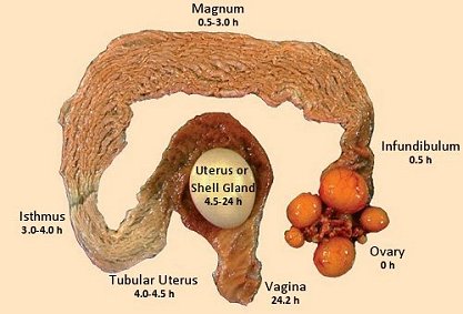
Calcium particle size is important for eggshell quality. Fine calcium carbonate particles pass through the gastrointestinal tract in 2-3 hours, whereas particles above 2mm are retained in the gizzard and will slowly solubilize, delaying the calcium assimilation. Eggshell formation takes 12 to 14 hours and occurs mainly during the night period. Providing a high amount of large calcium particle size before the night, when birds are sleeping, will help laying hens to produce a strong eggshell.

The ratio of coarse to fine calcium particles will increase with bird age as below. Changing the particle size ensures that more calcium will be available at night from the diet instead of from the bone.

Calcium particle size recommendations

Particle size Starter, Grower, Developer Pre-Lay Weeks

17-37

Weeks

38-48

Weeks

49-62

Weeks

63+

Fine (<2mm) 100% 50% 40% 35% 30% 25%
Coarse (2-4mm) 50% 60% 55% 70% 75%


The solubility of limestone may differ according to the source. Calcium with high solubility will not be stored for a long time in the gizzard, negating the particle size effect. Dietary calcium levels may need to be adjusted based on the solubility of your limestone. The in vitro solubility of your limestone source can easily be checked on the farm, with a simple technique using hydrochloric acid. The target is to recover 3-6% of the supplemented limestone.

Water

It’s impossible to have good eggshell quality if you don’t have good water intake and good quality water. For example, excessive salt levels in drinking water can cause persistent damage to shell quality.

Conclusion: invest in the rearing phase

Good nutrition and management practices are key to good shell quality. The rearing period is a key developmental time for future success during the laying period – it is an investment phase.

***

EW Nutrition’s Poultry Academy took place in Jakarta and Manila in early September 2023. Vitor Arantes, Global Technical Services Manager and Global Nutritionist, Hy-Line International, was a distinguished guest speaker in this event.


Housing and management strategies to mitigate heat stress in layers

Dr Daniel Valbuena, Global Manager of Technical Services, Hy-Line International – Conference Report

Heat stress is one of the major environmental stressors in the poultry industry, especially in regions with high temperatures and high humidity. In EW Nutrition’s Poultry Academy in September, the topic was approached in a comprehensive and practical presentation.

A layer’s normal body temperature is about 40° C. Hens are comfortable with an ambient temperature 18° C to 24° C. When that temperature gets above 32° C, the more serious consequences of heat stress occur.

To mitigate the negative effects of heat stress on bird welfare, production, and profitability, essential best practices include those listed below.

Ventilation

Airflow at the birds’ level is key to removing bird heat. Naturally-ventilated barns are particularly at risk of heat stress. Increase the movement of air in open houses with stir fans. Ensure a minimum velocity of 1.8–2 meters/second in the bird areas.

Clean and ensure function of fan louvers. Fan belts should be tightened or changed to avoid slipping or breaking during periods of high temperature. Poorly maintained fans will operate at 50% reduced efficiency.

Air inlets must be adequate to supply the airflow needed to ventilate the house during warm weather. Inadequate inlet space will throttle down the fans and decrease airflow. Inlets should be kept clean and free of anything that might restrict the flow of incoming air. Use baffle boards to direct incoming air onto the birds. Thermostats should be checked for accuracy. An auxiliary power system must be in place in case of a power outage during hot weather.

In addition to running fans throughout the day when it’s hot, fans should run overnight and early morning to bring in cooler air. Air inlets should be adjusted to achieve uniform airflow throughout the building.

In houses equipped with evaporative cooling systems, the pads should be cleaned or replaced when they become clogged. Water flow over the pads should be uniform with no dry areas. Air will flow preferentially through dry areas since there is less resistance. Clean spider webs and dust from window screens frequently to improve ventilation inside the house.

Fans
Fans increase air velocity within the house and create a cooling effect.

Foggers

Fogging or misting is effective at low humidity (<60% relative humidity). Excess moisture in the air from using foggers or misters at high humidity can worsen heat stress conditions.

Foggers or misters need to be checked routinely and should run about 2 minutes out of every 10 if the humidity is low, however, run times can be adjusted based on house temperature and humidity. Fogging the inlet air in negative pressure ventilation systems has a good cooling effect.

Fogging systems should have water filters (to keep nozzles from clogging) and have a positive shutoff to prevent dripping causing wet litter.

Foggers
Foggers should mist, not irrigate, and not operate if the humidity exceeds 60%.

Roof design and cooling

Insulated roofs (R28 is recommended) reduce the radiation and conduction of solar heat through the roof to the interior of the house. Reflective roofing materials or light-coloured materials are recommended.

Ensure to provide ridge vents at roof level to allow hot air to exit so that fresh air may enter the house through side openings. The roof should have an overhang (minimum 60 cm) to reduce direct and indirect sunlight getting into the house.

Thatching can provide cost-effective insulation, but may need to be replaced every few years, and is difficult to clean, and may harbor vermin.

Shade Netting

Thatching

Use of shade netting (left) or thatching material such as palm fronds, paddy straw, corn stalks, sugarcane tops (right) to reduce solar heating of the roof.

Using roof sprinklers during times of extremely high temperature can remove heat from the roof and cool the inside of the house.

Curtains

Adjustable, porous side-wall curtains can be used control the flow of air into the house, and protect birds from direct, hot winds. Water dripping onto side curtains can reduce the house temperature.

Window Shades
Porous window shades block direct sunlight from entering the house but allow air to pass through.

Bird handling

Management practices that require bird handling, such as beak trimming, transfer, and vaccinations, should be done in the early morning hours, or in the evening, when it’s not so hot. Heat-stressed birds have decreased immune function and may not respond as well to vaccination. Alternatively, if birds are panting, they may breathe in too much of a spray vaccine or ingest too much of a water-administered vaccine. In both situations, birds may exhibit signs of the disease that the vaccines are intended to prevent.

Stocking density

It’s critical not to overstock cages. If stocking density is high, the radiant heat between the birds accumulates and the temperature increases. Birds need to be able to spread their wings to increase airflow around their bodies. Caged birds are more susceptible to heat stress because they are unable to seek a cooler place and there is less opportunity for conductive heat loss in cages. The temperature within a cage can be much higher than the measured air temperature in the walkway. Increased air velocity within the cages increases the convective heat loss and removes trapped air between birds.

Manure management

Manure allowed to accumulate reduces ventilation in cages. Remove manure from the house before the hot season, if practical. Heat produced during the decomposition of manure contributes to the heat load in the house. The presence of large amounts of manure in shallow pit houses or under cage batteries restricts the movement of air.

Be prepared and anticipate

  • The key to minimizing the effects of heat stress is to be prepared and anticipate periods of high environmental temperatures, and implementing appropriate management measures prior to the rise in temperatures. Implement cooling systems, such as evaporative cooling pads, misting systems, or fans. Provide natural or artificial shading within the layer house to reduce direct sunlight exposure. Shade structures, curtains, or baffles can help protect the hens from excessive heat.
  • Farm personnel should be trained to recognize and respond to heat stress promptly. You should also have an emergency plan in place for extreme heat events.


Feed and water management strategies to mitigate heat stress in layers

Dr Daniel Valbuena, Global Manager of Technical Services, Hy-Line International – Conference Report

Feed and water management strategies are essential to help mitigate the negative effects of heat stress on bird welfare, production, and profitability. In EW Nutrition’s Poultry Academy in September, the topic was approached in a comprehensive and practical presentation.

Feed management

Feed consumption of the flock should be closely monitored during hot weather. It is important to rebalance the diet for critical nutrients, particularly amino acids, calcium, sodium and phosphorous according to the birds’ productivity demand (i.e., stage of production) and the observed feed intake. Insufficient amino acid intake is the primary reason for productivity loss during hot weather. Several strategies may be employed to help to manage elevated temperatures and maintain higher levels of feed intake:

  • Withdrawing feed from birds 6 hours before peak hot temperatures in the afternoon can lower the risk of heat stress. Encourage as much consumption as possible in the early morning or evening. Using lighting for midnight feeding encourages feed intake.
  • One third of the daily feed ration should be given in the morning and two thirds in the late afternoon. An additional advantage is the availability of calcium in the digestive system during shell formation at night and in the early hours of the morning. This will improve shell quality and reduce the birds from depleting bone calcium.
  • Normally a maximum 1 hour for feeder clean-out time is recommended, but this can be extended to 3 hours when the temperature exceeds 36°C.
  • Consider adding a 1-2-hour midnight feeding.
  • Alter feed particle size, either by increasing it or by feeding a crumble diet. With crumble diets in laying flocks, a supplementary source or presentation of large particle limestone is recommended.
  • Formulate diets using highly digestible materials, particularly protein sources. Metabolism of excess protein is particularly heat-loading on the bird. Formulate to digestible amino acid targets and do not apply a high crude protein minimum in the formula. Synthetic amino acids can reduce crude protein in the diet without limiting amino acid levels.
  • Increasing the proportion of energy contribution from highly digestible lipid, rather than starches or proteins, will reduce the body heat production resulting from digestion. This is known as heat increment and is lowest with the digestion of dietary fat.
  • The bird’s metabolizable energy requirement decreases as ambient temperature increases to above 21°C, resulting from a reduction of energy requirements for maintenance. The energy requirement will decrease with the rise of temperature up to 27°C, above which it will start to increase again since the bird needs additional energy for panting to reduce body heat.

Stress Management Schedule

Management schedule during times of heat stress

Water management

During periods of high environmental temperature, birds have a high demand for drinking water. The water-to-feed consumption ratio is normally 2:1 at 21°C but increases to 8:1 at 38°C. Adequate drinking water must be available to heat-stressed flocks. Ensure that drinkers have sufficient water flow (>70 mL/minute/nipple drinker). If water flow is less the lines need to be checked for flow restriction. If there’s a build-up of iron and other minerals, it needs to be removed. Don’t forget to routinely check water filters and replace them as needed.

It’s easy to overlook a non-functioning drinker here and there; drinkers must be systematically checked to make sure they’re all working. For floor-reared flocks, providing additional drinkers can help accommodate the increased water consumption.

During hot weather, you need to ensure your water system can accommodate the bird’s increased water consumption, and the additional water demands for foggers, evaporative cooling systems and roof sprinklers. The availability of drinking water to a heat-stressed flock should never be compromised.

Cool water temperatures (<25oC) will encourage the birds to drink and reduces the birds’ core temperature. Flush water lines and waterers routinely to keep the water fresh and cool, increasing water consumption, and sustaining egg production. If available, ice can also be added to header tanks. When mechanical cooling systems fail, water flushing can serve as an emergency measure during heat stress.

Drinking water from overhead water tanks can become hot if exposed to direct sunlight. These water tanks should be a light color, insulated and covered to avoid direct sunlight. Water tanks are ideally placed inside the house or underground. Water pipes in the house should not be installed close to the roof to avoid heat from the roof warming up the water in the pipes.

Eggs

Water Cooler
Having the water tank inside the house (above) or light-colored and covered to avoid direct sunlight (below) keeps the water cooler

Use vitamin (A, D, E and B complex) and electrolyte supplements in the drinking water to replenish the loss of sodium, chloride, potassium, and bicarbonate in the urine. Electrolyte supplements are best used in anticipation of a heat stress period and can be added to drinking water for up to 3 days.


Coping with evolutions in the performance and nutritional requirements of layers

Dr. Vitor Arantes, Global Technical Services Manager and Global Nutritionist, Hy-Line International – Conference Report

The layer industry has gone through significant changes during the past decades and has a remarkable capacity to cope with new challenges. Dr Vitor Arantes, Global Technical Services Manager and Global Nutritionist, Hy-Line International, noted that increased egg production, improved feed efficiency, and adaptation of egg quality and bird welfare to consumer preferences have contributed significantly to the success of the egg industry. However, continuous improvement in egg production per hen housed is the most important selection criteria in layer breeding.

Egg producers needs include:

  • More saleable eggs,
  • Eggshell quality,
  • Easier behaviour
  • Housing systems
  • Egg size specifics
  • Sanitary / environmental challenges
  • Profits through productivity

Primary breeders can deliver these producer needs through:

  • Having the correct product for each country
  • Constant follow up
  • Local presence, trust relationship
  • Accurate data collection
  • Critical data analysis
  • Understand the company’s goals
  • Customized technical services according to each customer needs

How has genetics changed?

Examples of genetic progress in layers from 1984 to 2022 cited by Dr Arantes include:

  • Higher persistency (+30 weeks >90%)
  • Higher egg mass (+5.5 kg/hen housed)
  • Smaller hen (-21% mature body weight)

Dr Arantes states the record clutch size, defined as the unstopped length of individual egg production on a daily basis, was an amazing 474 days for a White Plymouth Rock hen. This genetic progress necessitates adjustments in nutrition and management.

Layers weeks

As shown below, growth and organ development occur at various ages. “There is no margin for mistakes – a lack of growth during a stage could have a detrimental impact on pullet quality and subsequent production,” stressed Dr Arantes.

Multi-phasic growth and development during rearing and start of lay

System Age (weeks) Consequence
Gastrointestinal 0-6 Shorter intestinal tract/reduced nutrient absorption
Immune 0-6 Flocks more susceptible to disease challenges
Skeleton 6-12 Shorter frames/less calcium reserves
Muscle 6-12 Impact in persistency of production
Fat >12 Excess can lead to fatty liver, prone to prolapse and mortality

 0-6 weeks of age

Most of the development of the organs of the digestive tract and the immune system occurs during the first 6 weeks of age. Problems that occur during this period can have negative effects on the function of these systems. Birds stressed during this period may have lifelong difficulties in digesting and absorbing feed nutrients. Immunosuppression may also result from problems during this period, leaving the bird more susceptible to diseases and less responsive to vaccinations.

6-12 weeks of age

Most of the adult structural components – muscles, bones and feathers are obtained during the period of rapid growth that occurs at 6-12 weeks of age. Growth deficiencies during this period will prevent the bird from obtaining sufficient bone and muscle reserves, which are necessary to sustain a high level of egg production and to maintain good eggshell quality. About 95% of the skeleton is developed at the end of the bird’s 13 weeks of life. At this time, the plates of the long bones become calcified and further growth in bone size cannot occur.

12-18 weeks of age

During this period, the growth rate slows, and the reproductive tract matures and prepares for egg production. Muscle development continues and the proliferation of fat cells takes place. Excessive weight gain during this period can result in an excessive amount of abdominal fat. Low body weight and stressful events at this time can delay the start of egg production. From 7-10 days before oviposition of the first egg, the medullary bone that is located within the cavities of the long bones can be increased by feeding the bird a pre-laying ration with higher levels of calcium than the development stage.

Bodyweight is a key factor for flock management as this will influence future performance of birds. Consequently, bodyweight should be controlled during the whole life of the layer flocks. Management, in particular nutrition and lighting programs, can help to control bodyweight so birds can achieve their genetic potential.

Uniformity

Uniformity is the most important KPI in our business. However, with the trend towards larger flocks, maintaining uniformity is becoming more challenging. With larger flocks, it is difficult to source one unique flock which thus usually comprises multiple breeding flocks of different ages. Inevitably, uniformity will be poor, hence the need for tools to address unexpected issues. Lack of uniformity becomes a self-perpetuating cycle – dominant versus dominated.

Many egg producers use average body weights compared to the breeder recommendations as a guide to flock status. However, knowing if you have good body weight uniformity is another valuable management tool. In any flock some birds are lighter or heavier than the average body weight. Poor uniformity makes management decisions, such as lighting, feed amounts or diet phase more difficult.

Ideally, the body weight coefficient of variation (CV) should be +/-10% of the mean, increasing the likelihood that your management decision will be appropriate for most of the flock. Inappropriate diet changes, bird handling, vaccination and transfer can reduce uniformity. Flocks should be at 90% uniformity at the time of transfer to the laying facility. Body weight at point of lay significantly affected egg production and eggshell quality.

Grading into 2 or 3 sub-populations of different average bodyweights may be necessary so that each group can be managed in a way that will achieve good whole flock uniformity at the point of lay. The best predictor of future laying performance is the pullet’s body weight and body type at the point of lay.

Vision egg

Vision Egg is a custom diagnostic tool used to analyze data and emphasize flock performance to achieve the highest genetic potential from Hy-Line layers with recommendations connected to customer profitability. This growing, robust database includes data from over 1 billion hens strengthens our flock performance diagnostic tool for improved profitability for Hy-Line customers.

Hy-Line customers can take advantage of this opportunity by sending flock data to their regional business manager or technical service specialist. The information shared with Hy-Line is kept completely confidential.

Summary

The challenge is not egg numbers, stated Dr. Arantes, but saleable eggs. Correct body weight and high uniformity of the flock at point of lay will result in good performance over the laying period, with high peak production and good persistency of production and the production of good quality eggs. Management is the key factor to regulation of body weight during rearing and at point of lay.


Feeding layers for longer laying cycles and optimized production

LAYERS
Conference report 

At the recent EW Nutrition Poultry Academy in Jakarta Indonesia, Dr Steve Leeson, Professor Emeritus, University of Guelph, Canada, commented that “genetic progress in layer breeding has been substantial in recent decades. Since 1995, the yearly change has included +1 egg, -0.01 feed/dozen eggs, -10g final bodyweight, 0.02% mortality, and +1 week at >90% egg production. This improved persistency of commercial laying hens enables egg producers to keep flocks longer in production, provided egg shell quality can be maintained.”

He noted that “the increase in hen-housed egg production is mainly due to longer clutch length and improved uniformity of layer flocks. No doubt, there is a trend in cage layers to longer production cycles. A popular commercial goal is 500 eggs in one cycle with no moult, although this has already been surpassed in many flocks. The modern layer is capable of laying 150 eggs per clutch.”

Dr Leeson, however, stressed that “genetic progress and longer laying cycles have consequences. Long laying cycle programmes start during pullet rearing – you can’t make decisions at 72 weeks of age. Instead, you must start with your end goals, such as persistency, egg size and shell quality, in mind. You can then develop a life-cycle approach to feeding, lighting, nutrition, and general management.” Important issues to manage include:

Body weight control – early and late

Mature body weight dictates subsequent egg size. In the past, the common goal was being at, or above, management guide weight recommendations. For extended lay, a larger body weight results in too large an egg past 70 weeks of age, and so it is more difficult to maintain egg shell quality. Now the goal is to grow a slightly smaller pullet, and emphasis changes to achieving adequate early egg size from this smaller bird. This makes pre-lay nutrition for these slightly smaller pullets even more important.

The scheduling of rearing diets is more important than diet formulation. Dr Leeson’s guidelines are:

  • Starter diet – 19-20% CP, 2,850-2,900 kcal ME/kg from day old to target pullet body weight
  • Grower diet – 17-18% CP, 2,800-2,900 kcal ME/kg from target body weight to mature body size
  • Pre-lay diet (or layer diet?) – 16-18% CP, 2,800-2,900 ME/kg, mature body size to first egg

All nutrients are important, but energy is usually limiting for egg number, whereas protein/amino acids influence egg size (and feathering).

There is now even more emphasis on pullet growing to ensure adequate fat reserves through peak production, so birds are in a positive energy balance. The establishment of an energy reserve occurs during the rearing phase and has a significant effect on the bird’s body composition at point of lay.

Egg size control – early and late

The obvious solution to manage body weight (and egg size) is to light-stimulate a smaller pullet, or at least to not light-stimulate a heavy pullet. This achieves a balance between accepting reduced early egg size, versus limiting an increase in egg size late in the production cycle.

Egg size can be increased in smaller early-lay pullets by:

  • Reducing environmental temperature, if possible, to stimulate feed intake
  • Midnight feeding 19-29 weeks
  • Adequate amino acid nutrition intake, tailored to feed intake, especially methionine
  • Increased number of feedings/day and increased feed particle size (pellets)

Shell strength is negatively correlated with egg size. To temper egg size late in the cycle, Dr Leeson recommended:

  • Body weight control
  • Controlled day length: longer day length = increased feed intake, 14 hours maximum day length in controlled-environment houses
  • Warmer temperature – 26oC is ideal
  • Reduce number of feedings and particle size
  • Temper amino acid nutrition (with caution). Low crude protein/high amino acid diets limit the increase in egg size.

Midnight feeding provides about 1-hour extra light per day and therefore stimulating feed consumption in the middle of the dark period. Having access to feed during this period improves eggshell quality via the supply of calcium during the time when shell calcification takes place. The extra light period is perceived by the bird to be part of the night. The dark period after the light period must be longer than the initial dark period, as the bird perceives the start of the day is the end of the longest period of darkness. Removing midnight feeding should be done gradually – 15 minutes per week, advised Dr Leeson.

Preventing calcium depletion

Also known as cage layer fatigue, calcium depletion is becoming more common in all strains due to high sustained egg output. Calcium deficiency in the feed leads to loss of medullary or long bone (a reservoir of about 4g of calcium) and increased bone fragility. It is commonly seen at 35-40 weeks of age, with a 1-2% occurrence. If the incidence is more than 2%, seek advice for your pre-lay nutrition.

The development of the medullary bones takes about 10 days and requires additional calcium. Pre-lay rations support a smooth transition from developer feed to layer feed, with 2-2.5% calcium, while the other nutrients are similar to a layer feed. Pre-lay rations help the birds to adapt to the high calcium content of layer feed and to maintain sufficient daily feed intake.

To prevent calcium depletion, Dr Leeson suggested:

  • Optimise pre-lay calcium (Ca) and phosphorous (P) nutrition
  • Intake of 1.5g Ca, 350-450mg available P/day for at least 7 days prior to first egg
  • During early lay, ensure 3.5-4 g Ca and 420 mg available P/day
  • Consider vitamin D3 water treatment (150 IU/day, twice weekly)

Pre-lay diets provide the bird with the opportunity to deposit medullary bone. This bone deposition coincides with follicular maturation and is under the control of both estrogens and androgens. The latter hormone seems essential for medullary bone growth, and its presence is manifested in the growth and reddening of the comb and wattles. Consequently, there will be little medullary deposition, regardless of diet calcium level, if the birds are not showing comb and wattle development and this stage of maturity should be the cue for increasing the bird’s calcium intake.

Liver health

Excess energy relative to needs results in excess fat accumulation that is prone to oxidation. This is why you never see fatty liver haemorrhagic syndrome (FLHS) in poor-producing flocks. Layers normally have a very fatty liver, as 100% of egg yolk synthesis occurs in the liver.

The lower the fat content of the diet, the greater the stress/need to fat synthesis in the liver. With a low energy/low fat/carbohydrate diet FLHS is almost universal to varying degrees. One treatment is to add fat to the diet! Haemorrhage (not always FLHS) is inevitable with dietary omega-3s that are very prone to oxidation.

Dr Leeson recommended prevention/control for FLHS, which usually starts about weeks 36-40, including:

  • +1.0 kg choline
  • +0.5 kg methionine
  • +100 IU vitamin E
  • +30% does Hy-D because of impaired liver metabolism of vitamin D3 (that can also impact calcium absorption)
  • Add 2% dietary fat without change in diet energy level


***

EW Nutrition’s Poultry Academy took place in Jakarta and Manila in early September 2023. Dr. Steve Leeson, an expert in Poultry Nutrition & Production with nearly 50 years’ experience in the industry, was the distinguished keynote speaker.

Dr. Leeson had his Ph.D. in Poultry Nutrition in 1974 from the University of Nottingham. Over a span of 38 years, he was a Professor in the Department of Animal &Poultry Science at the University of Guelph, Canada. Since 2014, he has been Professor Emeritus at the same University. As an eminent author, he has more than 400 papers in refereed journals and 6 books on various aspects of Poultry Nutrition & Management. He also won the American Feed Manufacturer’s Association Nutrition Research Award (1981), the Canadian Society of Animal Science Fellowship Award (2001), and Novus Lifetime Achievement Award in Poultry Nutrition (2011).


Eggshell quality: 3 solutions for older laying birds

many eggs

by  Ruturaj Patil, Product Manager Phytogenic Liquids, EW Nutrition

Older laying birds are still a valuable asset, as long as they are managed for performance and productivity. Eggshell quality is one of the elements that, without proper management, can quickly deteriorate. It is therefore essential that the egg producer takes into account all the necessary elements for the formation of high-quality eggs.


The eggshell, in a nutshell

The eggshell represents ten percent of the entire egg, by weight[i]. For instance, a 60-gram egg contains approximately 6 g of shell. Out of this particular shell, approximately 95% is CaCO3[ii], with a total of 2.3 g of Calcium (Ca).

But where does the calcium in the eggshell come from?

The Ca required for the eggshell is obtained, in variable proportions, directly from the feed or water additives (absorbed from the gut and transported via the blood to the shell gland), or from the bone (resorbed by osteoclasts and the Ca transported to the blood to the shell gland).

Maintaining eggshell quality and bone calcium: Mission Impossible?

Eggshell quality is often negatively correlated to bone strength[iii], most probably because body calcium is redirected to the shell to the detriment of the bones and the other way around. This impacts the long-term health of the skeleton; however, modern laying hens can maintain shell quality while preserving bone mineralization[iv].

60 to 75% of shell Ca is derived from the diet on shell-forming days

Approximately 60 to 75% of shell Ca is derived directly from the diet on shell-forming days[v]. This means that the greater the proportion of Ca coming directly from the feed or water additives, the better the eggshell quality can be. Therefore, the factors that can improve shell quality will also reduce the need to mobilize bone Ca and can also help to maintain skeletal health.

In old laying birds, generally after peak production, the ability to deposit Ca onto the shell remains relatively constant[vi], so an increase in egg size after peak production will tend to result in reduced shell quality. Dietary requirements for Ca tend to increase and those for phosphorus (P) tend to decrease as hens age.

Also, as hens age, the efficiency of Ca metabolism decreases[vii]. Increases in dietary Ca and a widening of the Ca:available P ratio are intended to counter this issue. Excess dietary P can also reduce shell quality[viii].

Because of its importance in Ca and P absorption from the gut, adequate dietary vitamin D activity must also be provided[ix]. Feeding of the vitamin D metabolite 25-OH vitamin D3 can help to maintain skeletal and shell quality in high-producing laying hens[x].

Ca metabolism is a complex game

Ca metabolism is regulated by various hormones such as calcitonin, 1,25-dihydroxyvitamin D3 (calcitriol), and parathyroid hormone. Estrogen, androgens, and prostaglandins also appear to have an important role in avian Ca metabolism.


Source: Ricardo (2008)

Egg formation and Ca requirements


Source

A hen ovulates approximately 15 to 75 minutes following oviposition[xi], and the ovum takes approximately 4.25 hours to reach the shell gland[xii], at which point calcification takes approximately 17 hours[xiii]. Hens generally lay eggs in the morning and early in the afternoon[xiv]. The hen can use the Ca and P made available through diet to recover medullary bone losses during the next 5 hours after oviposition.

Once the ovum reaches the shell gland, the demand for calcium naturally increases greatly as eggshell formation progresses. The highest eggshell mineral accretion takes place 5 – 15 hours after the egg enters the shell gland[xv], which normally happens later in the afternoon and during the night preceding egg laying.

Hourly Ca requirements for eggshell calcification


Ca dietary requirements vary with species, age, breeding status, and dietary levels of vitamin D. Egg-laying birds and growing birds require more Ca than adult non-breeding birds.

Common eggshell quality problems and causes

In many cases, the source of eggshell problems can be detected by recognizing the specific markers. For instance, cracked, soft-shelled or corrugated eggs can be caused by saline water, or the impact of mycotoxins; shell-les eggs can be caused by improper amounts of Ca, P, Mn or vitamin D3, as well as by infectious bronchitis or Newcastle disease IB. However, among the main causes of eggshell quality issues is heat stress.

In hot temperatures, increased respiration rates can cause an increase in CO2 loss. The reduction of the pool of bicarbonate ions can result in respiratory alkalosis and an increase in blood pH[xvi]. A reduction in bicarbonate ions in the shell gland reduces the formation of CaCO3 and decreases shell quality.

Under heat stress, birds will also tend to decrease their feed intake during the day to reduce diet-induced thermogenesis. Calcium intake is therefore also reduced, and shell quality decreases as a consequence.

3 solutions for eggshell quality in older layers

Midnight feeding in hot climates

At midnight, when temperatures are typically cooler, the addition of one to two hours of light can help the birds increase feed consumption[xvii]. Midnight feeding can also have the benefit of providing a dietary source of Ca to support eggshell formation during the night and reduce reliance on bone reserves[xviii].

Nutrition supplements

Along with Calcium, some micro-minerals can also influence eggshell quality. Zinc, Manganese and Copper act as cofactors of enzymes involved in the mineralization process during eggshell formation. Although European Union legislation restricts the use of high levels of these minerals, several studies in layers indicate increased egg shell resistance by increasing the dietary concentrations of microminerals. Using organic forms of Zinc, Manganese and Copper appears to be an alternative way to increase the absorption of these minerals, as organic forms appear to be more digestible than inorganic forms. Considering the high cost of organic minerals, a mix of organic and inorganic forms of critical minerals could be a better option.

Liquid Ca supplements

If a hen is fed a diet containing only a small-particle Ca sources, such as finely ground limestone, the intestine will be deprived of a source of Ca during the night, when demand for Ca is highest. At that point, the hen will be entirely reliant on bone Ca to support eggshell formation. A combination of Ca supplementation through water additives can be a good alternative as readily available Ca to the hen to support high Ca requirements during the late afternoon and through the night. Liquid Ca additives also offer further precise and user-friendly    application.

Stimuvital IP: a liquid solution from EW Nutrition

Stimuvital IP (formerly Shellimprover) is a liquid nutritional additive for laying hens, supporting the quality of eggshells and bone health. It contains a cocktail of Ca and vitamins whose benefits in laying birds are well proven through field studies, existing literature, and years of market experience.

Benefits proven in Australia field trial

22,500 layer birds were split into two equal-size groups, one of which (11250 birds) was supplemented with Stimuvital IP for 3 days every two weeks, starting from age 53rd to 63rd week. Improvements in eggshell thickness and strength could be noticed after the application of Shellimprover. Egg weight was consistent in Stimuvital IP -supplemented birds.3 days every fortnight by using the Easy@ system. In total, the 11250 birds received (2 (feed lines) x 3 (times per days) x 265ml x 3 (days) x 6 (week 53, 55, 57, 59, 61, 63) 28620mll of Stimuvital IP.


Benefits proven in China field trial

The field trial was carried out on a commercial layer farm. A control group and Stimuvital IP (Shellimprover) group had 50,000 birds each. Stimuvital IP was supplemented for 3 days every two weeks, starting from age 57th to 62nd week. The Stimuvital IP supplementation improved eggshell quality, including eggshell thickness, laying rate, and number of saleable eggs during the trial period.


Optimizing quantity, quality, and overall profitability for layer producers

Ca concentration in the blood is controlled by many interacting feedback loops that involve Ca, phosphate, PTH, vitamin D3, and calcitonin. Supplementation of Vitamin D3 can help maintain skeletal and shell quality in high-producing laying hens[xxiv].

Stimuvital IP offers an essential cocktail that caters to the additional requirements of Ca and vitamins in older laying birds. It thus supports Ca metabolism and eggshell quality. And, in the end, better eggshell quality reduces broken egg percentage and optimizes the number of salable eggs and profitability for layer producers.


Notes

[i] Pelicia et al., 2009; Bello and Korver, 2019

[ii] Nys et al., 2004

[iii] Orban and Roland Sr, 1990

[iv] Bello and Korver, 2019

[v] Driggers and Comar, 1949

[vi] Roland Sr et al., 1975

[vii] Wistedt et al., 2019

[viii] Miles et al., 1983

[ix] Wen et al., 2019

[x] Silva, 2017; Akbari Moghaddam Kakhki et al., 2019

[xi] Beuving and Vonder, 1981

[xii] Roberts, 2004

[xiii] Hincke et al., 2012

[xiv] Samiullah et al., 2016; Hunniford et al., 2017

[xv] Hincke et al., 2012

[xvi] Franco-Jimenez et al., 2007

[xvii] van Staaveren et al., 2018

[xviii] Harms et al., 1996

[xix] Chowdhury, 1990

[xx] Leach and Gross, 1983

[xxi] Zhang et al., 2017

[xxii] Atteh and Leeson, 1983

[xxiii] Atteh and Leeson, 1985

[xxiv] Silva, 2017; Akbari Moghaddam Kakhki et al., 2019

Full references are available upon request.