INFOGRAPHIC – Target measurements for water quality

Water is a main nutrient and carrier for vaccines, medicine – including antibiotics, but also for pathogens
Chemistry


Microbiology


Water is a main nutrient and carrier for vaccines, medicine – including antibiotics, but also for pathogens
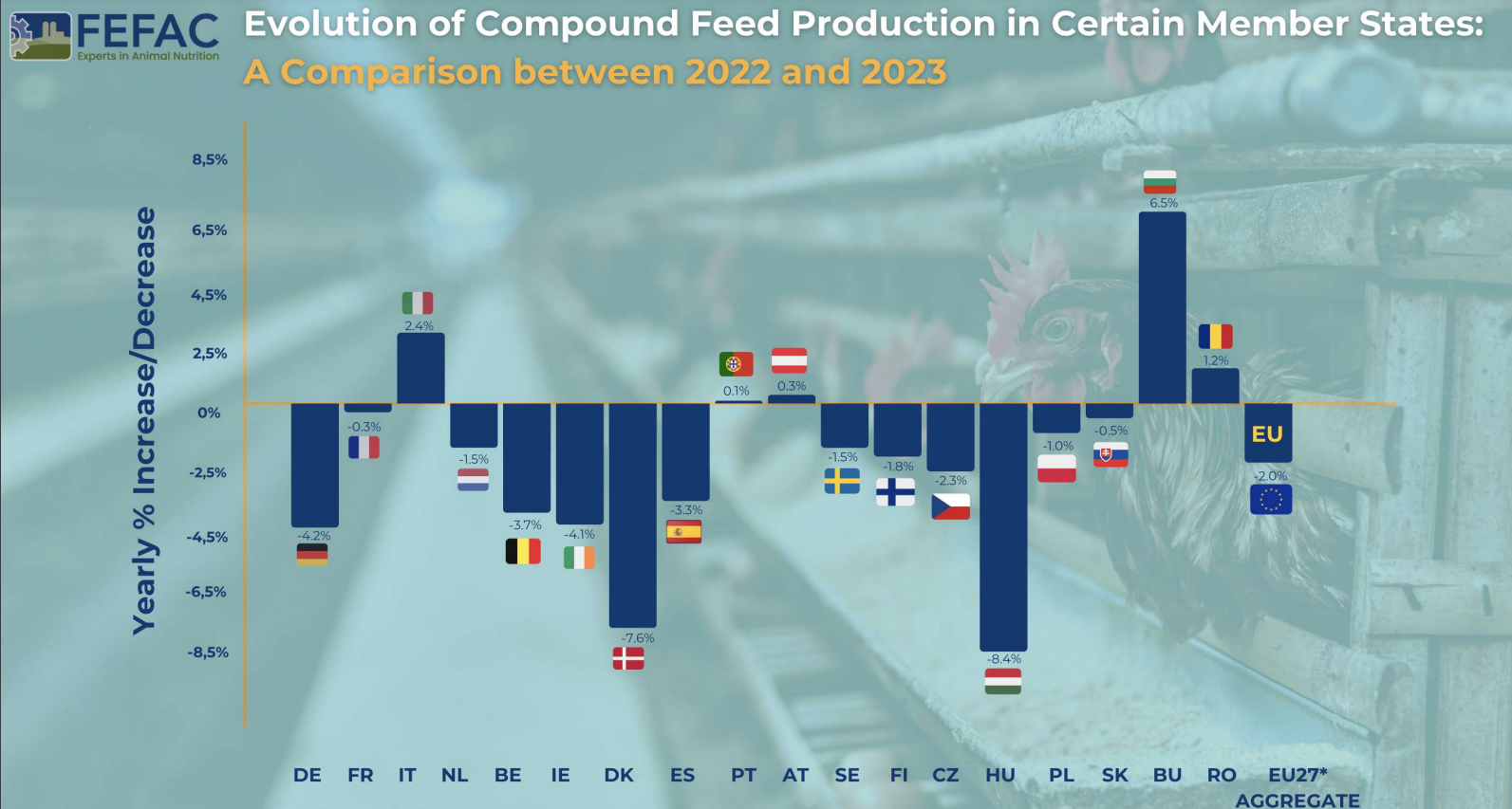
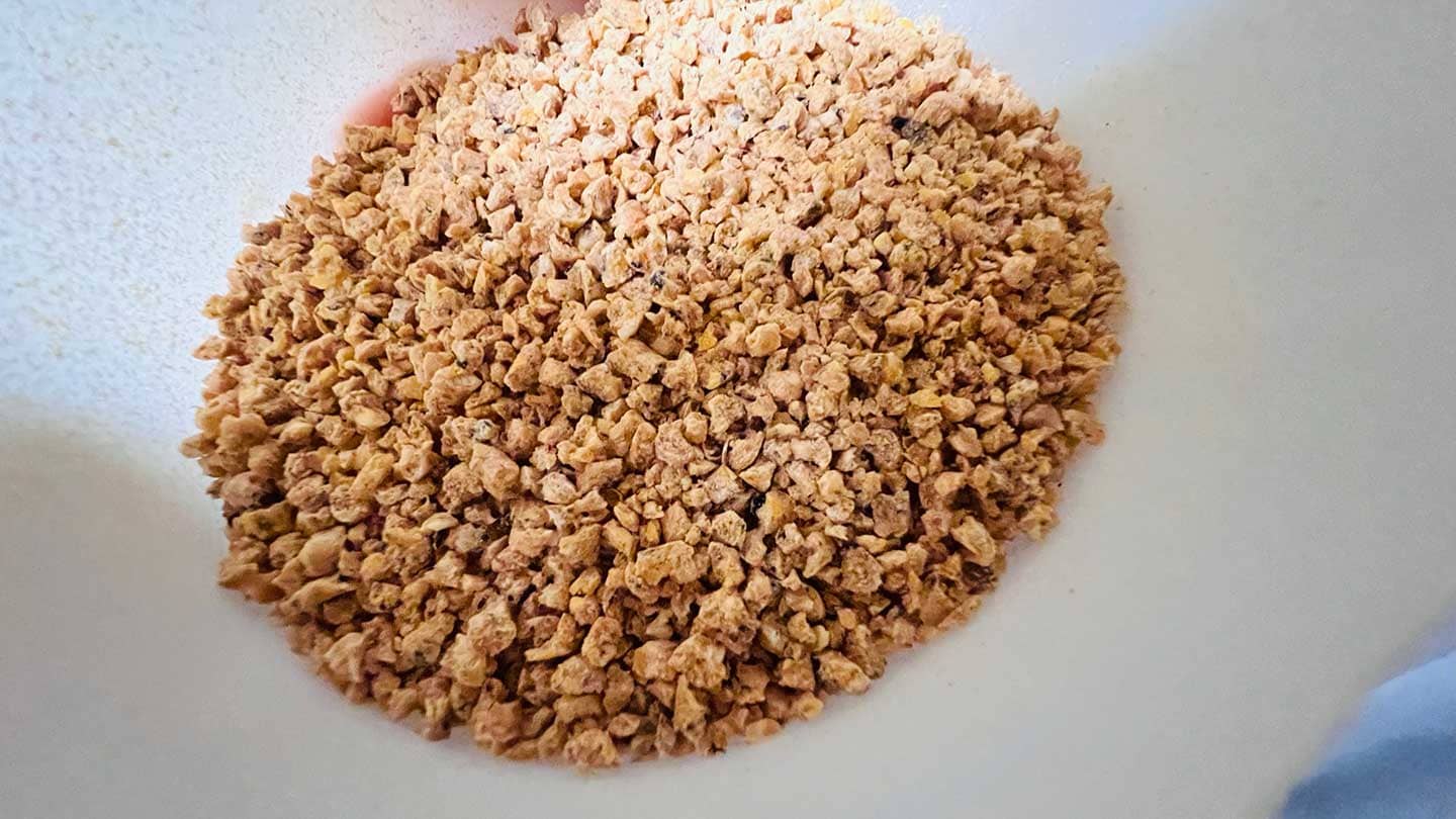
Total Production 2023: 144.3 million metric tons for farmed animals
Change from 2022: 2% decrease
Political and Market Pressures: Addressing crises and the shift towards sustainable feed.
Climate and Diseases: Effects of droughts, floods, Avian Influenza (AI), and African Swine Fever (ASF) on raw material supply and animal production.
National Policies: Initiatives for greenhouse gas and nitrate emission reduction.
Consumer Trends: Food price inflation impacting demand.
Production Variability: Different trends across EU Member States, with notable decreases in countries like Germany, Ireland, Denmark, and Hungary, and slight increases in Austria, Bulgaria, Italy, and Romania.

Pig Feed: Major decline of nearly 2.5 million tons. Key challenges included:
Poultry Feed: Increase by 0.9 million tons, yet still 700,000 metric tons below 2021 levels. Challenges included declines in Hungary and Czechia due to reduced broiler production.
Cattle Feed: Decrease of 0.8 million tons from 2022.
The EU’s compound feed production in 2023 faced numerous challenges, leading to an overall decrease. The pig feed sector was most severely hit, while poultry feed showed some recovery. The influence of environmental, economic, and policy factors played a significant role in shaping these trends. Despite the price of feed cereals falling back to the levels seen before Russia’s invasion of Ukraine, these challenges will continue to be felt in 2024.
Source: FEFAC
Certified Organic: (US, others) To be labeled as “Certified Organic” in the US, meat and poultry must come from animals that are raised in accordance with organic farming standards. These standards typically include restrictions on the use of synthetic pesticides, herbicides, antibiotics, and genetically modified organisms (GMOs). The animals are typically raised with organic feed and have access to the outdoors.
Chemical-free: (US) A product that contains no artificial ingredients or chemical preservatives.
Free-range or Free-roaming: (International) Poultry that has been allowed access to the outside.
Free-Range or Pasture-Raised: (US, others) These terms suggest that the animals had access to the outdoors or were raised on pasture, which can offer better living conditions than confined, industrial operations.
Fresh poultry: (US) Poultry that has never been below 26°F.
Frozen poultry: (US) Poultry that has been held at 0°F or lower.
Grain-Fed: (International) This label implies that the animals were primarily fed grains or other non-grass-based diets, which is common in many commercial meat production systems.
Grass-Fed: (International) “Grass-Fed” typically means that the animals were primarily fed a diet of grass or forage throughout their lives, although some supplemental grains may be allowed. This label does not necessarily imply organic or non-GMO practices.
Halal: (International) Halal meat is prepared following Islamic dietary laws. This includes specific slaughter methods and requirements for the handling and preparation of the meat.
Kosher: (International) Kosher meat is prepared according to Jewish dietary laws and involves specific slaughtering practices and inspections.
Mechanically separated meat: (US) A paste-like meat product produced by forcing bones, with attached edible meat, under high pressure through a sieve or similar device to separate the bone from the edible meat tissue.
Natural: (US) A product containing no artificial ingredient or added color and is only minimally processed (a process which does not fundamentally alter the raw product). The label must explain the use of the term natural (such as “no added colorings or artificial ingredients; minimally processed”).
No antibiotics: (US) The terms “no antibiotics added” may be used on labels for meat or poultry products if sufficient documentation is provided by the producer to the USDA demonstrating that the animals were raised without antibiotics. If an animal becomes sick and requires antibiotics, it cannot be sold as “no antibiotics added.”
No hormones (beef): (US) The term “no hormones administered” may be approved for use on the label of beef products if sufficient documentation is provided to USDA by the producer showing no hormones have been used in raising the animals.
No hormones (pork or poultry): (US) Federal regulations prohibit the use of hormones in raising hogs and poultry.
Non-GMO: (International) A “Non-GMO” label indicates that the animals were not fed genetically modified organisms. This label may apply to both feed and the animals themselves.
Organic: (International) Meat and poultry labeled as organic must come from animals fed organic – which also means non-GMO – feed, given fresh air and outdoor access, and raised without antibiotics or added growth hormones. Organic livestock must also have access to pasture for at least 120 days per year.
Protected Designation of Origin (PDO) and Protected Geographical Indication (PGI): (EU) These labels are used to protect and promote regional and traditional foods. Meat labeled with PDO and PGI must come from specific regions and meet particular quality and production standards.
Raised without Antibiotics or Antibiotic-Free: (International) This label indicates that the animals were not treated with antibiotics during their lifetime. However, this label does not necessarily mean the animals were raised in organic or free-range conditions.
Sustainably Sourced: (International) This label may indicate that the meat was produced with a focus on environmental and ethical considerations, such as minimizing ecological impact and promoting fair labor practices.
The authorization and marketing of feed additives in the European Union is currently governed by Feed Additives Regulation (EC) No 1831/2003, which came into effect in 2004. In 2021, the European Commission formalized an initiative to revise it, stating as reasons both the focus brought by the Farm to Fork Strategy, as well as inherent complexities in phrasing, process, and more. Representatives of the EC’s responsible unit, DG SANTE Unit G5, have now confirmed to EW Nutrition that, following consultations and analysis, the revision of the legislation on the authorisation of feed additives will not happen under the current Commission’s mandate.
The revision was initially deemed necessary on several grounds:
Near the end of the two-year assessment process, however, the response of European governmental, supra-national, and non-governmental bodies appears to have been lukewarm. Overall, the conclusion of the EC unit overseeing the process was that “while a review of the framework would be useful, it does not appear necessary, considering the possibilities already granted by the existing legal framework.” In other words, applicants will have to use the existing mechanisms for applications, with no prospect for change in the near future.
Other strategies and regulations have also fallen through the cracks. For instance, the EU Animal Health Strategy 2007-2013 has not been updated in 10 years and there are no plans to renew the initiative. This is likely because the Green Deal and the flurry of new or upcoming regulations related to it are expected to supplant the framework for protein production in the European Union.
As the mandate of the current EC ends in 2024, there is a slim chance that the feed additive authorization process might be made less cumbersome once a new commission takes over.
By Dr Merideth Parke BVSc, Regional Technical Manager Swine, EW Nutrition
We care for our animals, and antibiotics are a crucial component in the management of disease due to susceptible pathogens, supporting animal health and welfare. However, the administration of antibiotics in pig farming has become a common practice to prevent bacterial infections, reduce economic losses, and increase productivity.
All antibiotic applications have collateral consequences of significance, bringing a deeper consideration to their non-essential application. This article aims to challenge the choice to administer antibiotics by exploring the broader impact that antibiotics have on animal and human health, economies, and the environment.
Antibiotics do not specifically target pathogenic bacteria. By impacting beneficial microorganisms, they disrupt the natural balance of microbial communities within animals. They reduce the microbiota diversity and abundance of all susceptible bacteria – beneficial and pathogenic ones… many of which play crucial roles in digestion, brain function, the immune system, and respiratory and overall health. Resulting microbiota imbalances may present themselves in animals showing health performance changes associated with non-target systems, including the nasal, respiratory, or gut microbiome 7, 8, 14. The gut-respiratory microbiome axis is well-established in mammals. Gut microbiota health, diversity, and nutrient supply directly impact respiratory health and function13. In pigs specifically, the modulation of the gut microbiome is being considered as an additional tool in the control of respiratory diseases such as PRRS due to the link between the digestion of nutrients, systemic immunity, and response to pulmonary infections11.
The collateral effect of antibiotic administration disrupting not only the microbial communities throughout the animal but also linked body systems needs to be considered significant in the context of optimal animal health, welfare, and productivity.
The consideration of the pathogenesis of individual bacteria is critical to mitigate potential for direct collateral effects associated with antibiotic administration. For example, in cases of toxin producing bacteria, when animals are medicated either orally or parenterally, mortality may increase due to the associated release of toxins when large numbers of toxin producing bacteria are killed quickly2.
Numerous animal studies have investigated the modulatory role of intestinal microbes on the gut-brain axis. One identified mechanism seen with antibiotic-induced changes in fecal microbiota is the decreased concentrations of hypothalamic neurotransmitter precursors, 5-hydroxytryptamine (serotonin), and dopamine5. Neurotransmitters are essential for communication between the nerve cells. Animals with oral antibiotic-induced microbiota depletion have been shown to experience changes in brain function, such as spatial memory deficits and depressive-like behaviors.
Anaerobic treatment technology is well accepted as a feasible management process for swine farm wastewater due to its relatively low cost with the benefit of bioenergy production. Additionally, the much smaller volume of sludge remaining after anaerobic processing further eases the safe disposal and decreases the risk associated with the disposal of swine waste containing residual antibiotics4.
The excretion of antibiotics in animal waste, and the resulting presence of antibiotics in wastewater, can impact the success of anaerobic treatment technologies, which already could be demonstrated by several studies 6, 11. The degree to which antibiotics affect this process will vary by type, combination, and concentration. Furthermore, the presence of antibiotics within the anaerobic system may result in a population shift towards less sensitive microbes or the development of strains with antibiotic-resistant genes1, 12.
Regulatory authorities specify detailed withdrawal periods after antibiotic treatment. However, residues of antibiotics and their metabolites may persist in animal tissues, such as meat and milk, even after this period. These residues can enter the human food chain if not adequately monitored and controlled.
Prolonged exposure to low levels of antibiotics through the consumption of animal products may contribute to the emergence of antibiotic-resistant bacteria in humans, posing a significant public health risk.
As mentioned, the administration of antibiotics to livestock can result in the release of these compounds into the environment. Antibiotics can enter the soil, waterways, and surrounding ecosystems through excretions from treated animals, inappropriate disposal of manure, and runoff from agricultural fields. Once in the environment, antibiotics can contribute to the selection and spread of antibiotic-resistant bacteria in natural bacterial communities. This contamination poses a potential risk to wildlife, including birds, fish, and other aquatic organisms, as well as the broader ecological balance of affected ecosystems.
One of the widely researched concerns associated with antibiotic use in livestock is the development of antibiotic resistance. The development of AMR does not require prolonged antibiotic use and, along with other collateral effects, also occurs when antibiotics are used within recommended therapeutic or preventive applications.
Gene mutations can supply bacteria with abilities that make them resistant to certain antibiotics (e.g., a mechanism to destroy or discharge the antibiotic). This resistance can be transferred to other microorganisms, as seen with the effect of carbadox on Escherichia coli5 and Salmonella enterica2 and the carbadox and metronidazole effect on Brachyspira hyodysenteriae15. Additionally, there is an indication that the zinc resistance of Staphylococcus of animal origin is associated with the methicillin resistance coming from humans3.
Consequently, the effectiveness of antibiotics in treating infections in target animals becomes compromised, and the risk of exposure to resistant pathogens for in-contact animals and across species increases, including humans.
To successfully minimize the collateral effects of antibiotic administration in livestock, a unified strategy with support from all stakeholders in the production system is essential. The European Innovation Partnership – Agriculture9 concisely summarizes such a process as requiring…
In general, implementing responsible antibiotic stewardship practices is paramount. This includes limiting antibiotic use to the treatment of diagnosed infections with an effective antibiotic, and eliminating their use as growth promotors or for prophylactic purposes.
While antibiotics play a crucial role in ensuring the health and welfare of livestock, their extensive administration in the agricultural industry has collateral effects that cannot be ignored. The development of antibiotic resistance, environmental contamination, disruption of microbial communities, and the potential transfer of antibiotic residues to food pose significant challenges.
Adopting responsible antibiotic stewardship practices, including veterinary oversight, disease prevention programs, optimal animal husbandry practices, and alternatives to antibiotics, can strike a balance between animal health, efficient productive performance, and environmental and human health concerns.
The collaboration of stakeholders, including farmers, veterinarians, policymakers, industry and consumers, is essential in implementing and supporting these measures to create a sustainable and resilient livestock industry.
References
By Technical Team, EW Nutrition India
Consumer demand drives egg production. With 10 billion people on the planet by the year 2050 (1), producers are under more pressure to provide more protein of higher quality. Modern production practices help extend the laying cycle of commercial flocks to 90–100 weeks. The volume of eggs produced worldwide has thus increased by more than 100% since 1990. Consumers are pushing not just for more eggs, but also for larger eggs. Due to these shifting requirements, farmers and integrators are under pressure to meet demand. As a result, the birds are under metabolic stress to meet needs, which can compromise eggshell quality, laying consistency, and gut health.
Gut health is a key factor in achieving maximum productive potential and laying rate, not only because it’s a key factor for digestion and the absorption of nutrients but also because it’s an essential component of the bird’s immune system.
In today’s layer production, when the cycle is increasing and overall demand to limit the use of antibiotics is growing, laying persistency, eggshell quality, and gut health are critical topics. But what does a laying hen’s healthy gut mean?
Birds need a healthy gut to maximize production. Genetics, nutrition, management and biosecurity all affect production parameters. A gut with a diverse pH and healthy microbiota prevents infections. Gut health is affected by Goblet cells, paneth cells, endocrine cells, absorptive enterocytes, tight junctions, GALT, and mucus. To deal with potential challenges and ensure optimal bird performance, a complex approach is needed, consisting of optimal carbohydrates, proteins, amino acids, minerals, vitamins, enzymes, organic acids, and management strategies.
Vital amino acids, Zn, Vit E, Se, etc. must be supplemented according to the production status and environment to establish good immunity. Maximum production requires a stress-free, hormonally balanced, clean environment, as well as optimal nutrition. Especially given the push for reduced antibiotics and rising welfare and food standards, particularly from the expansion in cage-free farming, producers need to pay considerable attention to the issues of maintaining a healthy gut with these added challenges. Several aspects must be considered when it comes to gut health.
Both vectors create a connection between the external and internal environment of the hen, increasing the possibility of a negative effect on the intestinal balance.
Some common influences:
 Fig. 1: Effects of dietary mycotoxins on histopathology of the duodenum, jejunum, and ileum Adapted from Zhao et al., 2021 (3)
Nowadays the gut health of the layer matters more than ever. In many countries, consumer preferences have been shifting towards eggs produced in non-cage environments and, in these new housing systems, birds are in closer contact with the litter and are more prone to the proliferation of gut pathogens.
Traditionally, layers were housed in cages, which benefited egg producers by making better use of available space and increasing productivity. This resulted in more birds per house, more automated operations, better management, improved hygiene, decreased incidence of infectious diseases, and cheaper feed consumption and production costs.
Cages, on the other hand, pose other health and welfare concerns. They limit or prevent mobility, ground scratching, wing-flapping, and soaring. As a result, there is increasing pressure for birds to be cage-free and, eventually, free-range. The European Commission stated that, by the end of 2023, a legislative proposal will phase out, and eventually prohibit, the use of cages for a variety of farm animals, resulting in an increase in the number of layers reared in a cage-free system.
According to the Egg Track Report (2021), 219 egg farmers, retailers, food service firms, and hotel chains have pledged to transition completely to cage-free eggs by 2025, with 47 of these companies expanding their commitments to encompass their global supply. This means that farmers and integrators will face increased pressure to migrate from a cage system to a cage-free system. As a result, it is necessary to consider new issues or challenges that may be exacerbated by transitioning to a cage-free production system.
Water (70%), proteins (10%), and lipids (20%) make up egg yolks. The yolk lipids are triglyceride-rich lipoproteins that are produced in the liver and transferred to the ovary. Cholesterol transported to the egg yolk by lipoproteins is also deposited there, demonstrating the importance of the liver in egg formation.
The gut plays an important role in preventing liver damage by acting as a barrier against dangerous viruses and toxins that could enter the bloodstream and reach this key organ. Efficient feed digestion and absorption of nutrients are essential for the hen to obtain the “material” for maintenance, growth, and egg production.
The Association of Veterinarians of Egg Production in the US found in 2014 that gastrointestinal difficulties cause 40% of health issues during the pullet phase and 50% during production. Coccidiosis, necrotic enteritis, and feed passage were the biggest threats from these gastrointestinal illnesses.
Aging reduces digestive health, causing nutrient digestion and absorption problems and immunological issues. As a result, eggs produced by older hens show increased micro-cracks, gross cracks, and a higher number of dirty eggs.
Poor intestinal physiology can impair mineral absorption, notably calcium. When this happens, hens utilise the calcium from their bones, but if the problem persists, these stores may diminish and thin-shelled eggs may appear, increasing the percentage of broken eggs. Shell-less eggs are possible.
In continuation with what was described in the previous point, the bird’s skeletal system weakens due to the use of calcium reserves of the bones, which leads to bone fractures, such as the head of the femur, and other locomotor problems of similar pathogenesis.
Deficient intestinal physiology may also cause intestinal flora imbalance. Certain germs proliferate excessively, harming the mucosa and affecting faeces consistency. This raises the number of dirty eggs, which harms consumers due to cross-contamination.
Due to the alteration of the nutritional function of the intestine, feed digestion and nutrient absorption is affected, and this leads to a decrease in their concentration in the egg. This deficiency causes yolk pigmentation problems, poorer egg nutritional value, and worsening of the Haugh Units, among other issues.
Related to the previous point, the alteration of the nutritional functions of the intestine will also decrease the percentage of egg laying. This is because the bird will not absorb enough nutrients and minerals to cover the needs for egg production (both for the metabolic process and to form the egg). The mentioned problems, derived from inadequate intestinal physiology, lead to poor qualitative and quantitative egg production, which is, in most cases, very difficult to reverse in the short term, and that leads to significant economic losses.
During the production cycle, the gastrointestinal health of laying hens has a substantial impact on both efficiency and profitability. During peak egg production, chickens often cannot consume enough feed to meet their protein and calcium requirements. This stress can disrupt the gut microbiota, resulting in pathogenic bacterial outbreaks. Infections with Escherichia coli and Clostridium perfringens are prevalent in laying hens. Antibiotics would be administered to the birds to deter severe mortality.
Antibiotics, on the other hand, have hidden costs because eggs produced during antibiotic treatment and withdrawal cannot be marketed for human consumption. Furthermore, antibiotic misuse, such as using too little or for too short a time, might contribute to the development of antibiotic resistance.
A variety of non‐drug substances have been promoted as aids to enhance gut health and to mitigate the risks of coccidiosis and necrotic enteritis in antibiotic‐free production. These products include phytogenic additives, probiotics, prebiotics, organic acids, yolk immunoglobulins, bacteriophages, yeast products, and others.
Probiotics and competitive exclusion (CE) cultures are available for hatchery application, usually by spray, and most of the alternatives are available for feed or water administration. Because of logistical issues, producers usually prefer feed‐administered products, especially if intended for large‐scale applications for prevention.
Many gut health solutions can be added to water, included in feed at the feed mill, or top-dressed at the farm. Gut health supplements work differently, making selection challenging. Some gut health products encourage beneficial bacteria, gut tissue formation, digestion, or pathogen inhibition. Thus, while choosing a gut health product, it’s important to determine the root cause of the challenge and make sure the product can address the problem.
The right products are also effective in antibiotic reduction initiatives. However, their prophylactic use should be considered as an alternative option. A strategic method is to deliver a gut-friendly substance at key periods in the chicken’s life.
Development, transition, and maintenance are three primary stages in the gut development chain (see Figure 2).
Understanding the needs of the gut at different points in the bird’s life and the main goals of gut health support at these times is important when designing gut health strategies.
 Fig2: Gut need assessment and management strategy
Multiple scientific studies highlight phytomolecules as one of the key elements in antibiotic-free production. These substances support digestion and improve the utilization of nutrients, resulting in a higher daily weight gain, uniform flock, and better feed utilization.
They also have a proven anti-inflammatory effect, as shown in Figure 3. NF-κB is a critical regulator for the expression of genes involved in inflammation. It has been demonstrated that NF-κB plays a novel role in the mechanism of increased epithelial permeability induced by inflammatory factors (including LPS and TNF-α) (5).
Phytomolecules, when combined with effective delivery and synergistic value inside the animal, also have a proven antimicrobial effect and help prevent the development of resistance. Various forms of stresses and insults from feed water and the environment cause oxidative stress and thereby impaired tight junctions, resulting in a leaky gut. Leaky gut has multiple consequences, ranging from poor flock performance to wet litter and raised ammonia levels. Phytomolecules are well documented for their NF-κB inhibitory and anti-inflammatory properties (6). They also help curb oxidative stress and maintain gut integrity. The right product will also be mild on the beneficial flora, showing selective antimicrobial activity and preserving the balance of the gut microbiota.
Finding the right product (perfect formulation and technology to counter high volatility, offer high thermostability, and yet provide effective delivery inside the animal) is of paramount importance for desired results.
 Figure 3. NFkB activity with phytomolecule-based product Ventar D (EW Nutrition)
Optimal growth and FCR in food-producing animals depend on intestinal health. Researchers have studied gut flora, function, and immunity. Regional variations in chicken production, management styles, environment, disease challenge, and feed raw materials complicate gut health maintenance. Consequently, appropriate bird management techniques are essential to bird health, welfare, and performance.
Due to the recent focus on reducing or restricting antibiotic use, intestinal problems have increased, often resulting in productivity losses. This has led to the development of several feed additives that can improve intestinal microbiota, prevent pathogens from adhering to epithelial cells, and boost immune response.
Probiotics, prebiotics, organic acids, organic acid blends (protected or not), phytobiotics, and feed enzymes are everywhere. Feed additive performance depends on parameters like hen age, management, production method, genetics, etc. It also depends on additive formulation, a multi-layered mode of action, and on a coating technology that leads to effective release of ingredients in the GIT.
By Dr. Inge Heinzl, Editor, EW Nutrition
Nowadays, intensive livestock farming with high stocking densities causes stress in the animals and affects the immune system9, 13. The increase in respiratory diseases with associated losses and costs is only one of the consequences. Due to antimicrobial resistance, antibiotics should only be used in critical cases, so effective alternatives are requested to support the animals.
It already has a name: PRDC or the Porcine Respiratory Disease Complex describes the cooperation of viruses, bacteria, and non-infectious factors such as environmental conditions (e.g., insufficient ventilation), stocking density, management (e.g., all-in-all-out only by pens and not for the whole house) and pig-specific factors such as age and genetics, altogether causing respiratory issues in pigs. Non-infectious factors such as high ammonia levels weaken the immune system and lay the foundation for, e.g., mycoplasmas which damage the ciliated epithelial cells in the upper respiratory tract, the first line of defense, and pave the way for PRRS viruses. They, on their part, enter the respiratory tract embedded in inhaled dust. There, they harm the macrophages and breach a further barrier of defense. Another pathfinder is the Porcine Circovirus 2 (PCV2), which destroys specific immune cells and leads to a generally higher susceptibility to infectious agents. Bacteria such as Pasteurella multocida or Streptococcus suis further on can cause secondary infections7, 20, 22. Also, the combination of mycoplasma hyopneumoniae and porcine circovirus, both typically low pathogenic organisms, leads to severe respiratory disease15.
The main tasks of the respiratory tract are to take in oxygen from the air and to pump out the CO2 entailed by the catabolism of the tissue. In pigs, however, the respiratory tract is also responsible for thermoregulation, as pigs don’t have perspiration glands. The animals must get rid of excessive heat by rapid breathing. If the respiratory function is affected due to disease, thermoregulatory capacity is reduced. The resulting lower feed intake leads to decreased growth performance and less economic profit17. One of the first studies concerning this topic was conducted by Straw et al. (1989)21. They asserted that, with every 10 % more affected lung tissue, daily gain decreased by about 37g. This negative correlation between affected lung tissue and weight gain could be confirmed by Paz-Sánchez et al. (2021)18. They saw that animals with >10% lung parenchyma impacted by cranioventral bronchopneumonia needed a longer time to market (208.8 days vs. 200.8 days in the control), showed a lower carcass weight (74.1 kg vs. 77.7 kg in the control group) and, therefore, also a lower daily gain (500.8 g/day compared to 567.2 g/d). In another study, Pagot and co-workers (2007)16 observed 7000 pigs from 14 French farms. They saw a significant negative correlation (p<0.001) between the prevalence of pneumonia and growth and a weight gain loss of about 0.7 for each point of pneumonia increase.
People have always used herbal substances to cure illnesses, be it willow bark for pain, chamomile for anti-inflammation or an upset stomach. Ribwort and thyme are used as cough suppressants, and eucalyptus and menthol help you breathe better. What is good for humans can also be used for pigs. To use plant extracts efficiently, it is crucial to know their specific modes of action. Due to their volatile nature, essential oils can directly reach the target site, the respiratory tract, via inhalation1.
Many essential oils show some degree of antimicrobial activity. So, the oils of, e. g., oregano, tea tree, lemongrass, lemon myrtle, and clove are effective against a wide range of gram-positive and gram-negative bacteria. LeBel et al. (2019)12 tested nine different oils against microorganisms causing respiratory issues in pigs. They found the oils of cinnamon, thyme, and winter savory the most effective against Streptococcus suis, Actinobacillus pleuropneumoniae, Actinobacillus suis, Bordetella bronchiseptica, Haemophilus parasuis, and Pasteurella multocida, with MICs and MBCs from 0.01 to 0.156%.
Not only the direct bactericidal effect is important. 1,8 cineol, e.g., although often considered to have only marginal or no antimicrobial activity10, effectively causes leakage of bacterial membranes2 and allows other harmful substances to enter the bacterial cell. However, cineol possesses noted antiviral properties.
In the case of respiratory disease, mucolytic and spasmolytic characteristics of phytomolecules are decisive in allowing efficient respiration. Mucolytic substances dissolve the mucus, make it more liquid and facilitate the removal from the respiratory tract by the ciliated epithelium. As liquifying the mucus with essential oils or phytomolecules is related to local irritation, dosage and application form are of the highest importance5.
The “cleanup” is called mucociliary clearance. There are also substances that do not dissolve the mucus but stimulate the mucociliary apparatus itself and increase mucociliary transport velocity1.
Spasmolytic activity on airway smooth muscle is shown, for example, by menthol8 or the essential oil of eucalyptus tereticornis4. Menthol showed antitussive effects11.
If animals are suffering from a respiratory disease or are in danger of catching one, a supportive influence on the immune system is helpful. One thing is to make vaccination more effective. Mieres-Castro et al. (2021)14 figured out that the combined application of influenza vaccine and cineol to mice resulted in a longer survival time, less inflammation, less weight loss, a lower mortality rate, less pulmonary edema, and lower viral titers after a challenge with the virus seven days after the vaccination than the mice without cineol.
On the other hand, if the animals are already ill, strengthening their immune defense is essential. Li et al. (2012)13 showed that interleukin-6 concentration was lower (p<0.05) and the tumor necrosis factor-α level was higher (p<0.05) in the plasma of pigs fed a diet with 0.18% thymol and cinnamaldehyde than in the negative control group. Also, the lymphocyte proliferation for pigs fed the diet with thymol and cinnamaldehyde increased significantly compared with the negative control (p<0.05).
There are respiratory diseases in which reactive oxygen species (ROS) play an important role. In these cases, the antioxidant activity of phytomolecules is of interest. Here again, Li et al. (2012)13 asserted that a diet with 0.18% thymol and cinnamaldehyde increased the total antioxidant capacity level (p<0.05) in pigs compared to a negative control group.
Can Baser & Buchbauer (2010) described eucalyptus oil containing 1,8-cineole, the monoterpene hydrocarbons α-pinene (10–12%), p-cymene, and α-terpinene, and the monoterpene alcohol linalool, is used to treat diseases of the respiratory tract in which ROS play an important role.
High concentration of ammonia in the pig house stresses the pigs’ respiratory tract and makes them susceptible to disease. Ammonia develops when feces and urine merge and the enzyme urease degrades them. Yucca extract, containing a high percentage of saponins, can reduce ammonia emissions in animal houses. Ehrlinger (2007)5 supposes that the glyco-components of the saponins bind ammonia and other harmful gases. Another explanation can be the decreased activity of urease shown in a trial with rats19 or the reduction of total nitrogen, urea nitrogen, and ammonia nitrogen in sow manure3.
Due to their natural task – protecting the plant – essential oils typically do not show only one beneficial activity for us. Camphene, for example, in Thymus vulgaris, shows expectorant, spasmolytic, and antimicrobial properties and is used in treating respiratory tract infections. Menthol can be effectively used in cases of asthma due to its bronchodilatory activity on smooth muscle, its interaction with cold receptors, and the respiratory drive. Menthol acts antitussive in low concentration, gives the impression of decongestion and reduces respiratory discomfort and sensations of dyspnea.
Cineol, on its part, acts antimicrobial, antitussive, bronchodilatory, mucolytic, and anti-inflammatory. It promotes ciliary transport and improves lung function1, 6. Mucolytic, antioxidant, antiviral, and antibacterial activity is ascribed to thymol5.
A field study was conducted on a Philippine piglet farm with a history of chronic respiratory issues during the growing phase, with a morbidity of about 10-15%. In this study, a supplement for water containing phytomolecules that support animals against respiratory diseases (Grippozon) was tested. For the trial, 360 randomly selected 28-day-old pigs (average weight: 6.64±0.44 kg) were divided into two groups with 6 replications per group and 30 piglets per replication. All piglets came from sows raised antibiotic-free, and the piglets received antibiotics neither upon weaning except in case of symptoms (scouring: Baytril-1 mL/pig; respiratory disease: Excede – 1mL/pig). All piglets received the same feed and a regular water therapy regimen:
| Week 1 (1st week after weaning): |
|
| Week 2-10: |
|
Control group: no additional supplements
Grippozon group: Addition of 250 mL of Grippozon per 1000 L of water
As parameters, the incidence of respiratory disease, final weight, daily gain, FCR, and antibiotic cost, were recorded.

The phytomolecules-containing product reduced the incidence of respiratory diseases by 52 %, leading to a 53% lower cost for antibiotic treatment. The animals showed better growth performance (600 g higher average weight and 13 g higher average daily gain), altogether resulting in an extra cost-benefit of 1.76 US$ per pig.
Reduction in disease and medication ensures healthier pigs in the Grippozon-supplemented group, reflected by better performance.
Respiratory disease is a big problem in pigs. Due to the still high occurrence of antimicrobial resistance, it is essential to reduce antibiotic use as much as possible. Phytomolecules offer the possibility to strengthen the animals’ health so that they are less susceptible to disease or support them when they are already infected. With the help of phytomolecules, we can reduce antibiotic treatments and help keep antibiotics effective when their use is indispensable.
References

By Dr. Ajay Bhoyar, Global Technical Manager – Poultry, EW Nutrition
Rancidity testing is essential in the feed industry, as a key indicator of product quality and shelf life. It is conducted to determine the level of oxidation in samples of feed or feed ingredients and it can be performed through a number of analytical methods.
Rancidity is the process by which fats and oils in food become degraded, resulting into off-odor/flavor, taste, and texture. This process is caused by the oxidation of unsaturated fatty acids and can be accelerated by factors such as exposure to light, heat, and air. Rancidity can occur naturally over time, but it can also be accelerated by improper storage or processing of animal products. Fats are highly susceptible to degradation due to their chemical nature.
Oxidation occurs when an oxygen ion replaces a hydrogen ion within a fatty acid molecule and higher numbers of double bonds within the fatty acid increase the possibility of autoxidation. Oxidative rancidity results from the breakdown of unsaturated fatty acids in the presence of oxygen. Light and heat promote this reaction, which results in the generation of aldehydes and ketones – compounds which impart off-odors and flavors to food products. Pork and chicken fat demonstrate a higher degree of unsaturated fatty acids compared with beef fat and are therefore more prone for rancidity.
Fat/oil oxidation is a three-step process (Initiation, Propagation and Termination). Therefore, the oxidation products depend on the time. In the first phase, called Initiation, the formation of free radicals begins and accelerates.
Once the initial radicals have formed, the formation of other radicals proceeds rapidly in this second phase called Propagation. In this part of the process, a chain reaction of high energy molecules, which are variations of free radicals and oxygen, are formed and can react with other fatty acids. These reactions can proceed exponentially, if not controlled. Also in this phase, the rate of peroxide radical formation will reach equilibrium with the rate of decomposition to form a bell-shaped curve.
In the final phase, called Termination, the starting material has been consumed, and the peroxide radicals, as well as other radicals decompose into secondary oxidation by-products such as esters, short chain fatty acids, polymers, alcohols, ketones and aldehydes. It is these secondary oxidation by-products, which can negatively affect the growth and performance of animals.

Fig. 1: Oxidation: a three-phase series of reactions
Chemical antioxidants are used in the rendering industry to help preserve the quality of animal by-products. Synthetic antioxidants, such as BHA, BHT, and ethoxyquin, can help prevent the oxidation of these by-products, which can cause them to become rancid. These chemical antioxidants are added in small amounts to the raw materials prior to rendering or can be incorporated into the finished products to help extend their shelf life and maintain their nutritional value. It is important to note that the use of antioxidants in the rendering industry must be done in compliance with regulations and guidelines set forth by the FDA and other governing bodies.
Natural antioxidants like tocopherols, rosemary extract, ascorbyl palmitate, etc. are also used to prevent oxidation and maintain the freshness of rendered products, if the chemical antioxidants cannot be used.
Rancidity testing is the process of determining the level of rancidity in a product. Testing for level of rancidity is used widely as an indication of product quality and stability.
There are several methods used for rancidity testing, including:
Oxidation of fats and oils leads to a change in taste, smell, and appearance. Organoleptic testing involves using the senses (sight, smell, taste) to determine the level of rancidity. Trained testers will examine the product for visual signs of spoilage, such as discoloration or the presence of crystals, and will also smell and taste the product to detect any off-flavors or odors.
Chemical testing involves using chemical methods to measure the level of rancidity. One common method is the peroxide value test, which measures the amount of peroxides (indicators of rancidity) in the product. Another method is the p-anisidine test, which measures the level of aldehydes (another indicator of rancidity) in the product.
Peroxide Value (PV) testing determines the amount of peroxides in the lipid portion of a sample through an iodine titration reaction targeting peroxide formations. Peroxides are the initial indicators of lipid oxidation and react further to produce secondary products such as aldehydes. Because peroxide formation increases rapidly during the early stages of rancidification but subsequently diminishes over time, it is best to pair PV testing with p-Anisidine Value to obtain a fuller picture of product quality.

Fig.2: Oxidation products changes with time
p-AV is a determination of the amount of reactive aldehydes and ketones in the lipid portion of a sample. Both compounds can produce strong objectionable flavors and odors at relatively low levels. The compound used for this analysis (p-Anisidine) reacts readily with aldehydes and ketones and the reaction product can be measured using a colorimeter. Samples that are particularly dark may not be the most applicable for this analysis as the colorimeter may not be able to adequately measure the wavelength required.
Thiobarbituric acid reactive substances (TBARS) are a byproduct of lipid peroxidation (i.e. as degradation products of fats). This can be detected by the TBARS assay using thiobarbituric acid as a reagent. TBA Rancidity (TBAR) also measures aldehydes (primarily malondialdehyde) created during the oxidation of lipids. This analysis is primarily useful for low-fat samples, as the whole sample can be analyzed rather than just the extracted lipids.
The Instrumental testing involves using instruments to measure the level of rancidity.
One common method is the use of a gas chromatograph, which can detect the presence of volatile compounds that indicate rancidity.
FTIR method can detect changes in the chemical makeup of the product that indicate rancidity.
FFA testing determines the fatty acids that have been liberated from their triglyceride structure. A titration is performed on the extracted fat from a specific sample. The FFA content is then determined through a calculation of the amount of titrant used to reach the final result. Knowing what type of fat or fat containing product is being tested is important for this analysis to ensure that the appropriate calculation is applied. As the test does not differentiate between fatty acid types, samples with high palmitic or lauric fatty acid composition should have a different calculation factor applied so as to accurately represent the free fatty acid result.
OSI indicates how resistant a sample is to oxidation. Samples are subjected to heat while air is injected – a process which accelerates oxidation reactions. The samples are monitored, and the time required for the sample to reach an inflection point is determined. This test is useful when testing the efficacy of an antioxidant added to a product. Antioxidants should inhibit free radical propagation and thus increase a samples ability to hold up under the stressing conditions imposed by the OSI analysis. The measuring instrument, the Rancimat.
It is common to perform regular analytical testing in a rendering operation as a part of quality control and quality assurance program. There are several methods for testing rancidity in rendering operations. It is important to choose the appropriate method based on the type of product and the desired level of accuracy.
The results of rancidity testing are used to monitor and control the rendering process to prevent or minimize rancidity. This may involve adjusting processing conditions, using antioxidants, or implementing other measures to reduce oxidation.
| Test objective | Analysis | Remarks |
| Current state of oxidation |
|
|
| Potential for future oxidation | Oxidative Stability Index (OSI) | Analyze the stability of oil/fats |
| Residual antioxidant | Gas chromatography | Value decreases as the antioxidant gets sacrificed |
Table. 1: Analytical testing considerations for rendering
Rancidity is a common problem in rendered animal products. It can have detrimental effects on both the quality and safety of the product. It is caused by the oxidation of fats and oils, leading to the formation of harmful compounds such as free radicals and hydroperoxides. The best way to prevent rancidity is through proper storage, packaging, and handling techniques, as well as the use of antioxidants to slow down the oxidation process. It is important for manufacturers and consumers to be aware of the potential for rancidity in rendered animal products and take the necessary precautions to ensure the safety and quality of the product.
By Kouji Umeda, Production Director, EW Nutrition Japan
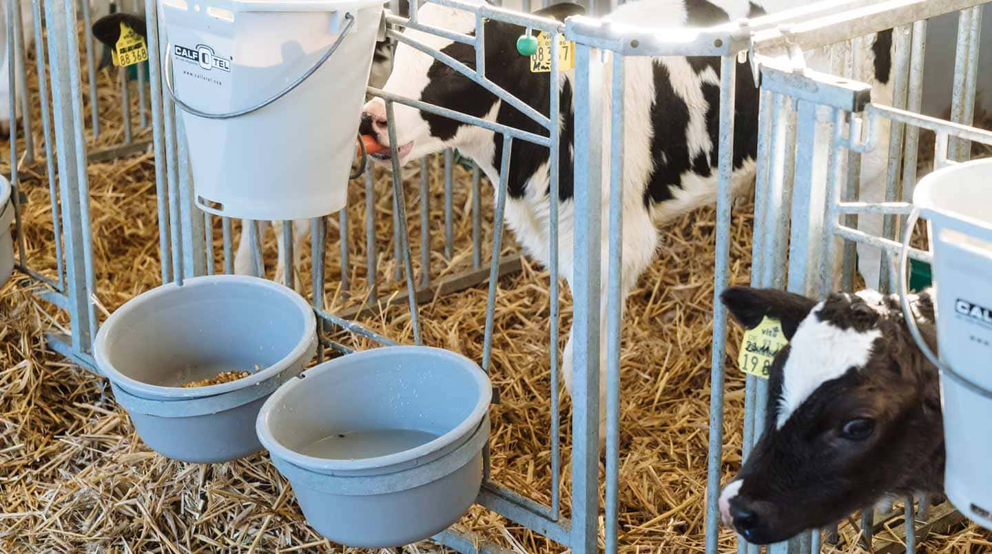
Calves are susceptible to infection by pathogens due to their immature congenital immunity. Bovine rotavirus and bovine coronavirus, pathogenic E. coli, Clostridium, Cryptosporidium, and Eimeria spp are the major pathogens of infectious diarrhea in calves less than one month of age. Bovine rotavirus, the most frequently detected in dairy and beef cattle, is responsible for approximately 40% of diarrhea cases. In addition, 60-70% of cases of diarrhea involving bovine rotavirus occur within the first two weeks of life. Symptoms include fever, anorexia, loss of energy, and acute yellow-white watery diarrhea after 12 to 36 hours post infection, which leads to dehydration and metabolic acidosis. In more severe cases, the disease can lead to death and is considered one of the most severe diarrhea-causing pathogens in newborn calves worldwide.
Rotaviruses belong to the family of Reoviridae and are classified into species A to J. The rotaviruses in bovines mainly belong to species A, B, and C, which are the leading infectious agents in cattle. Calf diarrhea is primarily caused by rotavirus A (RVA). This virus is transmitted orally through feces, bedding, utensils, or people contaminated with feces. Significant diarrhea caused by the virus is attributed to
Adult cattle and other host animals have an immune system that protects them from infection and the development of various pathogens. As RVA exists in different genotypes, the antibodies must be specifically against this genotype; otherwise, the virus-neutralizing activity, as well as protection against infection and pathogenesis, is significantly reduced.
Besides adequate sanitation in the production facilities, farmers try to “improve” the composition of the maternal colostrum by vaccinating the cow. For this purpose, the cows are inoculated with inactivated, previously isolated bovine RVA. However, the immunization of calves through colostrum may not be effective enough. It also may be difficult to prevent the spread of bovine RVA by barn hygiene alone due to the recent increase in the number of cattle being raised and moved from one farm to another.
In general, the three most common G genotypes of bovine RVA detected in calf diarrhea are G6, G8, and G10, and the three most common P genotypes are P[1], P[5], and P[11]. Based on the results of the genotyping survey in Japan from 1987 to 2000 (Fig. 1) and the one from 2017 to 2020 (figure 2) (Animal Health Research Division of the National Institute of Agrobiological Sciences (NIAH) together with IRIG), the bovine RVA genotypes identified as prevalent and endemic in Japan in recent years were G6P[5], G6P[11], and G10P[11]. However, the percentage of genotypes detected differed among cattle breeds (Fig. 3A, Fig. 3B, Fig. 3C).
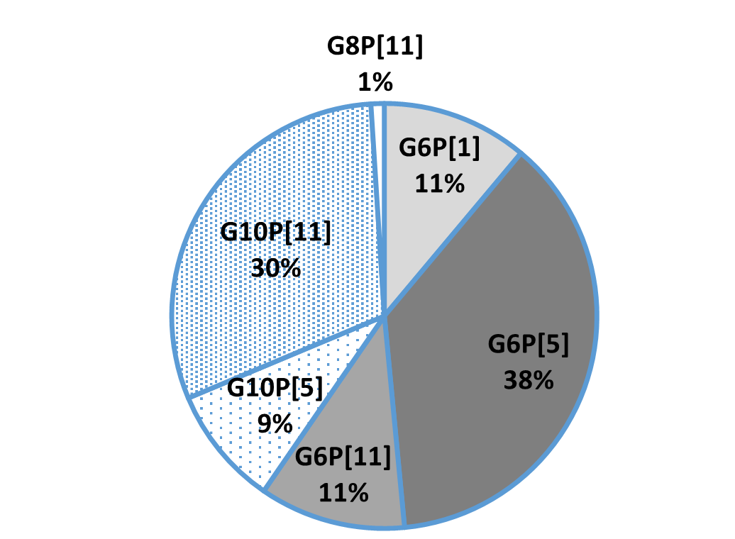 Fig.1: Genotyping results from 1987-2000
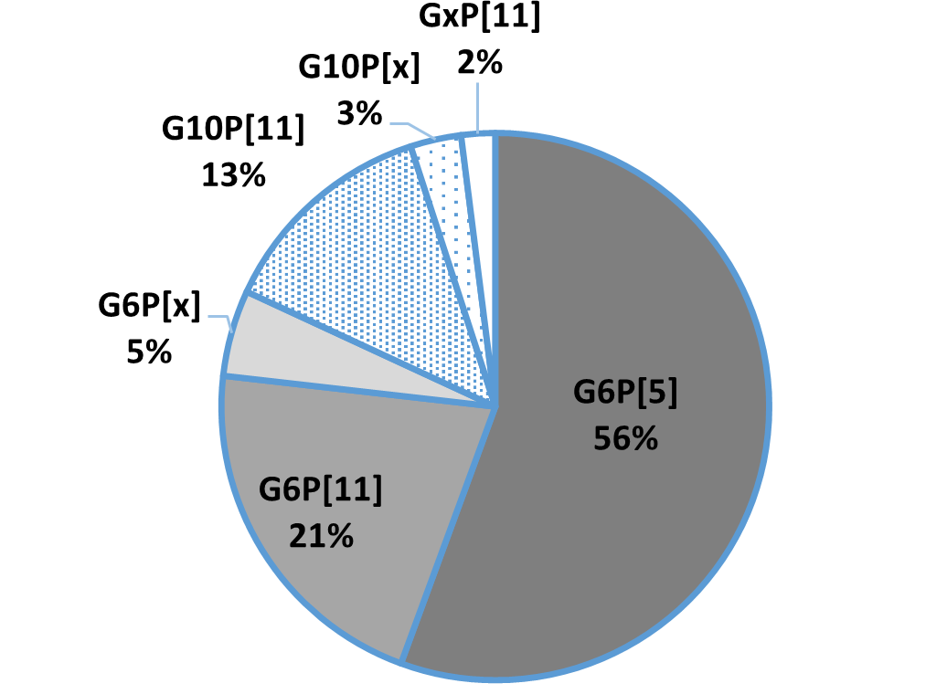 Fig.2: Genotyping results from 2017-2020
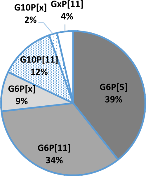 Fig. 3A:Percentage of detection in Holstein
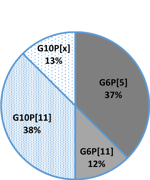 Fig. 3B: Detection rate in crossbreeds
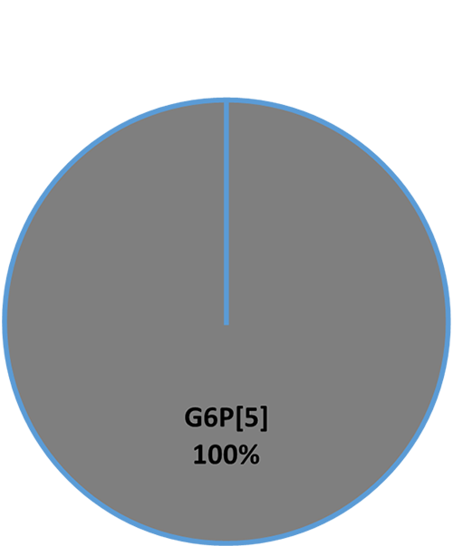 Fig. 3C: Detection rate in beef cattle (Wagyu)
A cow provides the calf with colostrum to ensure immunoglobulin delivery (passive immunity). In poultry, hens transfer immunoglobulins to the egg yolks and pass immunoglobulins to their chicks in this way. This biological mechanism of “immune transfer to the egg yolk” in birds can be used to arbitrarily produce yolk immunoglobulin (IgY) against pathogens of enteric infections in livestock (Ikemori et al., 1992; Ikemori et al., 1997; Yokoyama et al., 1998).
For this purpose, hens must get in contact with the respective pathogens. They produce antibodies against these pathogens – which also works with non-poultry-relevant pathogens such as bovine RVA – and transfer them to the egg (⇒IgY). The eggs with accumulated high levels of useful IgY can be collected almost daily. The immunoglobulins can be fed to livestock animals such as calves to protect them in critical times.
Continuous feeding of milk formulas containing IgY allows the IgY to remain in the intestinal lumen for a long time (Nozaki et al., 2019). There, they bind to the target pathogens and prevent infection by inhibiting their attachment to and cell invasion into intestinal epithelial cells.
A study verified that anti-bovine RVA IgY consisting of anti-G6P[1], anti-G6P[5], and anti-G10P[11] shows broad-spectrum virus-neutralizing activity against recent field isolates. Separate trials (see table 1) demonstrated that anti-G6 genotype IgY acted best against the RVA genotypes G6P[1] and G6P[5] and showed less activity against the G10 genotype. Anti-G10P[11] IgY worked optimally against the P[11] genotypes. The trials confirmed that either the G or the P genotype must match to achieve a sufficient virus-neutralizing activity. The IgY mixture is not helpful against bovine RVA strains that match neither the G nor the P genotypes (Odagiri et., 2020).
As the genotyping survey of 2017-2020 showed mainly G6 and G10 genotypes, a mixture of anti- bovine RVA G6P[1] IgY, G6P[5], and G10P[11] has strong virus neutralizing activity against bovine RVA that is currently prevalent and spreading in production sites.
Table 1: Virus-neutralizing activity of field-isolated bovine RVA against various genotypic strains
| IgY | Virus-neutralizing test strain | |||||||||
| SMN 1 | HKD 18 | SMN 35 | HKD 6 | HKD 7 | HKD 17 | KK-3 | OKY 31 | MYG 1 | Dai-10 | |
| 1978 | 2018 | 2018 | 2017 | 2017 | 2017 | 1983 | 2017 | 2017 | 2007 | |
| G6P[1] | G6P[5] | G6P[5] | G6P[11] | G6P[11] | G6P[11] | G10P[11] | G10P[11] | G8P[14] | G24P[33] | |
| anti-G6P[1] 1978 IgY | +++ | +++ | +++ | +++ | +++ | +++ | + | + | – | – |
| anti-G6P[5] 2018 IgY | +++ | +++ | +++ | ++ | ++ | ++ | + | + | + | – |
| Anti-G10P[11] 2017 IgY | + | + | + | + | ++ | ++ | +++ | +++ | – | – |
| Control IgY | – | – | – | – | – | – | – | – | – | – |
+++:Strong virus neutralizing activity; ++:Moderate virus neutralizing activity; +:Weak virus neutralizing activity; -:No virus neutralizing activity
To verify the protective effect of oral passive immunization with anti-bovine RVA IgY against bovine RVA infection, a trial with newborn calves was conducted.
Trial design: Eight calves were separated from their mothers immediately after birth without feeding colostrum and moved to a house with infected animals. From the first day, the calves were fed artificial milk supplemented with anti-bovine RVA IgY (n=4) or non-immune IgY (Control IgY; n=4) three times a day.
The parameters observed were fecal score, bovine RVA excretion, and weight gain; data were collected daily. The fecal score was calculated as the cumulative fecal score during the study period: 0 for normal stools, 1 for soft to muddy stools, and 2 for watery stools. Bovine RVA was isolated from daily fecal samples and evaluated by the total number of days of bovine RVA excretion.
Results: The anti-bovine RVA IgY group was found to be effective in reducing the incidence of diarrhea and shortening the duration of virus excretion in the infection test with the bovine RVA G6 genotype strain and the bovine RVA G10 genotype strain (tables 2 and 3).
Table 2: Efficacy of anti-bovine RVA IgY feeding in bovine RVA G6 genotype strain infection
| Test Group | Diarrhea incidence | Cumulative fecal score | Bovine RVA excretion days | Increase in body weight | |
| (n animals affected/n animals tested) | kg | % | |||
| Anti-bovine RVA IgY | 0% (0/4) | 0.0 ± 0.0* | 2.3 ± 0.5** | 1.3± 0.4** | 3.5 ± 0.7** |
| Control IgY | 100% (4/4) | 12.8 ± 4.8 | 7.8 ± 1.3 | – 3.3 ± 1.6 | – 7.6 ± 3.6 |
**: P<0.01; *: P<0.05
Table 3: Efficacy of anti-bovine RVA IgY feeding in bovine RVA G10 genotype strain infection
| Test Group | Diarrhea incidence | Cumulative fecal score | Bovine RVA excretion days | increase in body weight | |
| (n animals affected/n animals tested) | kg | % | |||
| Anti-bovine RVA IgY | 50% (2/4) | 2.3 ± 4.5** | 4.3 ± 1.3** | 1.1± 0.8** | 3.3 ± 3.1** |
| Control IgY | 100% (4/4) | 14.5 ± 3.7 | 7.3 ± 1.0 | – 4.2 ± 0.7 | – 11.1 ± 2.1 |
**: P<0.001
Newborn calves, susceptible to severe diarrhea caused by bovine RVA infection, require passive immunization with antibodies transferred from the colostrum of the mother cow. However, sometimes, calves don’t get enough antibodies which can be the case if
To compensate for this lack of immunity, calves have been fed milk formulas containing anti-bovine RVA IgY for some time. Continuous feeding of anti-bovine RVA IgY, which shows strong virus neutralizing activity against each genotype of bovine RVA isolated from recent cases of calf diarrhea, is expected to provide sufficient immunity and be an effective means of bovine RVA control.
In the case of disease outbreaks, it makes sense to utilize IgY with appropriate mechanisms of action in addition to improving the level of quarantine measures, including hygiene control and vaccination.
Ikemori, Yutaka, Masahiko Kuroki, Robert C. Peralta, Hideaki Yokoyama, and Yoshikatsu Kodama. “Protection of Neonatal Calves against Fatal Enteric Colibacillosis by Administration of Egg Yolk Powder from Hens Immunized with K99-Piliated Enterotoxigenic Escherichia Coli.” Amer. J. Vet. Res. 53, no. 11 (1992): 2005–8. PMID: 1466492.
Ikemori, Yutaka, Masashi Ohta, Kouji Umeda, Faustino C. Icatlo, Masahiko Kuroki, Hideaki Yokoyama, and Yoshikatsu Kodama. “Passive Protection of Neonatal Calves against Bovine Coronavirus-Induced Diarrhea by Administration of Egg Yolk or Colostrum Antibody Powder.” Veterinary Microbiology 58, no. 2-4 (1997): 105–11. https://doi.org/10.1016/s0378-1135(97)00144-2.
Nozaki, I., M. Itoh, F. Murakoshi, T. Aoki, K. Shibano, and K. Yamada. “Effect of an Egg Yolk Immunoglobulin(Igy)Product on Oocyst Shedding and Blood and Fecal Igy Concentrations in Cryptosporidium-Infected Calves.” Japanese Journal of Large Animal Clinics 10, no. 2 (2019): 68–72. https://doi.org/10.4190/jjlac.10.68.
Odagiri, Koki, Nobuki Yoshizawa, Hisae Sakihara, Koji Umeda, Shofiqur Rahman, Sa Van Nguyen, and Tohru Suzuki. “Development of Genotype-Specific Anti-Bovine Rotavirus a Immunoglobulin Yolk Based on a Current Molecular Epidemiological Analysis of Bovine Rotaviruses a Collected in Japan during 2017–2020.” Viruses 12, no. 12 (2020): 1386. https://doi.org/10.3390/v12121386.
Yokoyama, Hideaki, Robert C. Peralta, Kouji Umeda, Tomomi Hashi, Faustino C. Icatlo, Masahiko Kuroki, Yutaka Ikemori, and Yoshikatsu Kodama. “Prevention of Fatal Salmonelosis in Neonatal Calves, Using Orally Administered Chicken Egg Yolk Salmonella-Specific Antibodies.” Amer. J. Vet. Res. 59, no. 4 (1998): 416–20. PMID: 9563623.

By Dr. Inge Heinzl, Editor, and Dr. Ruturaj Patil, Global Product Manager – Phytogenics, EW Nutrition
For millennia, plants have been used for medicinal purposes in human and veterinary medicine and as spices in the kitchen. Since the ban of antibiotic growth promoters in 2006 by the European Union, they also came into focus in animal nutrition. Due to their digestive, antimicrobial, and gut health-promoting characteristics, they seemed an ideal alternative to compensate for the reduced use of antibiotics in critical periods such as brooding, feed change or gut-related stress.
To optimize the benefits of phytomolecules, it is crucial that
First step: Standardized phytomolecules
Essential oils and other phytogenics are sourced from plants. The composition of the plants substantially depends on genetic dissimilarity within accessions, plant origin, the site conditions, such as weather, soil, community, and harvest time, but also sample drying, storage, and extraction processes (Sadeh et al., 2019; Yang et al., 2018; Ehrlinger, 2007). For example, the oil extracted from thyme can contain between 22 and 71 % of the relevant phenol thymol (Soković et al., 2009; Shabnum and Wagay, 2011; Kowalczyk et al., 2020).
Modern technology enables the production of standardized phytomolecules with the highest degree of purity and lowest possible batch-to-batch variation for high-quality products. It also offers increased environmental and economic sustainability due to reliable and cost-effective sourcing technology.
Using such highly standardized phytomolecules enables the production of phytogenic-based feed supplements of consistently high quality.
Second step: Selection of the most suitable phytomolecules
Phytomolecules have different primary characteristics. Some support digestion (Cho et al., 2006, Oetting, 2006; Hernandez, 2004); others act against pathogens (Sienkiewitz et al., 2013; Smith-Palmer et al., 1998; Özer et al., 2007) or are antioxidants (Wei and Shibamoto, 2007; Cuppett and Hall, 1998). To optimize gut health in animal production, one of the main promising mechanisms is reducing pathogens while promoting beneficial microbes. The decrease of pathogens in the gut not only decreases the risk of enteritis incidence but also eliminates the inconvenient competitors for feed.
In order to find out the best combination serving the intended purpose, a high number of different phytomolecules need to be evaluated concerning their structure, chemical properties, and biological activities first. Availability and costs of the substances are further factors to consider. With the selection of the most suitable phytomolecules, different mixtures are produced and tested for their effectiveness. Here, it is essential to concern synergistic or antagonistic effects.
For an effective and efficient blend of phytomolecules, many steps of selection and tests are necessary – and as a result, possibly only a few mixtures can meet the requirements.
Third step: Protecting the ingredients
Many phytomolecules are inherently highly volatile. So, only having a standardized content of phytogenics in the product can not ensure the full availability of phytomolecules when used through animal feed. Some parts of the ingredients might already get lost in the feed mill due to the stringent feed hygienization process followed by feed millers to reduce pathogenic load. The heating is a significant challenge for the highly-volatile components in a phytomolecule-based product. So, protecting these phytomolecules becomes imperative to guarantee that the phytomolecules put into the feed will reach the animal.
A delicate balancing act is required to ensure the availability and activity of phytomolecules at the right site in the gut. The phytomolecules must not get lost during feed processing but must also be released in the intestine. A carrier with capillary binding of the phytomolecules together with a protective coating can be one of the available effective solutions. It protects the ingredients during feed processing, and ensures the release in the animal.
Ventar D is a latest generation phytomolecule-based solution for gut health optimization introduced by EW Nutrition, GmbH. A scientific study was conducted to compare the stability of Ventar D, in the pelleting process, with two leading phytogenics competitor feed supplements.
For this trial, feed with the different added phytogenic feed supplements had to undergo a conditioning and pelletization process. The active ingredients were analyzed before and after the pelletization process. All phytogenic feed supplements under testing were added to standard broiler feed at the producer’s recommended inclusion rate. The tests took place under conditioning times of 45, 90, and 180 seconds and pelleting temperatures of 70, 80, and 90°C (158, 176, and 194°F). After cooling, triplicate samples were collected and analyzed. The respective marker substance was analyzed through gas chromatography/mass spectrometry (GC/MS) analysis to measure the recovery rate in the finished feed. The phytomolecule content of the mash feed (before pelletization) found by the laboratory was used as a baseline and set to 100% recovery. The recovery rates of the pelleted feed were evaluated relative to this baseline.
The results are presented in figure 1. Ventar D showed the highest stability of active ingredients with recovery rates of 90% at 70°C/45 sec. or 80°C/90 sec and 84% at 90°C/180 sec. The modern production technology used for Ventar D ensures that the active ingredients are well protected throughout the pelletization process.
 Figure 1: Phytomolecule stability under processing conditions, relative to mash baseline (100%)
Another trial was conducted in a feed mill in the US. For this trial, ten samples were collected from different batches of mash feed where Ventar D was added at 110g/t. Conditioning of the mash feed was at 87.8°C (190°F) for 6 minutes and 45 seconds. After the pelleting process, ten samples from the pelleted feed were collected from the continuous flow with a 5 min gap between the samplings to determine Ventar D’s recovery.
The average recovery achieved for Ventar D was 92%.
Initial trials showed Ventar D’s complete release in digestion models. To examine the benefit in in-vivo conditions, Ventar D was tested in broilers at an inclusion rate of 100 g/MT.
Several in vitro studies proved the antimicrobial activity of Ventar D. One test also confirms that Ventar D could exhibit differential antimicrobial activity by having stronger activity against common enteropathogenic bacteria while sparing the beneficial ones (Heinzl, 2022). Moreover, Ventar D’s antioxidant and anti-inflammatory activity support better gut barrier functioning. Better gut health leads to higher growth performance and improved feed conversion, which could be demonstrated in several trials with broilers (figures 2 and 3). In the tests, a group fed Ventar D was compared to either a control group with no such feed supplement or groups supplied with competitor products at the recommended inclusion rates.
Compared to a negative control group, the Ventar D group consistently showed a higher average daily gain of 0.3-4.1 g (0.5-8.5 %) and a 3-4 points better feed conversion. Compared to competitor products, Ventar D provided 1-1.7 g (2-3 %) higher average daily gain and a 3 points better /1 point higher FCR than competitors 2 and 1.
 Figure 2: Average daily gain (g) – results of several trials conducted with broilers
 Figure 3: FCR – results of several trials conducted with broilers
Several in vitro and in vivo studies proved that Ventar D takes “phytomolecules’ power” to the next level: Combining standardized phytomolecules and optimal active ingredient protection leads to superior product stability during feed processing. The higher amount of active ingredients arriving in the gut improves gut health and increases the production performance of the animals. Ventar D shows how we can use phytomolecules more effectively and benefit from higher farm profitability.
References:
Cho, J. H., Y. J. Chen, B. J. Min, H. J. Kim, O. S. Kwon, K. S. Shon, I. H. Kim, S. J. Kim, and A. Asamer. “Effects of Essential Oils Supplementation on Growth Performance, IGG Concentration and Fecal Noxious Gas Concentration of Weaned Pigs”. Asian-Australasian Journal of Animal Sciences 19, no. 1 (2005): 80–85. https://doi.org/10.5713/ajas.2006.80.
Cuppett, Susan L., and Clifford A. Hall. “Antioxidant Activity of the Labiatae”. Advances in Food and Nutrition Research 42 (1998): 245–71. https://doi.org/10.1016/s1043-4526(08)60097-2.
Ehrlinger, M. “Phytogenic Additives in Animal Nutrition.” Dissertation, Veterinary Faculty of the Ludwig Maximilians University, 2007.
Heinzl, I. “Efficient Microbiome Modulation with Phytomolecules”. EW Nutrition, August 30, 2022. https://ew-nutrition.com/pushing-microbiome-in-right-direction-phytomolecules/.
Hernández, F., J. Madrid, V. García, J. Orengo, and M.D. Megías. “Influence of Two Plant Extracts on Broilers Performance, Digestibility, and Digestive Organ Size.” Poultry Science 83, no. 2 (2004): 169–74. https://doi.org/10.1093/ps/83.2.169.
Kowalczyk, Adam, Martyna Przychodna, Sylwia Sopata, Agnieszka Bodalska, and Izabela Fecka. “Thymol and Thyme Essential Oil—New Insights into Selected Therapeutic Applications.” Molecules 25, no. 18 (2020): 4125. https://doi.org/10.3390/molecules25184125.
Lindner, , U. “Aromatic Plants – Cultivation and Use.” Düsseldorf: Teaching and Research Institute for Horticulture Auweiler-Friesdorf, 1987.
Oetting, Liliana Lotufo, Carlos Eduardo Utiyama, Pedro Agostinho Giani, Urbano dos Ruiz, and Valdomiro Shigueru Miyada. “Efeitos De Extratos Vegetais e Antimicrobianos Sobre a Digestibilidade Aparente, O Desempenho, a Morfometria Dos Órgãos e a Histologia Intestinal De Leitões Recém-Desmamados.” Revista Brasileira de Zootecnia 35, no. 4 (2006): 1389–97. https://doi.org/10.1590/s1516-35982006000500019.
Sadeh, Dganit, Nadav Nitzan, David Chaimovitsh, Alona Shachter, Murad Ghanim, and Nativ Dudai. “Interactive Effects of Genotype, Seasonality and Extraction Method on Chemical Compositions and Yield of Essential Oil from Rosemary (Rosmarinus Officinalis L”.).” Industrial Crops and Products 138 (2019): 111419. https://doi.org/10.1016/j.indcrop.2019.05.068.
Shabnum, Shazia, and Muzafar G. Wagay. “Essential Oil Composition of Thymus Vulgaris L. and Their Uses”. Journal of Research & Development 11 (2011): 83–94.
Sienkiewicz, Monika, Monika Łysakowska, Marta Pastuszka, Wojciech Bienias, and Edward Kowalczyk. “The Potential of Use Basil and Rosemary Essential Oils as Effective Antibacterial Agents.” Molecules 18, no. 8 (2013): 9334–51. https://doi.org/10.3390/molecules18089334.
Smith-Palmer, A., J. Stewart, and L. Fyfe. “Antimicrobial Properties of Plant Essential Oils and Essences against Five Important Food-Borne Pathogens”. Letters in Applied Microbiology 26, no. 2 (1998): 118–22. https://doi.org/10.1046/j.1472-765x.1998.00303.x.
Soković, Marina, Jelena Vukojević, Petar Marin, Dejan Brkić, Vlatka Vajs, and Leo Van Griensven. “Chemical Composition of Essential Oils of Thymus and Mentha Species and Their Antifungal Activities”. Molecules 14, no. 1 (2009): 238–49. https://doi.org/10.3390/molecules14010238.
Wei, Alfreda, and Takayuki Shibamoto. “Antioxidant Activities and Volatile Constituents of Various Essential Oils.” Journal of Agricultural and Food Chemistry 55, no. 5 (2007): 1737–42. https://doi.org/10.1021/jf062959x.
Yang, Li, Kui-Shan Wen, Xiao Ruan, Ying-Xian Zhao, Feng Wei, and Qiang Wang. “Response of Plant Secondary Metabolites to Environmental Factors”. Molecules 23, no. 4 (2018): 762. https://doi.org/10.3390/molecules23040762.
Özer, Hakan, Münevver Sökmen, Medine Güllüce, Ahmet Adigüzel, Fikrettin Şahin, Atalay Sökmen, Hamdullah Kiliç, and Özlem Bariş. “Chemical Composition and Antimicrobial and Antioxidant Activities of the Essential Oil and Methanol Extract of Hippomarathrum Microcarpum (Bieb.) from Turkey”. Journal of Agricultural and Food Chemistry 55, no. 3 (2007): 937–42. https://doi.org/10.1021/jf0624244.