Mitigating Necrotic Enteritis through Natural Alternatives in Antibiotic-Free Production Systems

clostridium perfringens 1900

by EW Nutrition USA, Inc.


In the poultry industry, Necrotic Enteritis is of great interest due to the potential detrimental growth effects it may have in a flock, even at subclinical levels50. Coccidiostats and antibiotics have been used for a long time to get the disease-causing bacterium Clostridium perfringens under control, but with increasing antimicrobial resistance, alternative approaches are required. This article aims to give an overview of the disease and the measures against it.

Clostridium perfringens – a ubiquitous, highly resilient bacterium

Clostridium perfringens is a Gram-positive, spore-forming, anaerobic, rod-shaped bacterium50. This encapsulated, non-motile microorganism is fastidious in growth requirements59. Most often, complex media like cooked meat or thioglycolate broth are used as enrichment30.

It was Welch and Nuttall who first identified C. perfringens in 1892 as Bacillus aerogenes capsulatus18. In Great Britain, the bacterium was commonly known as C. welchii and sometimes called Frankel’s bacillus in Germany until designated C. perfringens by Bergey13.

Clostridium perfringens is the causal microorganism for Necrotic Enteritis (NE)14. In humans, it is one of the most common causes of foodborne illness20. The Centers for Disease Control and Prevention (CDC, 2012) estimates that nearly one million people are affected every year, making C. perfringens the third most frequent source of domestically acquired foodborne illness after Norovirus and Salmonella.

Clostridium perfringens can be found everywhere

Clostridium perfringens is found in soil, water, and other organic materials. As far as poultry facilities, C. perfringens has been isolated from litter, dust, walls, floors, fans, transportation coops, feeders, and feed89.

Additionally, C. perfringens is found in the GI tract of broiler chickens, humans, and other mammals47. When intestinal samples of broiler chickens were analyzed for C. perfringens, 75-95 % tested positive24. Drew and co-workers10 determined that C. perfringens is usually found at ~104 colony-forming units (CFU)/g of broiler digesta. These results agree with Jia et al.26, who stated that C. perfringens is present at low levels in healthy poultry. In humans, investigations in different parts of the world showed a prevalence of Clostridium perfringens between 57-94%32.

Different types of Clostridium perfringens with different toxins

There are five types (A-E) of C. perfringens, which can be identified through their toxin production (see table 1). All strains produce alpha-toxin. Furthermore, Clostridium perfringens has been described to produce eight other toxins, three (delta, theta, kappa) can be lethal, but these are seldom involved in disease origin37.

Table 1. Different types of Clostridium perfringens

    C. perfringens Type
A B C D E
Toxins Alpha x x x x x
Beta   x x    
Epsilon   x   x  
Iota         x
Enterotoxin x        
Diseases/animals18 Food-born disease/humans

NE/fowl

Dysentery/lambs

enterotoxaemia/ sheep, goats, guinea pigs

Food-born disease/humans

NE/fowl

Enterotoxaemia/

sheep

Pulpy kidney disease/lambs

Enterotoxaemia/ calves

Dysentery/sheep, guinea pigs, rabbits


High resilience gives an advantage against competitors

Since Clostridium perfringens is a spore-forming bacterium, it is very resilient to high temperatures, slight pH variations, and toxic chemicals43, 7.

Labbe et al.30 established that C. perfringens can reproduce at temperatures between 15-50 °C. Hence, proper refrigeration temperatures (below 10 °C) can be an effective means of control. The optimum range is between 37-47 °C, and at these temperatures, the mean generation time – the time required for the bacterial count to double – is approximately 10-12 minutes41. These short generation times allow the bacteria to outcompete other microorganisms that may need similar resources in a certain environment.

The optimum pH range of Clostridium perfringens is between 5.5-7.022. However, it can grow at a pH as low as 5 and as high as 9. In live broiler chickens, the pH in the small intestine has been determined to be between 6.00-7.78.


Necrotic enteritis in poultry

The disease necrotic enteritis was first described by Parish45, 46 in cockerels in England. Some of the symptoms include depression, reluctance to move, ruffled feathers, somnolence, diarrhea, loss of appetite, and anorexia21. Mortality ranges from 0-50% 6 have been reported in infected flocks. Since then, virtually every area that raises poultry has reported signs of necrotic enteritis.

Clostridium perfringens – How NE unravels

As already mentioned, 104 colony-forming units (CFU)/g of broiler digesta10 are normal and can be found in healthy birds. C. perfringens becomes problematic when counts reach 107-108 CFU/g6.

Necrotic enteritis is caused by types A and C of Clostridium perfringens, but normally, predisposing factors “set the stage”24, 48. This could be seen in an investigation where they wanted to create a model to reproduce NE in a laboratory setting. Researchers realized that inoculation of C. perfringens alone did not cause the disease found in the field48. Therefore, it was assessed that certain cofactors must play a significant role in the pathogenicity of C. perfringens. Williams57 reviewed concurrent infections of coccidiosis and necrotic enteritis in chickens (Figure 1). The copious interactions of these diseases with predisposing factors, control methods, sources of infection, and disease form is a testament to the complexity of this poultry industry matter.

Coccidiosis creates access

Shane et al.53 noted that several authors had considered coccidiosis to be a predisposing factor for NE. They proceeded to describe the pathogenesis of Eimeria acervulina, one of the protozoa responsible for coccidiosis in poultry. When the oocysts are ingested, they quickly attach to the intestinal wall causing lesions where the protozoa reproduce numerous times. These are the lesions to which C. perfringens attaches.

What happens in the animal?

Long et al.33 proposed the pathogenesis for NE: First, epithelial cells are vacuolated, and the epithelium lifts off the lamina propria, which is congested and edematous. These lesions can be caused by a combination of factors like toxin production and/or, as just mentioned, coccidiosis. Clostridium perfringens cells attach to the lamina propria, where they thrive. The tissue becomes necrotic as large numbers of heterophils, a type of phagocyte, flood the foci (sites of lesions).

A combination of disease-inducing factors such as bacteria proliferation, heterophil lysis, and villus’ necrosis seem to develop quickly. The inflammation zone then becomes riddled with mononuclear cells, cells containing lymphocytes, antigen-presenting cells, and eosinophilic-staining (proteinaceous) amorphous material. This necrotizing process moves from the tip of the villi to the crypt.

Chronic version

In chronic cases, villi may be found to have multiple cysts from recurrent necrosis. In birds that overcome the disease, injured epithelial cells are replaced by newly formed reticular structures. These new cells travel from the crypt to the tip of the villi and replace the old, damaged cells. The result is a short, flat villus with a reduced surface area for nutrient absorption44, 45, 34. These morphologically altered villi are the necrotic lesions found in the field and some C. perfringens challenge trials (Figure 2).

Acute form

The acute form of NE results in enlarged lesions along the gut wall, and the epithelium becomes eroded and detached; consequently, a diphtheritic membrane is formed. This yellow, green, or brownish pseudo-membrane is called the “Turkish towel,” which describes the appearance of the friable, gas-filled, foul-smelling GI tract57.

Subclinical form

Poultry producers are not only concerned with the acute form of NE. Recent studies have shown that the disease’s subclinical form can be as detrimental as the acute illness19. Lovland and co-workers35 stated that this symptomless disease is often overlooked at the farm, and the effects are only noticed at the processing facility.

Subclinical NE (SNE) can cause cholangiohepatitis, a condition where the liver is enlarged with pale reticular patterns and sometimes small, pale foci. In the United Kingdom, it was estimated that 4% of broiler carcasses and 12% of livers are condemned at processing plants due to clostridial infection; thereby, reducing profit36. Moreover, sparse lesions that may be found in a case of SNE may be enough to hinder growth performance; thus, resulting in an underproductive flock39.


Feeding Against Necrotic Enteritis

It has been reported that diet formulation has the greatest impact on the prevalence of C. perfringens in chicken GI tracts61. The poultry industry formulates diets on a least-cost basis, which may become problematic if nutritionists do not take into consideration the pathological consequences that some ingredients may have in the GI tracts of chickens. Every feed ingredient has a specific purpose in the diet. For instance, cereal grains are fed for their energy concentration as well as fiber. Also, some grain and animal/plant meals are used for their protein content. Since these ingredients are obtained from different sources, they are highly variable in macro and micronutrients1.

The diet provides the conditions for proliferation

There are multiple elements that affect the proliferation of C. perfringens in chicken intestines, one of the most critical factors being diet formulation5, 36. Some feed ingredients have been found to exacerbate the numbers of C. perfringens in chickens’ gastrointestinal tract. Diets formulated with wheat increased NE intestinal lesion scores compared to broiler chickens fed a corn-based diet4. In another study, Drew et al.10 investigated the effects of different protein sources on the intestinal populations of C. perfringens in broiler chickens. Diets were formulated to contain 230, 315, and 400 g/kg of fishmeal or soy protein concentrate (SPC). The numbers of C. perfringens in the ileum and ceca increased when the amount of protein increased from 230 to 400 g/kg.

Type of grain influences the occurrence of Clostridium perfringens

Authors have studied the effects of grain inclusion on gut microbiota, and it is well established that small cereal grains such as barley, rye, and wheat tend to increase the prevalence of C. perfringens in the GI tract. Shakouri et al.52 investigated the influence of barley, sorghum, wheat, and corn on counts of C. perfringens in the different intestinal segments. Corn and wheat had the lowest C. perfringens counts, followed by sorghum, while barley yielded the highest counts. These findings agree with Riddell and Kong51.

Other researchers have concluded that the increase in gut viscosity and increased chyme transit time elicit the overgrowth of C. perfringens in the intestines28. Grains like wheat and barley contain high amounts of non-starch polysaccharides (NSP), which increase viscosity26. Furthermore, it has been alleged that, since these grains are high in NSP, the bird cannot absorb nutrients as efficiently, thereby leaving them for microbes like C. perfringens to consume31.

Enzymes improve nutrient availability in the presence of C. perfringens

Shakori et al.52 and Jia et al.26 also studied the impact of several diets with the inclusion of a blend of carbohydrases such as glucanase and xylanase. Their findings suggested that enzyme addition did not affect counts of C. perfringens in the different intestinal sections. However, they did find an improvement in growth performance. They stated that enzymes improved chyme viscosity by degrading the encapsulation of nutrients in diets.

For this reason, researchers have investigated the use of enzymes in wheat and barley-based diets on the incidence of C. perfringens in chicken intestines. Jackson et al.25 studied the effect of beta-mannanase addition on flocks infected with Eimeria spp. and C. perfringens. They found that feeding this enzyme significantly reduced the impact of C. perfringens on the performance of infected flocks as well as intestinal lesion scores. Moreover, the authors explained that this might be due to beta-mannanase crossing the intestinal wall to provoke an immune response. They determined that this enzyme tended to ameliorate the symptoms of necrotic enteritis, but not significantly.

MOS may have a positive impact on immunity

Hofacre et al. 23 found similar results when birds were fed mannan-oligosaccharides. A marked effect was only found when mannan-oligosaccharides were included along with lactic acid-producing, competitive exclusion products (probiotics).

The feed form is decisive

Feed form has also been investigated on the incidence of C. perfringens. When birds were fed whole wheat compared to ground, researchers found reduced counts of C. perfringens in the gut2. These results can be extrapolated to the findings of Engberg et al.11. They found that when birds were fed coarse versus fine mash or pellets, C. perfringens counts were consistently higher in flocks fed mash diets. These authors concluded that feeding pellets or whole grains increases gizzard activity, which consequently triggers hydrochloric acid production and decreases pH in the GI tract. This drop in pH of approximately 0.5 units may be responsible for decreased C. perfringens counts.

Mind the protein source

Another well-established fact is that the C. perfringens population can be affected by the type of the protein source and the inclusion rates.

Potato is worse than fish

Palliyeguru et al. 42 studied the inclusion of protein concentrates (potato, fish, and soy) on subclinical NE. They determined that the potato-containing diet resulted in the highest incidence of C. perfringens in the gut, followed by fish and soy. Also, the potato-containing diet had the highest activity of trypsin inhibitors and lowest lipid content. Increased trypsin inhibition does not allow for the inactivation of alpha and beta toxins produced by C. perfringens, resulting in increased intestinal wall lesions.

Fish is worse than soy due to the amino acid composition

Drew et al.10 formulated diets containing fishmeal or a soy protein concentrate at different levels. Feeding dietary fishmeal resulted in a higher incidence of C. perfringens as compared to the soy protein diet. Furthermore, with increasing levels of soy and fishmeal diets, counts of C. perfringens increased as well. A notable difference in fishmeal protein concentrate compared to the soy protein concentrate was the amino acid ratio in this experiment; the methionine and glycine ratios were 1.3 times greater in fishmeal diets. Muhammed et al.40 determined that methionine was required for C. perfringens sporulation. This may be of interest to nutritionists since some authors have estimated that 10-20 % of synthetic amino acids are not absorbed and reach the lower intestinal tract, i.e., ceca; thereby, aiding in the proliferation of C. perfringens.

Fat source – animal fat is critical

The effects of fat sources on C. perfringens population remain largely unknown. Knarreborg et al.29 studied the bacterial microflora in chicken intestines after feeding different dietary fats (soy oil and a tallow and lard mix) in rations containing antibiotic growth promoters (AGP). When soy oil was fed, C. perfringens counts were significantly lower than diets containing animal fats. The authors stated that, since plant oils contain higher amounts of unsaturated fatty acids, the chyme in birds fed oil diets would have decreased viscosity, decreasing transit time. Furthermore, an additive effect was found when soy oil was provided along with AGP, which may be due to facilitated antibiotic dispersion caused by the oil’s lipophilic properties. Knarreborg et al. (2002) investigated the effects of fat sources on C. perfringens. They found that total anaerobic counts increased with animal fat addition. However, zinc bacitracin was included in their diets, specifically targeting Gram-positive microorganisms like C. perfringens; thus, potentially biasing their results.

Antibiotics and coccidiostats in the diet – helpful, but finite

Antibiotics and coccidiostats have been commonly included in poultry diets since the mid-1940s and 1950s61, 58.

Prescott et al.49 studied the inclusion of zinc bacitracin to prevent necrotic enteritis and concluded that it successfully controlled the C. perfringens challenge. Flocks in the antibiotic treatments were able to overcome disease and perform similarly to unchallenged birds. Multiple authors have replicated these results using different antibiotics such as virginiamycin and salinomycin17, 3, 11.

Improvements in flock performance with the inclusion of antibiotics and coccidiostats are well understood and omnipresent in the literature. However, the potential loss of subtherapeutic antibiotic usage in livestock in the United States due to increasing concerns over antimicrobial resistance and consumer demands makes research of viable alternatives to these compounds paramount.

So, what are your alternatives?

A lot of different approaches are possible. In general, these measures should act against Clostridium perfringens while supporting gut health.

Tested substances without the desired effects

Lastly, multiple options have been studied to control C. perfringens in poultry. Some researchers have studied the inclusion of complex carbohydrates and fibers like pine shavings, guar gum, and pectin with limited success4, 31. Another popular alternative is the use of competitive exclusion-based products such as prebiotics and probiotics27, 16. Still, these products failed to yield consistent results.

Other options that have been investigated are the addition of lactose and organic acids54, 38. Potassium diformate did not produce lowered counts of C. perfringens. Lactose reduced C. perfringens counts but resulted in undesirable ceca characteristics including, enlargement and increased fermentation54.

Essential oils alone or in combination may be a solution

Mitsch and coworkers39 investigated the efficacy of two blends of essential oils with positive effects on the reduction of C. perfringens from the gut and feces of broilers. Gaucher and coworkers15 compared growth performance and gut health of broilers fed a conventional (anticoccidials and AGPs) vs. ABF (Coccidiosis vaccine and essential oil blends) diet. They established that livability, age at slaughter, and percentage of condemnation did not change with diet type. However, average daily weight gain and FCR were negatively affected. Furthermore, NE was more prevalent in ABF flocks.  Still, many authors agree that a multifactorial approach is necessary if antibiotics should be completely replaced by these strategies36.

A contemporary study by Wati et al.56 aimed to compare AGPs to a commercial blend of essential oils fed to broilers. Authors found that chickens fed essential oils had body weights and FCRs that were statistically similar to the AGP treatment. Moreover, both AGP and essential oil treatments had statistically lower counts of Salmonella and E. coli after an oral challenge than the control group.

Conclusion

C. perfringens is a potential pathogen found in every place poultry is raised. Therefore, we must continue to identify strategies to control the development of Necrotic Enteritis. Since antibiotics alone may not always successfully control C. perfringens and have the potential for subtherapeutic use loss in the US, a multifactorial approach must be considered and investigated. Grain size, enzymes, feed form, animal protein source, fats, and feed supplements such as essential oils can affect the proliferation of C. perfringens. Nutritionists, veterinarians, and live production personnel must come together to develop the best approach for their specific complex circumstances.

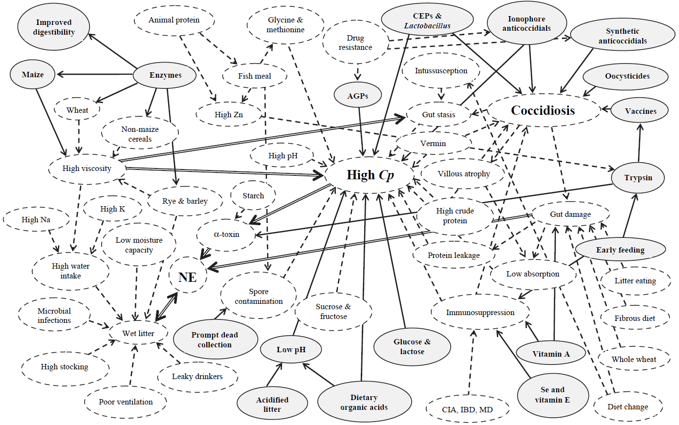
Figure 1. Interaction between coccidiosis and NE with environmental factors

Solid-line arrows are beneficial in controlling disease. Dashed-line arrows impart high disease risk factors. Double-line arrows depict major disease-risk factors. AGP, antibiotic growth promoter; CIA, chick infectious anemia; CEP, competitive exclusion product; Cp, Clostridium perfringens; IBD, infectious bursal disease; MD, Marek’s disease; NE, necrotic enteritis. (Williams, R.B. 2005)

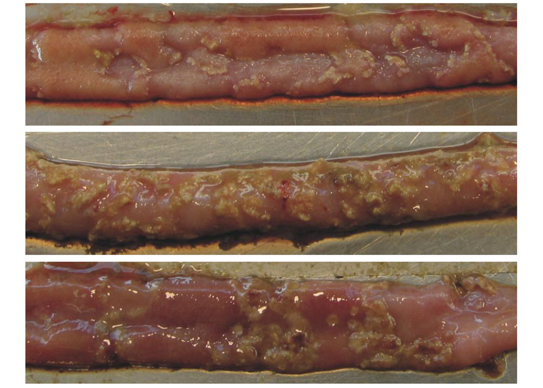
 Figure 2. Necrotic Enteritis lesions in chicken intestines

Yellowish necrotic lesions in three intestinal samples. Intestines A and C show a few marked lesions. Intestine B shows clusters of lesions typical of the “Turkish towel” syndrome. (Source: http://www.mdpi.com/2072-6651/2/7/1913/htm. Accessed: January 14, 2021).


References
  1. Bedford, M.R. 1996. Interaction Between Ingested Feed and the Digestive System in Poultry. Applied Poultry Science 5:86-95.
  2. Bjerrum, L., K. Pedersen, and R. M. Engberg. 2005. The Influence of Whole Wheat Feeding on Salmonella Infection and Gut Flora Composition in Broilers. Avian Disease 49:9-15.
  3. Bolder, N. M., J. A. Wagenaar, F. F. Putirulan, K. T. Veldman, and M. Sommer. 1999. The Effect of Flavophospholipol (FlavomycinR) and Salinomycin Sodium (SacoxR) on the Excretion of Clostridium perfringens, Salmonella enteritidis, and Campylobacter jejuni in Broilers After Experimental Infection. Poultry Science 78:1681-1689.
  4. Branton, S. L., B. D. Lott, J. W. Deaton, W. R. Maslin, F. W. Austin, L. M. Pote, R. W. Keirs, M. A. Latour, and E. J. Day. 1997. The Effect of Added Complex Carbohydrates or Added Dietary Fiber on Necrotic Enteritis Lesions in Broiler Chickens. Poultry Science 76:24-28.
  5. Choct, M. 2009. Managing Gut Health through Nutrition. British Poultry Science 50:9-15.
  6. Cooper, K., and J. G. Songer. 2009. Necrotic Enteritis in Chickens: A Paradigm of Enteric Infection by Clostridium perfringens Type A. Veterinary anaerobes and diseases 15:55-60.
  7. Craven, S. E., N. J. Stern, N. A. Cox, J. S. Bailey, and M. Berrang. 1999. Cecal Carriage of Clostridium perfringens in Broiler Chickens Given Mucosal Starter CultureTM. Avian Diseases 43:484-490.
  8. Craven, S. E., N. A. Cox, N. J. Stern, and J. M. Mauldin. 2001a. Prevalence of Clostridium perfringens in Commercial Broiler Hatcheries. Avian Diseases 45:1050-1053.
  9. Craven, S. E., N. J. Stern, J. S. Bailey, and N. A. Cox. 2001b. Incidence of Clostridium perfringens in Broiler Chickens and Their Environment during Production and Processing. Avian Diseases 45:887-896.
  10. Drew, M. D., N. A. Syed, B. G. Goldade, B. Laarveld, and A. G. Van Kessel. 2004. Effects of Dietary Protein Source and Level on Intestinal Populations of Clostridium perfringens in Broiler Chickens. Poultry Science 83:414-420.
  11. Engberg, R. M., M. S. Hedemann, Leser, T.D., and Jensen, B.B. 2000. Effect of Bacitracin and Salinomycin on Intestinal Microflora and Performance of Broilers. Poultry Science 79: 1311-1319.
  12. Engberg, R. M., M. S. Hedemann, and Jensen, B.B. 2002. The Influence of Grinding and Pelleting of Feed on the Microbial Composition and Activity in the Digestive Tract of Broiler Chickens. British Poultry Science 44:569-579.
  13. Freeman, B.A. 1979. Burrows Textbook of Microbiology. W.B. Saunders Company, Philadelphia, Pennsylvania, USA.
  14. Fukata, T., Y. Hadate, E. Baba, T. Uemura, and A. Arakawa. 1988. Influence of Clostridium perfringens and its Toxin in Germ-free Chickens. Research in Veterinary Science 44:68-70.
  15. Gaucher, M.L., Quessy, S., Letellier, A., Arsenault, J., and M. Boulianne. 2015. Impact of a drug-free program on broiler chicken growth performances, gut health, Clostridium perfringens and Campylobacter jejuni occurrences at the farm level. Poultry Science 94: 1791-1801.
  16. Geier, M. S., L. L. Mikkelsen, V. A. Torok, G. E. Allison, G. C. Olnood, M. Boulianne, R. J. Hughes, and M. Choct. 2010. Comparison of Alternatives to In-feed Antimicrobials for the Prevention of Clinical Necrotic Enteritis. Journal of Applied Microbiology 109:1329-1338.
  17. George, B. A., C. L. Quarles, and D. J. Fagerberg. 1982. Virginiamycin Effects on Controlling Necrotic Enteritis Infection in Chickens. Poultry Science 61:447-450.
  18. Hatheway, C. L. 1990. Toxigenic Clostridia. Clinical Microbiology Reviews 3:66-98.
  19. Heier, B. T., A. Lovland, K. B. Soleim, M. Kaldhusdal, and J. Jarp. 2001. A Field Study of Naturally Occurring Specific Antibodies against Clostridium perfringens Alpha Toxin in Norwegian Broiler Flocks. Avian Diseases 45:724-732.
  20. Heikinheimo, A., M. Lindstrom, and H. Korkeala. 2004. Enumeration and Isolation of cpe-Positive Clostridium perfringens Spores from Feces. Journal of Clinical Microbiology 42:3992-3997.
  21. Helmboldt, C. F., and E. S. Bryant. 1971. The Pathology of Necrotic Enteritis in Domestic Fowl. Avian Diseases 15:775-780.
  22. Hickey, C.S., and Johnson, M.G. 1981. Effects of pH Shifts, Bile Salts, and Glucose on Sporulation of Clostridium perfringens NTCT 8798. Applied and Environmental Microbiology 41:124-129.
  23. Hofacre, C. L., T. Beacorn, S. Collett, and G. Mathis. 2003. Using Competitive Exclusion, Mannan-Oligosaccharide and Other Intestinal Products to Control Necrotic Enteritis. Journal of Applied Poultry Research 12:60-64.
  24. Immerseel, F. V., J. De Buck, F. Pasmans, G. Huyghebaert, F. Haesebrouck, and R. Ducatelle. 2004. Clostridium perfringens in Poultry: an Emerging Threat for Animal and Public Health. Avian Pathology 33:537-549.
  25. Jackson, M. E., D. M. Anderson, H. Y. Hsiao, G. Mathis, and D. W. Fodge. 2003. Beneficial Effect of B-Mannanase Feed Enzyme on Performance of Chicks Challenged with Eimeria and Clostridium perfringens. Avian Diseases 47:759-763.
  26. Jia, W., B. A. Slominski, H. L. Bruce, G. Blank, G. Crow, and O. Jones. 2009. Effects of Diet Type and Enzyme Addition on Growth Performance and Gut Health of Broiler Chickens During Subclinical Clostridium perfringens Poultry Science 88:132-140.
  27. Kaldhusdal, M., C. Schneitz, M. Hofshagen, and E. Skjerve. 2001. Reduced Incidence of Clostridium perfringens-Associated Lesions and Improved Performance in Broiler Chickens Treated with Normal Intestinal Bacteria from Adult Fowl. Avian Diseases 45:149-156.
  28. Klasing, K. C. 1998. Nutritional Modulation of Resistance to Infectious Diseases. Poultry Science 77:1119-1125.
  29. Knarreborg, A., M. A. Simon, R. M. Engberg, B. B. Jensen, and G. W. Tannock. 2002. Effects of Dietary Fat Source and Subtherapeutic Levels of Antibiotic on the Bacterial Community in the Ileum of Broiler Chickens at Various Ages. Applied and Environmental Microbiology 68:5918-5924.
  30. Labbe, R. G. 1991. Clostridium perfringens. Journal of the Association of Official Analytical Chemists 74:711-714.
  31. Langhout, D. J., J. B. Schutte, P. Van Leeuwen, J. Wiebenga, and S. Tamminga. 1999. Effect of Dietary High- and Low-methylated Citrus Pectin on the Activity of the Ileal Microflora and Morphology of the Small Intestinal Wall of Broiler Chicks. British Poultry Science 40:340-347.
  32. Lindstrom, M., A. Heikinheimo, P. Lahti, and H. Korkeala. 2011. Novel Insights into the Epidemiology of Clostridium perfringens Type A Food Poisoning. Food Microbiology 28:192-198.
  33. Long, J.R., Pettit, J.R., and Barnum, D.A. 1974. Necrotic Enteritis in Broiler Chickens II. Pathology and Proposed Pathogenesis. Canadian Journal of Comparative Medicine 38: 467-474.
  34. Long, J. R., and R. B. Truscott. 1976. Necrotic Enteritis in Broiler Chickens III. Reproduction of the Disease. Canadian Journal of Comparative Medicine 40:53-59.
  35. Lovland, A., and M. Kaldhusdal. 1999. Liver Lesions Seen at Slaughter as an Indicator of Necrotic Enteritis in Broiler Flocks. FEMS Immunology and Medical Microbiology 24:345-351.
  36. McDevitt, R. M., J. D. Brooker, T. Acamovic, and N. H. C. Sparks. 2006. Necrotic Enteritis; A Continuing Challenge for the Poultry Industry. World’s Poultry Science Journal 62:221-247.
  37. McDonel, J. L. 1986. Clostridium perfringens Toxins (type A, B, C, D, E). Pharmacology and Therapeutics 10:617-655.
  38. Mikkelsen, L. L., J. K. Vidanarachchi, G. C. Olnood, Y. M. Bao, P. H. Selle, and M. Choct. 2009. Effect of Potassium Diformate on Growth Performance and Gut Microbiota in Broiler Chickens Challenged with Necrotic Enteritis. British Poultry Science 50:66-75.
  39. Mitsch, P., K. Zitterl-Eglseer, B. Kohler, C. Gabler, R. Losa, and I. Zimpernik. 2004. The Effect of Two Different Blends of Essential Oil Components on the Proliferation of Clostridium perfringens in the Intestines of Broiler Chickens. Poultry Science 83:669-675.
  40. Muhammed, S. I., S. M. Morrison, and W. L. Boyd. 1975. Nutritional Requirements for Growth and Sporulation of Clostridium perfringens. Journal of Applied Bacteriology 38:245-253.
  41. Murray, P. R., K. S. Rosenthal, and M. A. Pfaller. 2009. Medical Microbiology. 6th ed. Elsevier Health Sciences, Philadelphia, PA, USA.
  42. Palliyeguru, M. W. C. D., S. P. Rose, and A. M. Mackenzie. 2010. Effect of Dietary Protein Concentrates on the Incidence of Subclinical Necrotic Enteritis and Growth Performance of Broiler Chickens. Poultry Science 89:34-43.
  43. Paredes-Sabja, D., Torres, J.A., Setlow, P., and Sarker, M.R. 2008. Clostridium perfringens Spore Germination: Characterization of Germinants and their Receptors. Journal of Bacteriology 190:1190-1201.
  44. Parish, W. E. 1961. Necrotic Enteritis in the Fowl (Gallus Gallus Domesticus). I. Histopathology of the Disease and Isolation of a Strain of Clostridium welchii. Journal of Comparative Pathology 71:377-393.
  45. Parish, W. E. 1961. Necrotic Enteritis in the Fowl. II. Examination of the Causal Clostridium welchii. Journal of Comparative Pathology 71:394-404.
  46. Parish, W. E. 1961. Necrotic Enteritis in the Fowl. III. The Experimental Disease. Journal of Comparative Pathology 71:405-414.
  47. Pedersen, K., L. Bjerrum, B. Nauerby, and M. Madsen. 2003. Experimental Infections with Rifampicin-resistant Clostridium perfringens Strains in Broiler Chickens Using Isolator Facilities. Avian Pathology 32:403-411.
  48. Pedersen, K., L. Bjerrum, O. Heuer, D. Wong, and B. Nauerby. 2007. Reproducible Infection Model for Clostridium perfringens in Broiler Chickens. Avian Diseases 52:34-39.
  49. Prescott, J. F., R. Sivendra, and D. A. Barnum. 1978. The Use of Bacitracin in the Prevention and Treatment of Experimentally-induced Necrotic Enteritis in the Chicken. Canadian Veterinary Journal 19:181-183.
  50. Rehman, H., W. A. Awad, I. Lindner, M. Hess, and J. Zentek. 2006. Clostridium perfringens Alpha Toxin Affects Electrophysiological Properties of Isolated Jejunal Mucosa of Laying Hens. Poultry Science 85:1298-1302.
  51. Riddell, C., and X. Kong. 1992. The Influence of Diet on Necrotic Enteritis. Avian Diseases 36:499-503.
  52. Shakouri, M. D., P. A. Iji, L. L. Mikkelsen, and A. J. Cowieson. 2008. Intestinal Function and Gut Microflora of Broiler Chickens as Influenced by Cereal Grains and Microbial Enzyme Supplementation. Journal of Animal Physiology and Animal Nutrition 93:647-658.
  53. Shane, S. M., J. E. Gyimah, K. S. Harrington, and T. G. Snider. 1985. Etiology and Pathogenesis of Necrotic Enteritis. Veterinary Research Communications 9:269-287.
  54. Takeda, T., T. Fukata, T. Miyamoto, K. Sasai, E. Baba, and A. Arakawa. 1995. The Effects of Dietary Lactose and Rye on Cecal Colonization of Clostridium perfringens in Chicks. Avian Diseases 39:375-381.
  55. Tschirdewahn, B., S. Notermans, K. Wernars, and F. Untermann. 1991. The Presence of Enterotoxigenic Clostridium perfringens Strains in Faeces of Various Animals. International Journal of Food Microbiology 14:175-178.
  56. Wati, T., Ghosh, T., Syed, B., and S. Haldar. 2015. Comparative efficacy of a phytogenic feed additive and an antibiotic growth promoter on production performance, caecal microbial population and humoral immune response of broiler chickens inoculated with enteric pathogens. Animal Nutrition 1(2015): 213-219.
  57. Williams, R.B. 2005. Intercurrent Coccidiosis and Necrotic Enteritis of Chickens: Rational, Integrated Disease Management by Maintenance of Gut Integrity. Avian pathology 34(3):159-180.
  58. Williams, R. B., R. N. Marshall, R. M. La Regione, and J. Catchpole. 2003. A New Method for the Experimental Production of Necrotic Enteritis and its Use for Studies on the Relationships Between Necrotic Enteritis, Coccidiosis and Anticoccidial Vaccination of Chickens. Parasitology Research 90:19-26.
  59. Wise, M. G., and G. R. Siragusa. 2005. Quantitative Detection of Clostridium perfringens in the Broiler Fowl Gastrointestinal Tract by Real-Time PCR. Applied and Environmental Microbiology 71:3911-3916.
  60. Wiseman, R.W., Bushnell, O.A., and Rosengerg, M.M. 1956. Effects of Rations on the pH and Microflora in Selected Regions of the Intestinal Tract of Chickens. Poultry Science 35:126-132.
  61. Yegani, M., and D. R. Korver. 2008. Factors Affecting Intestinal Health in Poultry. Poultry Science 87:2052-2063.


How phytomolecules support antibiotic reduction in pig production

swine schmidtkord

by  Merideth Parke, Regional Technical Manager, EW Nutrition

To contain and reverse antimicrobial resistance, consumers and government regulators expect changes in pork production with the clear goal to reduce antibiotic use. For healthy, profitable pig production with simultaneous antibiotic reduction, a holistic strategy is required: refocusing human attitudes and habits, optimal pig health and welfare, and applying potential antibiotic alternatives.

Corn is often contaminated with Aspergillus fungi that can produce poisonous mycotoxins

Pig producers need to manage pathogenic pressure while reducing antibiotics

Intensive pig production has stress points associated with essential husbandry procedures such as weaning, health interventions, and dietary modifications. Stress is widely accepted to have a negative impact on immune system effectiveness, enhancing opportunities for pathogenic bacteria to invade at a local or systemic level. The gastrointestinal and respiratory systems are highly susceptible to developing disease as a result of these combined factors. Interventions such as antibiotics are commonly implemented to reduce the impact of pathogens and manage pig health. Processes that minimize the number of pathogens in the environment are the foundation for a successful antibiotic reduction plan. The challenge is to smartly combine strategies to keep the gastrointestinal and respiratory tract intact and robust.

Phytomolecules, the specific active defense compounds found in plants, have been identified as capable of enhancing pig health through antimicrobial (Cimanga et al., 2002, Franz et al., 2010), antioxidative (Katalinic et al., 2006, Damjanovic-Vratnica et al., 2007, Lee et al., 2011), digestion-stimulating and immune-supportive functions. As many thousands of phytomolecules exist,  laboratory research has focused on identifying those with the capability of microbial management, facilitating the end goal of reducing the reliance on antibiotics for pig health and welfare and the production of safe pork (Zhai et al., 2018).

Which roles can phytomolecules play in reducing antibiotics?

The gastrointestinal tract benefits from applying phytomolecules such as capsaicin, carvacrol, and cinnamaldehyde, as they:

  • support a balanced and stable biome,
  • prevent dysbiosis, maintain tight junction integrity (Liu et al., 2018),
  • increase secretion of digestive enzymes, and
  • enhance gut contractility (Zhai et al., 2018).

Pigs most susceptible and in need of phytomolecule gastrointestinal supportive actions are piglets at weaning and pigs of all ages undergoing stress, pathogen challenges, and/or dietary changes.

Porcine respiratory disease is a complex multifactorial disorder. It frequently requires antibiotics to manage infection pressure and clinical disease to maintain pig health, welfare, and production performance. Causal pathogens may be transmitted by direct contact between pigs in saliva (Murase et al., 2018) or bioaerosols (LeBel et al., 2019), via the nasal or oral cavities (inhalation directly into the airways and lungs), or via an unhealthy gut. Phytomolecules such as carvacrol and cinnamaldehyde have antimicrobial properties. Hence, they may help contain respiratory pathogens in their natural habitat (the upper respiratory tract) or during transit through the oronasal cavity and gastrointestinal tract (Swildens et al., 2004, Lee et al., 2001).

In addition to supporting the gastrointestinal and respiratory systems, phytomolecules such as menthol and 1,8-cineole have been shown to enhance the physical and adaptive immune systems in multiple species (Brown et al., 2017, Barbour et al., 2013). When applied via drinking water, adherence to the oronasal mucosa facilitates the inhalation of the active phytomolecule compounds into the respiratory tract. There, they act as mucolytics, muscle relaxants, and enhancers of the mucociliary clearance mechanism (Başer and Buchbauer, 2020). Phytomolecules have also been documented to positively influence the adaptive immune system, promoting both humoral and cell-mediated immune responses (Awaad et al., 2010, Gopi et al., 2014, Serafino et al., 2008).

How phytomolecules feature in the holistic approach to antibiotic reduction

Antibiotic reduction programs positively enact social responsibility by reducing the risk to farmworkers of exposure to antimicrobial-resistant bacteria. They also help maintain or increase efficiency in safe pork production – pork with minimal risk of antibiotic residues.

Implementation of a successful health program with reduced antibiotic use will require:

  • application of strict internal and external biosecurity processes;
  • evaluation and monitoring of AMR bacteria;
  • partnerships with specialist nutritionists to target a lifetime healthy gut biome; and
  • phytomolecule-assisted health management (Figure 1).

Figure 1: The role of phytomolecules within EW Nutrition’s holistic Antibiotic Reduction program


A combination of in vitro and in vivo studies provides evidence that specific phytomolecules can support both enteric and respiratory systems through biome stabilisation and pathogen management (Bajabai et al., 2020). Antimicrobial activity of thymol, carvacrol, and cinnamaldehyde has been reported against respiratory pathogens including S. suis, A. pleuropneumoniae, and H. parasuis (LeBel et al., 2019); multi-drug resistant and ESBL bacteria (Bozin et al., 2006); enteric pathogens including E. coli, Salmonella enteritidis, Salmonella cholerasuis, and Salmonella typhimurium (Penalver et al., 2005); Clostridium spp., E. coli spp., Brachyspira hyodysenteriae (Vande Maelle et al., 2015); and Lawsonia intracellularis (Draskovic et al., 2018). These results have shown phytomolecules to be effective antimicrobial alternatives for incorporation into holistic pig health programs.

Additionally, the inclusion of phytomolecules into pig production systems also enhances production performance by reducing the negative impact of stress on the pig and increasing the positive effects on gut health and nutrient utilization (Franz et al., 2010). Phytomolecules that directly impact digestive actions include capsaicin, which optimizes the production of digestive enzymes and increases serotonin for gut contraction maintenance and improved digesta mixing (Zhai et al., 2018). Cineol’s antioxidative activities provide support during times of stress (Cimanga et al., 2002).

Phytomolecules are key to reducing antibiotics in pig production

The pig industry searches for alternatives to therapeutic, prophylactic, and growth-promoting antibiotic applications to keep available antibiotics effective for longer – and to address the social responsibility of mitigating AMR. This search for ways to produce safe pork has made it clear that only a combination of management and antibiotic alternatives can achieve these aligned goals.

Biosecurity, hygiene, stress reduction, and husbandry and nutritional advances form the foundation for the strategic application of specific phytomolecules (Zeng et al. 2016). Supporting pig production and health, this complete holistic solution (EIP-AGRI) moves the pig industry into a future where antibiotic reduction or removal, with equivalent or increased production of safe pork, becomes a reality.


References

Awaard M, Abdel-Alim G, Sayed K, Kawkab, Ahmed1 A, Nada A , Metwalli A, Alkhalaf A. “Immunostimulant effects of essential oils of peppermint and eucalyptus in chickens”. Pakistan Veterinary Journal (2010). 2:61-66. http://www.pvj.com.pk/

Bajagai YS, Alsemgeest J, Moore RJ, Van TTH, Stanley D. “Phytogenic products, used as alternatives to antibiotic growth promoters, modify the intestinal microbiota derived from a range of production systems: an in vitro model”. Applied Microbiology and Biotechnology (2020). 104:10631-10640. https://doi.org/10.1007/s00253-020-10998-x

Barbour EK, Shaib H, Azhar E, Kumosani T, Iyer A, Harakey S, Damanhouri G, Chaudary A, Bragg RR. “Modulation by essential oil of vaccine response and production improvement in chicken challenged with velogenic Newcastle disease virus”. Journal of Applied Microbiology (2013). 115, 1278-1286. https://doi:10.1111/jam.12334

Biljana Damjanovic-Vratnica, Tatjana Dakov, Danijela Sukovic, Jovanka Damjanovic. “Antimicrobial effect of essential oil isolated from Eucalyptus globulus Labill” (2011). Czech Journal of Food Science 27(3):277-284. https://www.agriculturejournals.cz/publicFiles/39925.pdf

Bozin B, Mimica-Dukic N, Smin N, Anackov G. “Characterization of the volatile composition of essential oils of some Lamiaceae spices and the antimicrobial and antioxidant activities of the entire oils” Journal of Agriculture and Food Chemicals (2006). 54:1822-1828 https://pubs.acs.org/doi/10.1021/jf051922u

Brown SK, Garver WS, Orlando RA. “1,8-cineole: An Underappreciated Anti-inflammatory Therpeutic” Journal of Biomolecular Research &Therapeutics (2017). 6:1 1-6  https://doi: 10.4172/2167-7956.1000154

Cimanga K., Kambu K., Tona L., Apers S., De Bruyne T., Hermans N., Totte J., Pieters L., Vlietinck A.J. “Correlation between chemical composition and antibacterial activity of essential oils of some aromatic medicinal plants growing in the Democratic Republic of Congo”. Journal of Ethnopharmacology (2002) 79: 213–220. https://doi.org/10.1016/s0378-8741(01)00384-1

Draskovic V, Bosnjak-Neumuller J, Vasiljevic M, Petrujkic B, Aleksic N, Kukolj V, Stanimirovic Z. “Influence of phytogenic feed additive on Lawsonia intracellularis infection in pigs” Preventative Veterinary Medicine (2018). 151: 46-51 https://doi.org/10.1016/j.prevetmed.2018.01.002

European Innovation Partnership Agricultural Productivity and Sustainability (EIP-AGRI). https://ec.europa.eu/eip/agriculture/en/european-innovation-partnership-agricultural

Franz C., Baser KHC, Windisch W. “Essential oils and aromatic plants in animal feeding-a European perspective. A review Flavour”. Flavour and Fragrance Journal (2010) 25:327-40. https://doi.org/10.1002/ffj.1967

Gopi M, Karthik K, Manjunathachar H, Tamilmahan P, Kesavan M, Dashprakash M, Balaraju B, Purushothaman M. “Essential oils as a feed additive in poultry nutrition”. Advances in  Animal and Veterinary Sciences (2014) 1:17.  https://doi.10.14737/journal.aavs/2014.2.1.1.7

Başer, Kemal Hüsnü Can, and Gerhard Buchbauer. Handbook of Essential Oils Science, Technology, and Applications. Boca Raton: CRC Press, 2020.

Hengziao Zhai, Hong Liu, Shikui Wang, Jinlong Wu, Anna-Maria Kluenter. “Potential of essential oils for poultry and pigs.” Animal Nutrition 4 (2018): 179-186.  https://doi.org/10.1016/j.aninu.2018.01.005

Katalinic V., Milos M., Kulisic T., Jukic M. “Screening of 70 medicinal plant extracts for antioxidant capacity and total phenols”. Food Chemistry (2006) 94(4):550-557.  https://doi.org/10.1016/j.foodchem.2004.12.004

LeBel G., Vaillancourt K., Bercier P., Grenier D. “Antibacterial activity against porcine respiratory bacterial pathogens and in vitro biocompatibility of essential oils”. Archives of Microbiology (2019) 201:833-840; https://doi.org/10.1007/s00203-019-01655-7

Lee KG, Shibamoto T. “Antioxidant activities of volatile components isolated from Eucalyptus species”. Journal of the Science of Food and Agriculture (2001). 81:1573-1597. https://doi.org/10.1002/jsfa.980

Liu SD, Song MH, Yun W, Lee JH, Lee CH, Kwak WG Han NS, Kim HB, Cho JH. “Effects of oral administration of different dosages of carvacrol essential oils on intestinal barrier function in broilers” Journal of Animal Physiology and Animal Production (2018) https://doi.org/10.1111/jpn.12944

Murase K, Watanabe T, Arai S, Kim H, Tohya M, Ishida-Kuroki K, Vo T, Nguyen T, Nakagawa I, Osawa R, Nguyen N, Sekizaki T. “Characterization of pig saliva as the major natural habitat of Streptococcus suis by analyzing oral, fecal, vaginal, and environmental microbiota”. PLoS ONE (2019). 14(4). https://doi.org/10.1371/journal.pone.0215983

Nethmap MARAN report 2018. https://www.wur.nl/upload_mm/7/b/0/5e568649-c674-420e-a2ca-acc8ca56f016_Maran%202018.pdf

Penalver P, Huerta B, Borge C, Astorga R, Romero R, Perea A. “Antimicrobial activity of 5 essential oils against origin strains of the Enterobacteriaceae family”. Acta Pathologica Microbiologica, et Immunologica Scandinavica (2005) 113:1-6. AromaticScience, LLC Antimicrobial activity of five essential oils against origin strains of the Enterobacteriaceae family.

Serafino A, Vallebona PS, Adnreola F, Zonfrillo M, Mercuri L, Federici M, Rasi G, Garaci E, Pierimarchi P. “Stimulatory effect of Eucalyptus essential oil on innate cell-mediated immune response” BioMed Central (2008). 9:17 https//:doi:10.1186/1471-2172-9-17

Swildens B, Stockhofe-Zurwieden N, van der Meulen J, Wisselink HJ, Nielen M. “Intestinal translocation of Streptococcus suis type 2 EF+ in pigs”. Veterinary Microbiology (2004) 103:29-33. https://doi: 10.1016/j.vetmic.2004.06.010

Vande Maele L, Heyndrickx M, Maes D, De Pauw N, Mahu M, Verlinden M, Haesbrouck F, Martel A, Pasmans F, Boyen F. “In vitro susceptibility of Brachyspira hyodysenteriae to organic acids and essential oil components”. Journal of Veterinary Medical Science (2016). 78(2):325-328.  https://doi.org/10/1292/jvms.15-0341

Zeng Z, Zhang S, Wang H, Piao X. “Essential oil and aromatic plants as feed additives in non-ruminant nutrition: a review”. Journal of Animal Science and Biotechnology (2015) 6:7. https://doi.org?10/1186/s40104-015-004-5


Dysbacteriosis and gut health management in poultry

broiler dsc02107 trat gross

by  Dr. Srinivasan Mahendran, Regional Technical Manager – India, EW Nutrition, and Dr. Ajay Bhoyar, Global Technical Manager – Poultry, EW Nutrition

The growing restrictions on the use of antibiotics growth promoters (AGPs), as well as the development of resistance to some routinely used antimicrobials in the recent past, have increased the incidence of dysbacteriosis within intensive poultry farming. What is the solution to maintaining gut health and animal performance in these circumstances?

poultry

What is dysbacteriosis?

Dysbacteriosis has been defined as the presence of a qualitatively and/or quantitatively abnormal microbiota in the proximal parts of the small intestine. This abnormal microbiota produces a cascade of reactions in the gastrointestinal tract, including reduced nutrient digestibility and impaired intestinal barrier function, increasing the risk of bacterial translocation and inflammatory responses (Panneman, 2000; Van der Klis, 2000 and Lensing, 2007).  Dysbacteriosis is not a specific disease but a secondary syndrome. Along the entire GI tract, there is a diverse microbial community comprised of bacteria, yeasts, archaea, ciliate protozoa, anaerobic fungi, and bacteriophages, commonly referred to as the intestinal microbiota.

Dysbacteriosis is an imbalance in the gut microbiota as a consequence of an intestinal disruption. The impact of dysbacteriosis can be separated into economic and welfare issues (Bailey, 2010). Dysbacteriosis can lead to very wet litter and caking issues. The prolonged contact of broilers with the caked litter can result in painful ulceration of the feet and hocks (pododermatitis and hock-burn), leading to a serious welfare issue and degradation of the carcass.

Apart from these issues, a major economic impact comes from reduced growth rates, FCR, and increased veterinary treatment costs (Kizerwetter-Świda and Binek, 2008).

Causes of dysbacteriosis

It is believed that both non-infectious and infectious factors can play a role in dysbacteriosis (DeGussem, 2007).

Non-infectious causes are:

  • Diet
  • Brooding
  • Biosecurity
  • Risk periods
  • Environmental conditions

Diet

Intestinal bacteria derive most of their energy from dietary compounds. Thus, diet has a major influence over the bacterial populations (Apajalahti et al., 2004). Any change in feed and feed raw materials, as well as the physical quality of feed, influence the balance of the gut microbiota. Processing significantly affects the characteristics of the feed as a substrate for the bacterial community. Perhaps the temperature and pressure of the conditioning process give its characteristic signature to the bacterial community structure.

Inappropriate brooding conditions

The provision of optimal brooding conditions is essential for ensuring optimal gut microbiota development. Birds receiving appropriate brooding develop a gut that performs well and has a greater capacity to cope with the challenges of the broiler shed. Early access to feed and water is crucial. One of the most critical factors for the occurrence of dysbacteriosis is the lack of digesta. The microbiota can change in a period of hours when nutrients are not present. The quality of water is also essential to maintain normal intestinal function and digesta pH.

Faulty biosecurity

If clean-out and disinfection procedures are improperly conducted, pathogens will be introduced into the poultry shed, and exposure to these pathogens will influence gut health and development. It has been proven that litter management regimes affect chicken gastrointestinal tract (GIT) and microbiota (Wang et al., 2016)

Risk periods

There are times during poultry production when the bird will be challenged, for example, during feed changeovers, vaccination handling and transportation, overcrowding, or placement in new housing. During these periods, the gut microbiota can fluctuate and, in some cases, if management is sub-optimal, dysbacteriosis can occur.

Environmental conditions

Achieving optimal environmental conditions will promote good gut health. Any perturbation in gastroenteric physiology or immunity of the bird, caused by temperature stress or other environmental discomforts, can cause dysbacteriosis and/or enteritis. These are associated with lower absorption of nutrients by the host. Suzuki et al. 1983 demonstrated that overcrowding and heat stress, very commonly seen in intensive poultry farming, has a significant impact on the microbiota of chickens.


Infectious agents that potentially play a role in dysbacteriosis

  • Mycotoxins
  • Eimeria spp.
  • Clostridium perfringens
  • Other bacteria producing toxic metabolites

Mycotoxins

Many mycotoxins can stimulate the secretion of several antimicrobial molecules, which have positive effects on the maintenance of intestinal homeostasis. Fumonisins inhibit the growth of fungi, Fusarium toxins exhibit different antimicrobial defensive mechanisms, and aflatoxins exhibit a moderate antimicrobial activity against Escherichia coli, Bacillus subtilis, and Enterobacter aerogenes [Bevins et al. 1999 and Wan et al.2013]. Mycotoxins such as aflatoxins, trichothecenes, zearalenone, fumonisin, and ochratoxin can alter the normal intestinal functions, such as the barrier function and nutrient absorption. Some mycotoxins, like trichothecenes and ochratoxin, affect the histomorphology of the intestine (Winnie et al., 2018). Mycotoxicosis changes the population equilibrium, which can lead to dysbacteriosis.

Eimeria spp.

Coccidiosis caused by Eimeria spp. in chickens appears to be one of the principal destabilizing agents, causing the destruction of enterocytes and affecting the integrity of the intestinal mucosa and wall. The lesions that it causes, the inflammatory process, the reduced absorption and consequent excess of nutrients in the lumen all contribute to the proliferation of certain groups of bacteria. This situation clearly predisposes birds to intestinal dysbacteriosis and/or bacterial enteritis, and in particular to necrotic enteritis.

Clostridium perfringens

Clostridium perfringens is a natural part of the habitat in the hindgut that is not dangerous under normal circumstances. If it multiplies, the bacterium produces toxic substances that damage the intestinal mucosa and cause a condition called necrotic enteritis.  The disease is characterized by necrosis and inflammation of the GIT. Without treatment, this can escalate to perforation of the intestines, hemorrhages, and eventual death from septic shock.

Signs and consequences of dysbacteriosis

Dysbacteriosis can have profound effects on the host. Dysbacteriosis alters the GIT environment and favors the growth of pathogenic bacteria. Pathogenic bacteria produce toxins that increase intestinal motility or cause alterations in the amounts of mucus produced or in its composition. They also result in modifications of gastric acidity, reduction in the production of bacteriostatic peptides in the pancreas, and reduced immunoglobulin (IgA) secretion.

Toxins released by entero-pathogens damage intestinal villi, resulting in focal ulcerations of the mucosa, tissue necrosis, and shifts in gut microorganism numbers and metabolism. The costliest condition for animal production is the chronic inflammatory response of the animal to constant minor dysbacteriosis. These chronic responses can reduce weight gain and cause low feed conversion efficiency. Coccidiosis infections and any other enteric disease can be aggravated when dysbacteriosis is prevalent. Generally, animals with dysbacteriosis have high concentrations of Clostridium that generate more toxins, leading to necrotic enteritis.

In broilers, the syndrome is generally seen between 20 and 30 days of age (Wilson et al., 2005). Clinically, the main signs are:

  • pale, glistening or orange droppings with undigested food particles
  • wet and greasy droppings and hence dirty feathers
  • sometimes foamy caecal droppings
  • reduced physical activity
  • increased water intake
  • decrease in feed intake with a check in weight or reduced gain rates
  • increased feed conversion

(Wilson et al., 2005; De Gussem, 2007)

Wet litter is also a general outcome of dysbacteriosis that may affect the air quality of the house, leading to a higher incidence of respiratory problems.

Additionally, foodborne pathogens such as Salmonella spp. and E.coli proliferate more in the dysbiotic intestine and can become persistent residents of the hindgut.

At necropsy, the main observations are

  • a thin, fragile, often translucent intestinal wall
  • watery or foamy intestinal contents
  • frequent orange mucus and undigested particles in the intestines
  • ballooning of the gut
  • intestinal inflammation

(Pattison, 2002; De Gussem,2007)


Prevention of dysbacteriosis

The most important factors to prevent dysbacteriosis are

  • Minimizing environmental stress
  • Maintaining good water quality
  • Improving feed digestibility
  • Avoiding antinutritional factors, mycotoxins, and rancidity
  • Feed additives that could modulate microbial component and avoid dysbacteriosis

Growth-promoting antibiotics are well known for the inhibition of undesired microbiota and the negative effects of their metabolites, and selection for beneficial bacteria. However, the adverse result is that they diminish the natural diversity of the gut microbiota. Antibiotics can also result in animals developing bacterial resistance.

Other products have been proposed as alternatives to growth promotion, taking into consideration the increasing bacterial resistance to some antibiotic categories.

Alternate feed additive technologies that have a promising role in controlling dysbacteriosis are:

  • Probiotics
  • Prebiotics
  • Enzymes
  • Organic acids
  • Essential oils and phytomolecules

Probiotics

The post-hatch period is very critical for the chicks’ intestine development. Exposure to the environment in hatchery and farm affects microbial colonization in the intestine tract. The use of selective probiotics in day-old chicks at the hatchery and on the farm immediately after placement in broiler house reduces the risk of dysbacteriosis. Probiotics work by competitive exclusion, thereby prevent the colonization of potentially pathogenic bacteria. Probiotics prevent enteric diseases, improves intestine development and digestion process.

The benefits include enhanced growth and laying performance, improved gut histomorphology, immunity, and an increase in beneficial microbiota (Rajesh Jha et al., 2020)

Prebiotics: Mannan Oligosaccharide

(MOS) mimics the properties of the cells on the gut wall to attract and bind with harmful bacteria. Rather than allowing the bad bacteria to attach to the gut wall, the MOS acts as a sticky sponge, clearing up the harmful bacteria and removing them from the digestive system. MOS play an important role in gut functionality and health, through enhanced nutrient digestibility and improved gut barrier function and local defenses. MOS is also related to long villi and shallow crypts in the intestine, so a larger surface area helped with the absorption of nutrients and improved animal performance (Chand et al., 2016b)

Enzymes

Careful choice of feed enzymes will reduce nutrients available for pathogenic bacterial growth and improve gut health. Bacterial Xylanase is showing promise by digesting both soluble and insoluble arabinoxylans and reducing the viscosity of intestinal content. It maintains gut motility, improves nutrients digestibility, and impairs the growth of pathogenic bacteria in the hindgut.

Organic acids

Organic acids ameliorate the conditions of the GIT through the reduction of GIT pH, promoting proteolytic enzyme activity, intensifying pancreatic secretions. They encourage digestive enzyme activity and nutrient digestibility. Organic acids are creating stability of the microbial population by stimulating the growth of beneficial bacteriaPapatisiros et al., 2013).

Phytomolecules

Multiple scientific studies have proven the positive effects of phytomolecules (also known as phytogenics or secondary plant compounds) on the gut health of livestock animals. These substances support digestion and improve the utilization of nutrients. This results in higher daily weight gain and better feed conversion. In addition, phytomolecules have a proven antimicrobial effect, based on different biological modes of action.

EW Nutrition offers standardized phytomolecule-based solutions (Activo® and Activo® Liquid) that positively influence gut health and subsequent performance parameters in poultry. In scientific studies, the Activo® product line has shown a positive effect on gut pathogenic bacteria, reducing necrotic enteritis (Fig 1) and improving production performance.

Necrotic enteritis score with Activo

Conclusion

Dysbacteriosis can have profound effects on the host. Acute dysbacteriosis can result in the proliferation of pathogenic microorganisms that become enteropathogenic. Pathogenic bacteria can produce toxins and metabolites that increase gut motility, increase fermentation with gas production, change gut pH, irritate the mucosa, cause inflammation, and increase mucous secretion. This process reduces the digestibility and absorption of nutrients.

Maintaining the equilibrium of the gut ecosystem is key to avoiding dysbacteriosis. Improving feed digestibility and using feed additives that modulate gut microflora help to maintain more stable gut ecosystems, even during periods of intestinal stress preventing dysbacteriosis. Effective prevention and control of dysbacteriosis help increase poultry operations’ economic profitability by way of improved performance, health, and welfare, and reduce foodborne pathogens and environmental impact of poultry production.


References

Apajalahti, J., Kettunen, A., and H. Graham. 2004. Characteristics of the gastrointestinal microbial communities, with special reference to the chicken. World Poultry Sci J 60:223- 232.

Bailey, Richard A. 2010. Intestinal microbiota and the pathogenesis of dysbacteriosis in broiler chickens. PhD thesis submitted to the University of East Anglia. Institute of Food Research, United Kingdom.

Bevins, C. L.; Martin-Porter, E.; Ganz, T. Defensins and innate host defence of the gastrointestinal tract. Gut, 1999, 45, 911–915.

De Gussem , M. 2007. Coccidiosis in poultry: review on diagnosis, control, prevention and interaction with overall gut health . In Proceedings of the XVI European  Symposium on Poultry Nutrition (pp. 160 169 . Strasbourg , France.

Gurrre, Philippe. 2020. Review Mycotoxin and Gut Microbiota Interactions. Toxins, 12, 769.

Jha, Rajesh, Razib Das, Sophia Oak, and Pravin Mishra, 2020. Probiotics (Direct-Fed Microbials) in Poultry Nutrition and Their Effects on Nutrient Utilization, Growth and Laying Performance, and Gut Health: A Systematic Review. Animals (Basel). 10(10): 1863.

Kizerwetter-Świda, M., and M. Binek. 2008. Bacterial microflora of the chicken embryos and newly hatched chicken. Journal of Animal and Feed Sciences 17:224-232

Panneman, H. 2000 . Clostridial enteritis/dysbacteriosis, fast diagnosis by T-RFLP, a novel diagnostic tool. In Proceedings of the Elanco Global Enteritis Symposium. Cork Ireland.

Papatisiros VG, Katsoulos PD, Koutoulis KC, Karatzia M, Dedousi A, Christodoulopoulos G. Alternatives to antibiotics for farm animals. CAB Rev Ag Vet Sci Nutr Res. (2013) 8:1–15. doi: 10.1079/PAVSNNR20138032.

Pui-Pui, Winnie, and Sabran Mohd-Redzwan. 2018. Mycotoxin: Its Impact on Gut Health and Microbiota. Frontiers in Cellular and Infection Microbiology, 8:60.

Rebel, J.M.J., Balk, F.R.M., Post, J., Van Hemert, S., Zekarias, B. and Stockhofe, N. 2006. Malabsorption syndrome in broilers. World’s Poultry Science Journal, 62: 17–29.

Saeed, Mohammad, Fawwad Ahmad, Mohammad Asif Arain, Mohamed E Abd El-Hack, Mohamed Emam, Zohaib Ahmed Bhutto and Arman Moshaven, 2017. Use of Mannen – Oligosaccharides (MOS) As a Feed Additive in Poultry Nutrition. J. World Poult. Res. 7(3): 94-103.

Suzuki, K., R. Harasawa, Y. Yoshitake, and T. Mitsuoka. 1983. Effects of crowding and heat stress on intestinal flora, body weight gain, and feed efficiency of growing rats and chicks. Nippon Juigaku Zasshi 45:331-8.

Van der Klis, J.D. and Lensing, M. 2007. Wet litter problems relate to host–microbiota interactions. World Poultry, 23: 20–22.

Wan, M. L.; Woo, C. S.; Allen, K. J.; Turner, P. C.; El-Nezami, H. Modulation of porcine-defensins 1 and 2 upon individual and combined fusarium toxin exposure in a swine jejunal epithelial cell line. App. l. Environ. Microbiol., 2013, 79(7), 2225-2232

Wang L, Lilburn M, Zhongtang Y. 2016. Intestinal microbiota of broiler chickens as affected by litter management regimens Front. Microbiol (2016).

Wilson, J., Tice, G., Brash, M.L. and St Hilaire, S. 2005. Manifestations of Clostridium perfringens and related bacterial enteritides in broiler chickens. Worlds Poultry Science Journal, 61: 435–449.


From sub-acute ruminal acidosis to endotoxins: Prevention for lactating cows

udder 562980 1920 pixabay

by Technical Team, EW Nutrition

Sub-acute acidosis (SARA) is linked to high levels of ruminal LPS. The LPS cause inflammation and contribute to different metabolic conditions and diseases. Various strategies and solutions can be applied to modulate the rumen microbiota and prevent this risk.

lactating cows

In sub-acute rumen acidosis (SARA), the quantity of free lipopolysaccharides (LPS) coming from Gram- bacteria increases considerably. These LPS cross the ruminal wall and intestine, passing into the bloodstream. The negative consequences on the health of the animal are then reflected in decreased productive and reproductive performance.

The LPS are released during the lysis of GRAM- bacteria which die due to the low pH, and these bacteria are mainly responsible for the production of propionic acid for the energy yield that is obtained. It is essential to preserve ruminal balance between Gram+ and Gram- such that there is no excess of LPS.

Nutritional needs of lactating cows with SARA

In the first phase of lactation (from 1 week after calving to 80 – 100 days of lactation), the cow needs a high energy level to meet the large demand for milk production. This energy demand is often not fully satisfied and feed intake falls short. This deficit leads to the need to provide as much energy as possible per feed ration.

Imagine a 650 kg live weight cow, producing about 35 kg of milk per day with a fat percentage of 3.7 and a protein percentage of 3.2. To achieve this production level and fulfill its maintenance requirements, this animal needs a feed intake of 22 kg of dry matter (DM) per day, with an energy level of 21 UFL equal to 36,000 Kcal/day of NE l (Net Energy Lactation)).

To obtain an energy supply of this type, it is necessary to provide rations with a high content of cereals rich in nonstructured carbohydrates (NSC). This will allow the animals to obtain the maximum efficacy in getting the NE I from the metabolizable energy  (ME) expressed as kl*.

*kl expresses the effectiveness in passing from EM to EN l net of the heat dissipated by the animal, therefore kl = ENl/EM (Van Es 1978).

Compared to a diet rich in NDF (Neutral Detergent Fiber), this type of diet promotes and stimulates certain strains of bacteria to the detriment of others, shifting the balance towards a greater population of bacteria that produce propionic acid instead those which produce acetic acid. This change also determines a greater share of Gram- compared to Gram+.

What is rumen acidosis?

Rumen acidosis is that “pathology” whereby the volume of SCFA (Short Chain Fatty Acids) produced by the rumen bacteria is greater than the ability of the rumen itself to absorb and neutralize them. Rumen acidosis is mainly caused by the amylolytic and saccharolytic bacteria (Streptococcus bovis; Selenomonas ruminantium, Bacteroides amylophilus, Bacteroides ruminicola and others) responsible for the production of lactic acid. Unlike the other most representative volatile fatty acids (acetic, butyric and propionic), lactic acid has a lower pKa: 7 (3.9 versus 4.7). This means that for the same amount of molecules produced, lactic acid releases a number of ions H+ in the fluid ten times greater than other VFAs, with evident effects on the pH.

Ruminal acidosis can be characterized as acute or subacute. During acute ruminal acidosis, the pH in the rumen drops below 4.8 and remains low for an extended period of time. Acute acidosis leads to complete anorexia, abdominal pain, diarrhea, lethargy, and eventually death. However, the prevalence of acute acidosis in dairy is very low.

Consequences of rumen acidosis

In such situations, a series of negative consequences can be triggered in the lactating cow. Investigations (for instance, using fistulated cows) can reveal, among others, the following alteration in the rumen:

  • Shift in total microbiome rumen profile (density; diversity; community structure)
  • Shift in protozoa population (increase in ciliates protozoa after 3 weeks of SARA; increase in the GNB population)
  • Shift in fungi population (decreasing the fungi population with high fibrolytic enzymes, which are sensitive to low pH)
  • Rise in LPS rumen concentration (increasing the GNB strain and their lysis)
  • Influence on the third layer of Stratified Squamous Epithelium (SSE) (desmosomes and tight junctions)
  • Lower ruminal fiber degradation (reduction in the number of cellulolytic bacteria which are less resistant to acid pH)
  • Reduction of the total production of fatty acids (propionic, acetic, butyric), therefore less available energy
  • Lower rumen motility (also as a consequence of the smaller number of protozoa)
  • The increased acid load damages the ruminal epithelium
  • Acid accumulation increases the osmotic pressure of the rumen inducing an higher flux of water from the blood circulation into the rumen, causing swelling and rupture of rumen papilla as well as a greater hemoconcentration

The last points are extremely important, as it enables an easier passage of fluids from the blood to the pre-stomachs, greatly influencing the fermentation processes.

Furthermore, with diets low in NDF, the level of chewing and salivation is certainly lower, with a consequent lower level of salivary buffers that enter the rumen and which would maintain an appropriate pH under normal conditions.

Rumen sub-acute and acute acidosis: a fertile ground for LPS

Studies inducing SARA in dairy cows have shown that feeding high levels of grain causes the death and cell lysis of Gram- bacteria, resulting in higher concentration of free LPS in the rumen. In a trial conducted by Ametaj et al., in 2010 (Figure 1), a lower ruminal pH and an increase in the concentration of LPS in the rumen fluid -measured as ng / ml (nanograms / milliliter)-, was the result of increasing of NSC present in the diet (% of grains).

Rumen endotoxins
Figure 1. The increase in the level of endotoxins in the rumen is directly correlated with an increase in ration concentrates


In the rumen, the presence of Gram- is very significant, however the dietary changes towards high energy concentrates, reduce the substates necessary for them to thrive, leading to their lysis and favoring gram-positive bacteria (Gram+). Gram+ also produce bacteriocins against a wide variety of bacteria, including many Gram-. Figure 2 shows the influence of ruminal pH in the population of different bacteria, many of which are are crucial for the production of SCFA and therefore of energy. 

Gram bacteria influenced by pH
Figure 2. Activity of main bacteria in the rumen in function of pH (Daniele Cevolani Edizioni Agricole di New Business Media srl 2020)


It is therefore necessary to pay close attention to the energy level of the ration as an energy input (generally around 1500 – 1700 Kcal/kg of DM intake). At the same time, we need to ensure that the animal does receive and ingest that daily amount of DM. If ingestion is negatively influenced by acidosis (clinical or sub-clinical), this can lead to endotoxemia, with harmful consequences for the animal’s health and production performance.

We can therefore note that the level of LPS (endotoxins) present in the rumen is directly correlated with the pH of the rumen itself and with a symptomatologic picture dating back to SARA. This occurs when the mortality and lysis of Gram- bacteria (GNB) is high and through the consequent imbalance created with diets containing excess fermentable starches, compared to diets with higher fiber content.

In fact, it was shown that the transition from a concentrated fodder ratio of 60:40 to a more stringent ratio of 40:60 caused the level of free LPS in the rumen to go from 410 to 4.310 EU / ml.

Endotoxemia: Pathological consequences in dairy cows

Once the LPS enter the bloodstream, they are transported to the liver (or other organs) for the detoxification. However, sometimes this is not enough to neutralize all the endotoxins present in blood. The remaining excess can cause issues such as the modification of the body’s homeostasis or cause that cascade of inflammatory cytokines responsible for the most common pathologies typical in cows in the first phase of lactation. The most common symptoms are the increase of somatic cells in milk or claws inflammation.

Pro-inflammatory cytokines as TNF, IL6 and IL8 induced by LPS-related inflammation are able to stimulate the production of ACTH (adrenocorticotropic hormone).

ACTH, together with cortisol and the interleukins, inhibit the production of GnRH and LH, with serious effects on milk production. The productivity and the fertility of the animal are thus compromised.

Moreover, prostaglandins are as well stimulated by LPS, and are linked with fever, anorexia and ruminal stasis. This not only limits the amount of energy available for production and maintenance functions, but also induces a higher susceptibility to disease and adds-up to the emergence of other metabolic conditions, such as laminitis and mastitis.

Preventing rumen acidosis

The solution to these massive risks is a prudent and proactive approach by the nutritionist towards all situations that can cause a rapid increase of Gram- in the rumen. It is therefore necessary to avoid cases of clinical and sub-clinical acidosis (SARA) in order to avoid the issues listed above. This would also help avoid stressful conditions for the animal that would lead to decreased performance and health.

To maintain balance and a healthy status of the animal, the use of additives such as phytomolecules and binders is suggested in the first phase of lactation, starting from 15 days before giving birth.

Activo Premium (a mix of phytogenic substances) has given excellent results in decreasing the acetic/propionic acid ratio, while safeguarding the population of Gram+ bacteria. This is in contrast to treatments with ionophores, which, as is well known, interfere with the Gram+ population.

Case study. Acetic acid:propionic acid ratio with Activo Premium

In a study conducted at the the University of Lavras and the Agr. Res. Comp. of Minas Gerais (both Brazil), 30 Holstein cows were allocated to two groups considering parity and milk production. One group was fed the standard feed (control), the other group received standard feed containing 150mg of Activo Premium/kg of dietary dry mass (DM). The following parameters were measured or calculated: intake of DM and milk production, milk ingredients such as fat, protein, lactose every week, body weight and body condition score every two weeks, and ruminal constituents (ph and SCFAs) through oesophaeal samples at day 56.

Activo Premium was able to decrease the ratio between acetic acid and propionic acid, and at the same time maintain the level of Gram+ bacteria in the rumen, thus reducing the risk of endotoxins. The same trial carried out at the University of Lavras demonstrated how the performance of the animals was superior in the group fed with Activo Premium compared to the control group (see below).

Figure 3. Effect of Activo Premium on ruminal constituents


Figure 4. Effect of Activo Premium on animal performance


Solution: Preserve Gram+ bacteria levels while decreasing free LPS

We have therefore seen how important it is to decrease the acetic:propionic ratio in the rumen to obtain a greater share of available energy. However, the level of endotoxins in the rumen must remain low in order to avoid those problems of endotoxemia linked to very specific pathologies typical of “super productive cows”. These pathologies (always linked to inflammatory manifestations) can be prevented by decreasing the level of free LPS in the rumen with a product that can irreversibly bind the LPS and thus make them inactive.

In a trial with porcine intestinal cells (IPEC-J2) challenged by E. coli LPS, a decrease in the intensity of inflammation was observed when Mastersorb Gold was added. This decrease could be shown through a lower amount of phosphorylated NF-kB in an immunofluorescence trial, as well as through the reduced production of Interleukin (IL)-8 in the cells measured by ELISA.  

The fact that pig intestine tissue was used does not affect the adsorption concept. In this case, these intestinal cells are only a vehicle to demonstrate that in an aqueous solution containing 50 ŋg of LPS / ml and in the same solution with the addition of Mastersorb Gold, the level of LPS actually active is decreased, as a part of the LPS was tied up by Mastersorb. The solution with a lower level of LPS gave minor “inflammatory” reactions to intestinal cells, and this can be statistically reported in dairy cows.

Immunofluorescence in PEG-J2
Figure 5. Immunofluorescence in PEG-J2: Challenge with LPS without (in the middle) and with Mastersorb Gold (right)


IL-8 AP secretion
Figure 6. IL-8 AP secretion after incubation with LPS 0111:B4 for 24h without and with Mastersorb Gold


Conclusions

To demonstrate how the decrease in the level of LPS in the rumen is directly correlated with inflammatory states in general, a trial with a total of 60 dairy cows shows that the inclusion of 25g of Mastersorb Premium/animal/day increases milk yield and improves milk quality by decreasing somatic cell count. Adsorbing substances contained in Mastersorb Premium tie up the LPS produced in the rumen in different cow lactation phases.

Normally, the rise in the level of somatic cells in milk depends on etiological agents such as Streptococcus spp, Staphylococcus spp, mycoplasma and more. LPS stress is not the sole agent responsible for raising somatic cell counts, but also other factors among which:

  • Lactation stage and age of the animal
  • Season of the year (in summer the problem is increased)
  • Milking plant (proper maintenance)
  • General management and nutrition

 However, by reducing the level of LPS, Mastersorb provides an important aid to decrease somatic cell count.

somatic cell count
Figure 7. Effect of Mastersorb Premium on somatic cell count


Prevent escalation with rumen balance

In the end, ruminant producers are, like all livestock operations, interested in producing healthy animals that can easily cope with various stressors. Ensuring a proper diet, adjusted to the energy requirements of various production stages, is a first step. Providing the animal with the ingredients that modulate the microbiota and reduce the negative impact of stress in the rumen is the next essential step in efficient production.


Phytomolecules: Boosting Poultry Performance without Antibiotics

Photo 1 la salud intestinal y la alimentacion 1024x681

Boosting poultry performance

Antimicrobial resistance (AMR) is a major threat to global public health. It is largely caused by the overuse of antibiotics in human medicine and agriculture. In intensive poultry production most antibiotics are used as antimicrobial growth promoters and/or used as prophylactic and metaphylactic treatments to healthy animals. Reducing such antibiotic interventions is crucial to lowering the incidence of AMR. However, antibiotic reduction often results in undesirable performance losses. Hence alternative solutions are needed to boost poultry performance. Phytomolecules have antimicrobial, digestive, anti-inflammatory and antioxidant properties, which could make them key to closing the performance gap.

Poultry performance depends on intestinal health

Poultry performance is to a large extent a function of intestinal health. The intestines process nutrients, electrolytes and water, produce mucin, secrete immunoglobulins and create a barrier against antigens and pathogens.

In addition, it is an important component of the body’s immune defense system. The intestine has to identify pathogens and reject them, but also has to tolerate harmless and beneficial microorganisms. If the intestines do not function properly this can lead to food intolerance, dysbiosis, infections and diseases. All of these are detrimental to feed conversion and therefore also to animal performance.

Antibiotics reduce the number of microorganisms in the intestinal tract. From a performance point of view this has two benefits: first, the number of pathogens is reduced and therefore also the likelihood of diseases; second, bacteria are eliminated as competitors for the available nutrients. However, the overuse of antibiotics not only engenders AMR: antibiotics also eliminate probiotic bacteria, which negatively impacts the digestive tracts’ microflora.

Products to boost poultry performance may be added to their feed or water. They range from pre- and probiotics to medium chain fatty acids and organic acids to plant extracts or phytomolecules. Especially the latter have the potential to substantially reduce the use of antibiotics in poultry farming.

Phytomolecules are promising tools for antibiotic reduction

Plants produce phytomolecules to fend off pathogens such as moulds, yeasts and bacteria. Their antimicrobial effect is achieved through a variety of complex mechanisms. Terpenoids and phenols, for example, disturb or destroy the pathogens’ cell wall. Other phytomolecules inhibit their growth by influencing their genetic material. Studies on broilers show that certain phytomolecules reduce the adhesion of pathogens such as to the wall of the intestine. Carvacrol and thymol were found to be effective against different species of Salmonella and Clostridium perfringens.

There is even evidence that secondary plant compounds also possess antimicrobial characteristics against antibiotic resistant pathogens. In-vitro trials with cinnamon oil, for example, showed antimicrobial effects against methicillin resistant Staphylococcus aureus, as well as against multiresistant E. coli, Klebsiella pneumoniae and Candida albicans.

Importantly, there are no known cases to date of bacteria developing resistances to phytomolecules. Moreover, phytomolecules increase the production and activity of digestive enzymes, they suppress the metabolism of pro-inflammatory prostaglandins and they act as antioxidants. Their properties thus make them a promising alternative to the non-therapeutic use of antibiotics.

Study design and results

In order to evaluate the effect of phytomolecules on poultry performance, multiple feeding studies were conducted on broilers and laying hens. They were given a phytogenic premix (Activo®, EW Nutrition GmbH) that contains standardized  amounts of selected phytomolecules.

To achieve thermal stability during the feed processing and a targeted release in the birds’ gastrointestinal tract, the product is microencapsulated. For each , the studies evaluated both the tolerance of the premix and the efficacy of different dosages.

Study I: Evaluation of the dose dependent efficacy and tolerance of Activo© for broilers
Animals:             400 broilers; age: 1-35 days of age
Feed:                  Basal starter and grower diets
Treatments:
– No supplement (negative control)
– 100 mg of Activo® /kg of feed
– 1.000 mg of Activo® /kg of feed
– 10.000 mg of Activo® /kg of feed
Parameters:       weight gain, feed intake, feed conversion ratio, health status, and blood parameters

Results: The trial group given the diet supplemented with 100 mg/kg Activo® showed significant improvements in body weight gain during the starter period (+4%) compared to the control group. Additional significant improvements in feed conversion ratio (FCR) in the growing period (+4%) resulted in an overall improvement in FCR of 3%. At a 1.000 mg/kg supplementation, a significant improvement in FCR of 6% was observed over the entire feeding period. Hematological parameters were within the reference range of healthy birds when feeding up to 10,000 Activo®/ kg of feed.

Study II: Evaluation of the dose depending efficacy and tolerance of Activo© for laying hens

Animals:             200 hens; age: 20 to 43 weeks
Feed:                  basal diet for laying hens
Treatments:
– No supplement (negative control)
– 100 mg of Activo®/ kg of feed
– 250 mg of Activo®/ kg of feed
– 500 mg of Activo®/ kg of feed
– 5.000 mg of Activo®/ kg of feed
Parameters:      weight gain, feed intake, feed conversion ratio, health status, and blood parameters

Results: Inclusion levels from 100 mg/kg of Activo® onwards improved laying performance, egg mass and egg weight and reduced FCR compared to the control group. Results recorded for hematological parameters were within the reference range of healthy birds when feeding up to 5.000 mg Activo®/ kg of feed.

Study III: Evaluation of the dose-dependent effects of Activo© for coccidiosis vaccinated broilers

Animals:             960 broiler chickens; age: 42 days
Feed:                  Standard starter and finisher feed
Treatments:
– No supplement (negative control)
– 50 g of Activo® /US ton of feed
– 100 g of Activo® /US ton of feed
– 150 g of Activo® /US ton of feed
– 200 g of Activo® /US ton of feed
– 250 g of Activo® /US ton of feed
– Antibiotic growth promoter (AGP)(positive control)
Parameters:      weight gain, feed efficiency
Specific:           In order to represent field conditions, the birds were challenged with used, homogenized litter.

Results: A clear dose response for both body weight gain and feed efficiency was observed (see Figure 1): the more phytogenic premix given, the better the birds’ performance. The group with 200g of Activo® /US ton of feed showed similar performance levels than the positive control group supplemented with AGP.

Figure 1: Dose-dependent effects of for coccidiosis vaccinated broilers

Study IV:  Evaluation of the dose-dependent effects of Activo© for laying hens

Animals:           40 hens; age: week 20 to 43
Feed:                basal diet for laying hens
Treatments:
– No supplement (negative control)
– 100 mg of Activo®/ kg of feed
– 250 mg of Activo®/ kg of feed
– 500 mg of Activo®/ kg of feed
– 5.000 mg of Activo®/ kg of feed
Parameters:      weight gain, feed intake, egg production, feed conversion ratio, health status
Duration:         168 days of feeding period

Results: The laying hens showed a higher laying rate when fed with a higher concentration of phytomolecules (Figure 2). Similarly improved results were observed for the feed efficiency. The more phytogenic premix added to their diet the better feed efficiency (Figure 3).

Figure 2: Dose-dependent effects of Activo© on laying rate in laying hens

Figure 3: Dose-dependent effects of Activo© on feed efficiency in laying hens

In conclusion, all four studies indicate that the inclusion of phytomolecules in broilers’ and laying hens’ diet improves their performance. Increasing levels of a phytogenic premix (Activo®) significantly increased the production parameters for both groups. These improvements might bring performance in antibiotic-free poultry production on par with previous performance figures achieved with antimicrobial growth promoters.

The studies also showed that microencapsulated phytogenic premixes are safe when used in dose ranges recommended by the suppliers. No negative effects on animal health could be observed even at a 100 fold / 50 fold of the recommended inclusion rate in diets for broiler or laying hens, respectively. Thanks to their positive influence on intestinal health, phytomolecules thus boost poultry performance in a safe and effective way.

By Technical Team, EW Nutrition

Literature

Alanis, Alfonso J. “Resistance to Antibiotics: Are We in the Post-Antibiotic Era?” Archives of Medical Research 36, no. 6 (October 08, 2005): 697-705. doi:10.1016/j.arcmed.2005.06.009.

Borda-Molina, Daniel, Jana Seifert, and Amélia Camarinha-Silva. “Current Perspectives of the Chicken Gastrointestinal Tract and Its Microbiome.” Computational and Structural Biotechnology Journal 16 (March 15, 2018): 131-39. doi:10.1016/j.csbj.2018.03.002.

Diaz-Sanchez, Sandra, Doris Dsouza, Debrabrata Biswas, and Irene Hanning. “Botanical Alternatives to Antibiotics for Use in Organic Poultry Production.” Poultry Science 94, no. 6 (June 2015): 1419-430. doi:10.3382/ps/pev014.

Du, Encun, Weiwei Wang, Liping Gan, Zhui Li, Shuangshuang Guo, and Yuming Guo. “Effects of Thymol and Carvacrol Supplementation on Intestinal Integrity and Immune Responses of Broiler Chickens Challenged with Clostridium Perfringens.” Journal of Animal Science and Biotechnology 7, no. 19 (March 22, 2016). doi:10.1186/s40104-016-0079-7.

Gao, Pengfei, Chen Ma, Zheng Sun, Lifeng Wang, Shi Huang, Xiaoquan Su, Jian Xu, and Heping Zhang. “Feed-additive Probiotics Accelerate Yet Antibiotics Delay Intestinal Microbiota Maturation in Broiler Chicken.” Microbiome 5, no. 1 (August 03, 2017). doi:10.1186/s40168-017-0315-1.

Khan, Rosina, Barira Islam, Mohd Akram, Shazi Shakil, Anis Ahmad Ahmad, S. Manazir Ali, Mashiatullah Siddiqui, and Asad Khan. “Antimicrobial Activity of Five Herbal Extracts Against Multi Drug Resistant (MDR) Strains of Bacteria and Fungus of Clinical Origin.” Molecules 14, no. 2 (February 04, 2009): 586-97. doi:10.3390/molecules14020586.

Manafi, Milad, Mahdi Hedayati, Saeed Khalaji, and Mohammad Kamely. “Assessment of a Natural, Non-antibiotic Blend on Performance, Blood Biochemistry, Intestinal Microflora, and Morphology of Broilers Challenged with Escherichia Coli.” Revista Brasileira De Zootecnia 45, no. 12 (December 2016): 745-54. doi:10.1590/s1806-92902016001200003.

Photo source: Aviagen