By Vaibhav Gawande, Assistant Manager Technical Services, Dr. Inge Heinzl, Editor, and Technical Team, EW Nutrition
The utility value of feed consists of the nutritional value and the quality. The first covers all characteristics concerning the essential nutrients and is important for feed formulation and the adequate supply of the animals.
Feed quality comprises all characteristics of a feed influenced by treatment, storage, conservation, hygiene, and its content of specific substances. For many factors, guidance and threshold values are available which should be met to guarantee animal health and welfare, as well as to protect public health, since some undesirable substances can be transferred to animal products such as meat, eggs, and milk.
In this article, we will focus on feed hygiene. We will talk about the consequences of low feed quality, how to understand it, its causes, and possible solutions.
What are the effects of deficient feed hygiene?
The consequences of deficient feed hygiene can be divided into two parts, impurities and spoilage.
Impurities comprise:
the presence of soil, sand, or dust
contamination with or residues of heavy metals, PCB, dioxins, pesticides, fertilizers, disinfectants, toxic plants, or banned feed ingredients
In the case of spoilage, we see:
degradation of organic components by the action of molds and bacteria
growth of pathogens such as E. coli, salmonella, etc.
accumulation of toxins such as mycotoxins or bacterial toxins (Hoffmann, 2021)
Bad feed hygiene can also negatively impact the feed’s nutritional value by leading to a loss of energy as well as decreasing the bioavailability of vitamins A, D3, E, K, and B1.
But, how can all signs of deficient feed hygiene be recognized? Soil, sand, and probably dust can be seen in well-taken samples and impurities can be analyzed. But is it possible to spot spoilage? In this case, agglutinated particles, rancid odor, moisture, and discoloration are indicators. Sometimes, also the temperature of the feed or ingredient increases. However, spoilage is not always obvious and an analysis of the feed can give more information about the spoilage-related organisms present. It also helps to decide if the feed is safe for the animals or not. In the case of obvious alterations, the feed should not be consumed by any animal.
Different organisms decrease feed quality and impact health
Several organisms can be responsible for a decrease in feed quality. Besides the visible pests such as rats, mice, or beetles, which can easily be noticed and combatted, there are organisms whose mastering is much more difficult. In the following part, the different harmful organisms and substances are described and solutions are presented.
Enteropathogens can cause diarrhea and production losses
In poultry, different bacteria responsible for high production losses can be transferred via the feed. The most relevant of them are Clostridium perfringens, Escherichia coli, and some strains of Salmonella.
Clostridium perfringens, the cause of necrotic enteritis
Clostridium perfringens is a Gram-positive, anaerobic bacterium that is extremely resistant to environmental influences and can survive in soil, feed, and litter for several years and even reproduce. Clostridium perfringens causes necrotic enteritis mainly in 2-16 weeks old chickens and turkeys, being more critical in 3-6 weeks old chicks.
There is a clinical and a subclinical form of necrotic enteritis. The clinical form can be detected very well due to clear symptoms and mortality rates up to 50%. The subclinical form, while harder to detect, also raises production costs due to a significant decrease in performance. The best prophylaxis against clostridia is the maintenance of gut health, including feed hygiene.
Clostridia can be found in animal by-products, as can be seen in table 1.
Sr. No.
Sample details
Clostridium perfringens contamination
Total number of samples
Positivity %
Positive
Negative
1
Meat and bone meal
39
52
91
42.86
2
Soya meal
0
3
3
0
3
Rape seed meal
0
1
1
0
4
Fish meal
21
17
38
55.26
5
Layer Feed
21
71
93
22.58
6
Dry fish
5
8
13
38.46
7
De-oiled rice bran
0
2
2
0
8
Maize
0
2
2
0
9
Bone meal
13
16
29
44.83
Table 1: Isolation of Clostridium perfringens from various poultry feed ingredients in Tamil Nadu, India (Udhayavel et al., 2017)
Salmonella is harmful to animals and humans
Salmonella is a gram-negative enterobacterium and can occur in feed. There are only two species – S. enterica and S. bongori (Lin-Hui and Cheng-Hsun, 2007), but almost 2700 serotypes. The most known poultry-specific Salmonella serotypes are S. pullorum affecting chicks and S. gallinarum affecting adult birds. The other two well-known serotypes, S. enteritidis and S. typhimurium are the most economically important ones because they can also infect humans.
Salmonella enteritidis, in particular, can be transferred via table eggs to humans. The egg content can be infected vertically as a result of a colonization of the reproductive tract of the hen (De Reu, 2015). The other possibility is a horizontal infection, as some can penetrate through the eggshell from a contaminated environment or poor egg handling.
Salmonella can also be transferred through meat. However, as there are more production steps where contamination can happen (breeder and broiler farm, slaughterhouse, processing plants, food storage…), traceability is more complicated. As feed can be vector, feed hygiene is crucial.
Moreover, different studies have found that the same Salmonella types found in feed are also detected – weeks later – in poultry farms and even further in the food chain, as reviewed by Ricke and collaborators (2019). Other researches even imply that Salmonella contamination of carcasses and eggs could be significantly reduced by minimizing the incidence of Salmonella in the feed (Shirota et al., 2000).
E. coli – some are pathogenic
E. coli is a gram-negative, not acid-resistant bacterium and most strains are inhabitants of the gut flora of birds, warm-blooded animals, and humans. Only some strains cause disease. To be infectious, the bacteria must have fimbriae to attach to the gut wall or the host must have an immune deficiency, perhaps due to stress. E. coli can be transmitted via contaminated feed or water as well as by fecal-contaminated dust.
Escherichia coli infections can be found in poultry of all ages and categories and nearly everywhere in the bird. E. coli affects the navel of chicks, the reproductive organs of hens, several parts of the gut, the respiratory tract, the bones and joints, and the skin and are part of the standard control.
The feed microbiome can contribute to a balanced gut microbial community. The origins of pathogenic E. coli in a flock can also be traced to feed contamination (Stanley & Bajagai, 2022). Especially in pre-starter/starter feeds, E. coli contamination can be critical as the day-old chick’s gut is starting to be colonized. Especially in this phase, maintaining a low microbial count in feed is crucial.
Molds cause feed spoilage and reduce nutritional value
Molds contaminate grains, both in the field and during storage, and can also grow in stored feed and even in feed stored or accumulated in storage facilities in animal production farms.
The contamination of feed by molds and their rapid growth can cause heating of the feed. As molds also need nutrients, their growth results in a reduction of energy and the availability of vitamins A, D3, E, K, and B1, thus decreasing the feed’s nutritional value. This heating occurs in most feeds with a moisture content higher than 15 /16%. Additionally, mold-contaminated feed tends to be dusty and has a bad taste impacting palatability and, as a consequence, feed intake and performance.
Molds produce spores that can, when inhaled, cause chronic respiratory disease or even death if the animals are exposed to contaminated feed for a longer time. Another consequence of mold contamination is the production of mycotoxins by several mold species. These mycotoxins can affect the animal in several ways, from decreasing performance to severe disease (Esmail, 2021; Government of Manitoba, 2023).
With effective feed hygiene management, we want to stop and prevent mold growth, as well as all its negative consequences.
Prevention is better than treatment
It is clear that when the feed is spoiled, it must be removed, and animal health supporting measures should take place. However, it is better to prevent the consequences of low feed hygiene on animals. Proper harvest and adequate storage of the feed are basic measures to stop mold growth. Additionally, different tools are available to protect the animals from feed bacterial load and other risk factors.
Solutions are available to support feed hygiene
There are several solutions to fight the organisms which decrease feed quality. Some directly act against the harmful substances / pathogens, and others act indirectly, meaning that they change the environment to a non-comfortable one for the organism.
Formaldehyde and propionic acid – an unbeatable team against bacteria
A combination of formaldehyde and propionic acid is perfect to sanitize feed. Formaldehyde results in bacterial DNA and protein damage, and propionic acid is active against bacteria and molds. Together, they improve the microbiological quality of the feed and reduce the risk of secondary diseases such as necrotic enteritis or dysbiosis on the farm. In addition to the pure hygienic aspect, organic acids support digestion.
An in-vitro trial was conducted to evaluate the effect of such a combination (Formycine Gold Px) against common poultry pathogens. Poultry feed was spiked with three different bacteria, achieving very high initial contamination of 1,000,000 CFU/g per pathogen. One batch of the contaminated feed served as a control (no additive). To the other contaminated batches, 1, 2, or 4 kg of Formycine per ton of feed were added. The results (means of triplicates) are shown in figures 1 a-c.
Figures 1 a-c: Reduction of bacterial count due to the addition of Formycine
Formycine Gold Px significantly reduced the bacterial counts in all three cases. A clear dose-response-effect can be seen and by using 2 kg of Formycine / t of feed, pathogens could not be detected anymore in the feed.
A further trial showed the positive effects of feeding Formycine Gold Px treated feed to the animals. Also here, the feed for both groups was contaminated with 1,000,000 CFU of Clostridium/g. The feed of the control group was not treated and to the treatment group, 2 kg of Formycine per t was added.
Figure 2: Preventive effect of Formycine Gold Px concerning necrotic enteritis gut lesions
Figure 3a and 3b: Performance-maintaining effect of Formycine Gold Px
The trial showed that Formycine Gold Px reduced the ingestion of the pathogen, and thus could prevent the lesions caused by necrotic enteritis (Fig. 2). The consequence of this improved gut health is a better feed conversion and higher average daily gain (Fig.3a and 3b).
Products containing formaldehyde may represent a risk for humans, however, the adequate protection equipment helps to reduce/avoid exposure.
A combination of free acids and acid salts provides optimal hygienic effects
Additionally, another blend of organic acids (Acidomix AFG) shows the best effects against representatives of relevant feed-borne pathogens in poultry. In a test, 50 µl solution containing different microorganisms (reference strains of S. enterica, E. coli, C. perfringens, C. albicans, and A. niger; concentration 105 CFU/ml, respectively) were pipetted into microdilution plates together with 50 µl of increasing concentrations of a mixture of organic acids (Acidomix) After incubation, the MIC and MBC of each pathogen were calculated.
The test results show (figure 4, Minimal Bactericidal Concentration) that 0.5% of Acidomix AFG in the medium (≙ 5kg/t of feed) is sufficient to kill S. enterica, C. albicans, and A. niger and even only 2.5kg/t in the case of E. coli. If the pathogens should only be prevented to proliferate, even a lower amount of product is requested (figure 5, Minimal Inhibitory Concentration – MIC)
Figure 4: MBC of Acidomix AFG against different pathogens (%)
Figure 5: MIC of Acidomix AFG against different pathogens (%)
In addition to the direct antimicrobial effect, this product decreases the pH of the feed and reduces its buffering capacity. The combination of free acids and acid salts provides prompt and long-lasting effects.
Feed hygiene: a critical path to animal performance
Feed accounts for 65-70% of broiler and 75-80% of layer production costs. Therefore, it is essential to use the available feed to the utmost. The quality of the feed is one decisive factor for the health and performance of the animals. Proper harvesting and storage are in the hands of the farmers and the feed millers. The industry offers products to control the pathogens causing diseases and the molds producing toxins and, therefore, helps farmers save feed AND protect the health and performance of their animals.
References:
Dinev, Ivan. Diseases of Poultry: A Colour Atlas. Stara Zagora: Ceva Sante Animal, 2007.
Esmail, Salah Hamed. “Moulds and Their Effect on Animal Health and Performance.” All About Feed, June 17, 2021. https://www.allaboutfeed.net/all-about/mycotoxins/moulds-and-their-effect-on-animal-health-and-performance/.
Government of Manitoba. “Spoiled Feeds, Molds, Mycotoxins and Animal Health.” Province of Manitoba – Agriculture. Accessed March 16, 2023. https://www.gov.mb.ca/agriculture/livestock/production/beef/spoiled-feeds-molds-mycotoxins-and-animal-health.html.
Hoffmann, M. “Tierwohl Und Fütterung.” LKV Sachsen: Tierwohl und Fütterung. Sächsischer Landeskontrollverband e.V., January 25, 2021. https://www.lkvsachsen.de/fuetterungsberater/blogbeitrag/artikel/tierwohl-und-fuetterung/.
Ricke, Steven C., Kurt Richardson, and Dana K. Dittoe. “Formaldehydes in Feed and Their Potential Interaction with the Poultry Gastrointestinal Tract Microbial Community–A Review.” Frontiers in Veterinary Science 6 (2019). https://doi.org/10.3389/fvets.2019.00188.
Shirota, Kazutoshi, Hiromitsu Katoh, Toshihiro Ito, and Koichi Otsuki. “Salmonella Contamination in Commercial Layer Feed in Japan.” Journal of Veterinary Medical Science 62, no. 7 (2000): 789–91. https://doi.org/10.1292/jvms.62.789.
Stanley, Dragana, and Yadav Sharma Bajagai. “Feed Safety and the Development of Poultry Intestinal Microbiota.” Animals 12, no. 20 (2022): 2890. https://doi.org/10.3390/ani12202890.
Su, Lin-Hui, and Cheng-Hsun Chiu. “Salmonella: Clinical Importance and Evolution of Nomenclature.” Chang Gung Med J 30, no. 3 (2007): 210–19.
Udhayavel, Shanmugasundaram, Gopalakrishnamurthy Thippichettypalayam Ramasamy, Vasudevan Gowthaman, Shanmugasamy Malmarugan, and Kandasamy Senthilvel. “Occurrence of Clostridium Perfringens Contamination in Poultry Feed Ingredients: Isolation, Identification and Its Antibiotic Sensitivity Pattern.” Animal Nutrition 3, no. 3 (2017): 309–12. https://doi.org/10.1016/j.aninu.2017.05.006.
Beyond AGPs: Controlling necrotic enteritis through gut health optimization
Antibiotic growth promoters (AGPs) have routinely been used in intensive poultry production for improving birds’ performance. However, in recent years, reducing the use of antibiotics in animal production has become a top priority, due to concerns about the development of antibiotic-resistant bacteria and mounting consumer pressure. Multiple countries have introduced bans or severe restrictions on the non-therapeutic use of antibiotics, including in the US, where the Food and Drug Administration has implemented measures to curb the use of antibiotics since 2017.
However, the removal of AGPs poses challenges for poultry performance, including reduced feed efficiency, decreased daily weight gain, as well as higher mortality. Moreover, the withdrawal of AGPs in feed is widely recognized as one of the predisposing factors for necrotic enteritis (NE). NE is one of the most common and economically important poultry diseases, with an estimated global impact of US$ 5 to 6 billion per year. As a result of withdrawing AGPs, the usage of therapeutic antibiotics to treat NE has increased. To break out of this vicious cycle and to secure the efficiency of poultry production, alternatives are needed that combat NE where it starts: in the gut.
Necrotic enteritis: a complex disease
NE is caused by pathogenic strains of Clostridium perfringens (CP): ubiquitous, gram-positive, spore-forming anaerobic bacteria. The spores of CP can be found in poultry litter, feces, soil, dust, and contaminated feed. Low levels of different CP strains are naturally present in the intestines of healthy birds, kept in check by a balanced microbiome. However, when gut health is compromised, pathogenic strains can proliferate at the expense of unproblematic strains, resulting in clinical or sub-clinical NE.
Animals suffering from the clinical form show symptoms such as general depression, reluctance to move, and diarrhea, with mortality rates of up to 50%. Infected birds suffer from degenerated mucosa lesions in the small intestines. Even in its “mild”, subclinical form, which often goes unnoticed, the damage to the animals’ intestinal mucosa can result in permanently reduced performance and consequent economic losses for the producer.
Certain predisposing factors have been found to enable the proliferation of pathogenic strains in the gastrointestinal tract. Diet is a key example: the composition of the gut flora is directly linked to feed composition. High inclusion rates of cereals (barley, rye, oats, and wheat) that contain high levels of non-starch polysaccharides (NSPs), high levels of indigestible protein, and inclusion of proteins of animal origin (e.g. fishmeal) have been shown to predispose birds to NE.
A range of diseases (e.g. chicken infectious anemia, Gumboro, and Marek’s disease), but also other factors that have immunosuppressive effects, such as heat or cold stress, mycotoxins, feed changes, or high stocking density, render birds more susceptible to intestinal infections. The single most prominent predisposing factor for the occurrence of NE is the mucosal damage caused by coccidiosis.
Gut health is key to combating necrotic enteritis
To control NE, a holistic approach to optimizing the intestinal health of poultry is needed. It should take into account not only parameters such as diet, hygiene, and stress, but should also make use of innovative tools.
Phytomolecules, also known as secondary plant compounds, are essentially plants’ defense mechanisms against pathogens such as moulds, yeasts, and bacteria. Studies have demonstrated the antimicrobial effects of certain phytomolecules, including against antibiotic-resistant pathogens. Phytomolecules have also been found to boost the production of digestive enzymes, to suppress pro-inflammatory prostaglandins and have antioxidant properties. These features make them a potent tool for optimizing gut health, potentially to the point of replacing AGPs.
Can phytomolecules mitigate the impact of necrotic enteritis?
To study the impact of phytomolecules on the performance of broilers challenged with a NE-causing CP strain, a trial was conducted at a US-based research facility. In this 42-day study, 1050 male day-old Cobb 500 broiler chicks were divided into 3 groups, with 7 replicates of 50 chicks each.
On the first day, all animals were vaccinated against coccidiosis through a live oocyst spray vaccination. The experimental diets met or exceeded the National Research Council requirements, and were fed as crumbles/pellets. On days 19, 20, and 21, all pens, except the negative control group, were challenged with a broth culture of C. perfringens. A field isolate of CP known to cause NE (originating from a commercial broiler operation) was utilized as the challenge organism. On day 21, three birds from each pen were selected, sacrificed, group weighed, and examined for the degree of present NE lesions.
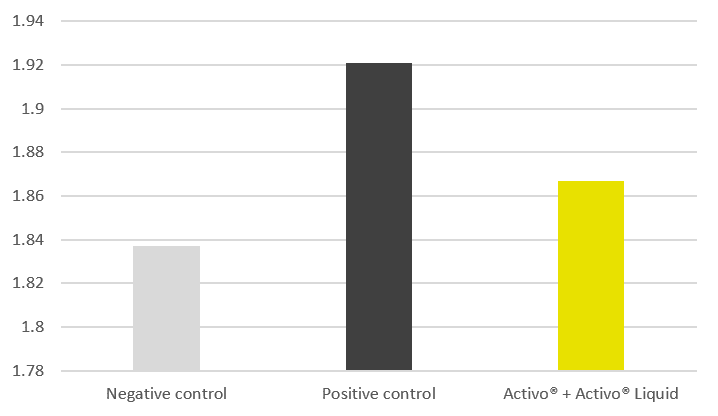
The positive control group received no supplements. The trial group received a synergistic combination of two phytogenic products containing standardized amounts of selected, microencapsulated phytomolecules: an in-feed phytogenic premix (Activo®, EW Nutrition GmbH) and a liquid complementary feed supplied via the drinking water (Activo® Liquid, EW Nutrition GmbH). The products were given at inclusion rates corresponding to the manufacturer’s baseline antibiotic reduction program recommendations (Figure 1):
Figure 1: Trial design
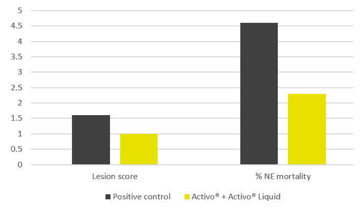
The trial results indicate that the addition of phytomolecules helps to mitigate the impact of NE on broilers’ performance. The group receiving Activo® and Activo® Liquid showed a better feed conversion (Figure 2) compared to the positive control group (NE challenge, no supplement). Also, better lesion scores were noted for animals receiving phytomolecules (0.7 and 1) than for the positive control group (1.6).
The most significant effect was observed concerning mortality: the group receiving Activo® and Activo® Liquid showed a 50% lower mortality rate than the positive control group (Figure 3). These results clearly indicate that phytomolecules can play an important role in mitigating losses due to NE.
Figure 1: Adjusted FCR
Figure 2: Lesion scores and mortality
Tackling necrotic enteritis in a sustainable way
In an age of AGP-free poultry production, a concerted focus on fostering animals’ gut health is key to achieving optimal performance. This study strongly demonstrates that, thanks to their antimicrobial, digestive, anti-inflammatory and antioxidant properties, phytomolecules effectively support birds’ intestinal health when challenged with NE. The inclusion of Activo® and Activo® Liquid, two phytogenic products designed to synergistically support birds during critical periods, resulted in improved feed conversion, better lesion scores, and 50% lower mortality.
In combination with good dietary, hygiene, and management practices, phytomolecules are therefore a potent tool for reducing the use of antibiotics: including Activo® and Activo® Liquid in their animals’ diets allows poultry producers to reduce the incidence of NE, to mitigate its economic impact in case of outbreaks, and therefore to control NE in a sustainable way.
Tang, Karen L., Niamh P. Caffrey, Diego B. Nóbrega, Susan C. Cork, Paul E. Ronksley, Herman W. Barkema, Alicia J. Polachek, Heather Ganshorn, Nishan Sharma, James D. Kellner, and William A. Ghali. “Restricting the Use of Antibiotics in Food-producing Animals and Its Associations with Antibiotic Resistance in Food-producing Animals and Human Beings: A Systematic Review and Meta-analysis.” The Lancet Planetary Health 1, no. 8 (November 6, 2017): 316-27. doi:10.1016/s2542-5196(17)30141-9.
Van Immerseel, Filip, Julian I. Rood, Robert J. Moore, and Richard W. Titball. “Rethinking Our Understanding of the Pathogenesis of Necrotic Enteritis in Chickens.” Trends in Microbiology 17, no. 1 (2009): 32-36. doi:10.1016/j.tim.2008.09.005.
byInge Heinzl, Marisabel Caballero, Ajay Bhoyar, EW Nutrition
Eliminating necrotic enteritis from your operations starts from a good understanding of what it is, how to prevent it, and how to mitigate its effects on your poultry production.
Necrotic enteritis is a poultry disease caused by an overgrowth of Clostridium perfringens type A, and to a lesser extent type C, in the small intestine. The toxins produced by C. perfringens also damage the intestinal wall. In general, it occurs in broiler chickens of 2-6 weeks of age. In subclinical forms, it is characterized by impaired digestion. Clinical forms lead to severe problems and increased flock mortality in a very short time.
Necrotic enteritis is the cause of USD 6 billion annual losses in global poultry production and this controllable disease is on the rise. One reason is the voluntary or legally required reduction of antibiotics in animal production. This trend is driven by the increasing occurrence of antimicrobial resistance, as well as by consumer demand. Another reason is the reduction of ionophores which, besides their activity against coccidia, also show efficacy against clostridia. When anticoccidial live vaccines are used, the application of these ionophores is not possible and clostridia / necrotic enteritis increase (Williams, 2005).
While this is a widespread problem in all poultry, for broilers in particular, necrotic enteritis and coccidiosis are the most significant health problem.
Clinical and subclinical forms of NE
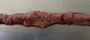
The clinical form
(c) Rob Moore
…is characterized by acute, dark diarrhea resulting in wet litter and suddenly increasing flock mortality of up to 1% per day after the first clinical signs appear (Ducatelle and Van Immerseel, 2010), sometimes summing up to mortality rates of 50% (Van der Sluis, 2013). The birds have ruffled feathers, lethargy, and inappetence.
Necropsy typically shows ballooned small intestines with a roughened mucosal surface, lesions, and brownish (diphtheritic) pseudo-membranes. There is a lot of watery brown, blood-tinged fluid and a foul odor during post-mortem examination. The liver is dark, swollen, and firm, and the gall bladder is distended (Hofacre et al., 2018).
In the case of peracute necrotic enteritis, birds may die without showing any preliminary signs.
The subclinical form
When birds suffer from the subclinical form, chronic damage to the intestinal mucosa and an increased quantity of mucus in the small intestine lead to impaired digestion and absorption of nutrients resulting in poor growth performance.
The deteriorated feed conversion and the resulting decreased performance become noticeable around day 35 of age. As feed contributes approximately 65-75% of the input cost to produce a broiler chicken, poor feed conversion increases production costs and significantly influences profitability. Often, due to a lack of clear symptoms, this subclinical disease remains untreated and permanently impacts the efficiency of production.
Pathogens
Responsible for necrotic enteritis are Gram-positive, anaerobic bacteria, specific strains of Clostridium perfringens type A and, to a lesser extent, type C (Keyburn et al., 2008).
Clostridia primarily occur in the soil where organic substances are degraded, in sewage, and the gastrointestinal tract of animals and humans. These bacteria produce spores, which are extremely resistant to environmental impact (heat, irradiation, exsiccation) as well as some disinfectants, and can survive for several years. Under suitable conditions, C. perfringens spores can even proliferate in feed or litter.
Clostridium perfringens is a natural inhabitant of the intestine of chickens. In healthy birds, it occurs in a mixture of diverse strains at 102-104 CFU/g of digesta (McDevitt et al., 2006). The disease starts when C. perfringens proliferates in the small intestine, usually due to a combination of factors such as high amount protein, low immunity, and an imbalance in the gut flora. Then, the number rises to 107-109 CFU/g of digesta (Dahiya et al., 2005).
NetB, a key virulence factor for NE
To establish in the host, Clostridium Spp. and other pathogens depend on virulence factors (see infobox). These virulence factors include, for example, “tools” for attachment, evasion or suppression of the host’s immune system, “tools” for getting nutrients, and “tools” for entry into intestinal cells. Over the years, the α-toxin produced by C. perfringens was assumed to be involved in the development of the disease and a key virulence factor. In 2008, Keyburn and coworkers found another key virulence factor by using a C. perfringens mutant unable to produce α-toxin, yet still causing necrotic enteritis.
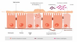
Thus, another toxin was identified occurring only in chickens suffering from necrotic enteritis: C. perfringens necrotic enteritis B-like toxin (NetB). NetB is a pore-forming toxin. Pore-forming toxins are exotoxins usually produced by pathogenic bacteria, but may also be produced by other microorganisms. These toxins destroy the integrity of gut wall cell membranes. The leaking cell contents serve as nutrients for the bacteria. If immune cells are destroyed, an immune reaction might be partially impacted (Los et al., 2013).
Additionally, pathogenic strains of C. perfringens produce bacteriocins – the most important being Perfrin (Timbermont et al., 2014) – to inhibit the proliferation of harmless Clostridium Spp. strains and to replace the normal intestinal flora of chickens (Riaz et al., 2017).
Examples of virulence factors
1. Adhesins
Enable the pathogen to adhere or attach within the target host site, e.g. via fimbria. Pili enable the exchange of RNA or DNA between pathogens.
2. Invasion factors
Facilitate the penetration and the distribution of the pathogens in the host tissue (invasion and spreading enzymes). For example: hyaluronidase attacking the hyaluronic acid of the connective tissue or flagella enabling the pathogens to actively move.
3. Toxins
Damage the function of the host cells or destroy them (e.g. endotoxins – lipopolysaccharides, exotoxins)
4. Strategies of evasion
Enable the pathogen to bypass the strategies of defense of the immune system (e.g. antiphagocytosis factors provide protection against an attack by phagocytes; specific antibodies are inactivated by enzymes).
A chicken with optimal gut health may be less susceptible to NE. Additional predisposing factors are necessary to allocate nutrients and make the gut environment suitable for the proliferation of these pathogens, enabling them to cause disease (Van Immerseel et al., 2008; Williams, 2005).
Predisposing factors
Feed: composition and particle size
The role of feed in the development of necrotic enteritis should not be underestimated. This is where substances creating an intestinal environment favorable for C. perfringens come into play.
Mycotoxin contamination
Mycotoxins harm gut integrity and create ideal conditions for the proliferation of Clostridium perfringens.
Mycotoxins do not have a direct effect on C. perfringens proliferation, toxin production, or NetB transcription. However, mycotoxins disrupt gut health integrity, creating a favorable environment for the pathogen. For example:
DON provides good conditions for proliferation of C. perfringens by disrupting the intestinal barrier and damaging the epithelium. The possibly resulting permeability of the epithelium and a decreased absorption of dietary proteins can lead to a higher amount of proteins in the small intestine. These proteins may serve as nutrients for the pathogen (Antonissen et al., 2014).
DON and other mycotoxins decrease the number of lactic acid producing bacteria indicating a shift in the microbial balance (Antonissen et al., 2016.).
Eimeria ssp.
An intact intestinal epithelium is the best defense against potential pathogens such as C. perfringens. Here, Coccidiosis comes into play. Moore (2016) showed that by damaging the gut epithelium, Eimeria species give C. perfringens access to the intestinal basal domains of the mucosal epithelium. Then, the first phase of the pathological process takes place and from there, C. perfringens invades the lamina propria. Damage to the epithelium follows (Olkowski et al., 2008). The plasma proteins leaking to the gut and the mucus produced are rich nutrient sources (Van Immerseel et al., 2004; Collier et al., 2008). A further impact of Coccidiosis is shifting the microbial balance in the gut by decreasing the number of e.g., Candidatus savagella which activates the innate immune defense.
Eimeria induce leakage of plasma proteins by killing epithelial cells
They enhance mucus production in the intestine
1+2 lead to an increase in available nutrients and create an environment favorable for the proliferation of C. perfringens.
Not only Eimeria Spp., also other pathogens (e.g. Salmonella Spp., Ascarid larvae, viruses) and agents, such as mycotoxins damaging the intestinal mucosa can pave the way for a C. perfringens infection.
Predisposing factors like wet litter, the moisture of which is essential for the sporulation of Eimeria Spp. oocysts, must also be considered as promoting factors for necrotic enteritis (Williams, 2005).
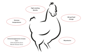
Immunosuppressive factors
Besides the already explained influencers feed, mycotoxins and coccidia, also other predisposing factors must be mentioned. In general, any factor which induces stress in the animals disrupts the balance of the intestinal flora. The resulting suppression of the immune system contributes to the risk of necrotic enteritis (Tsiouris, 2016). These factors include:
Bacteria: Shivaramaiah and coworkers (2011) investigated a neonatal Salmonella typhimurium infection as a predisposing factor for NE. The early infection causes significant damage to the gut (Porter et al., 1998) Additionally, Hassan et al. (1994) showed that the challenge with Salmonella typhimurium negatively impacted the development of lymphocytes which might also promote a colonization of Clostridium perfringens.
Viruses: Infectious Bursal Disease is known to increase the severity of infections with salmonella, staphylococci, but also clostridia. Another clostridia-promoting viral disease is Marek’s Disease.
Stress: The intestinal tract is particularly sensitive to any type of stress. This stress can be caused by e.g. too high temperatures, high stocking densities, an abrupt change of feed.
Treatment
In acute cases, the farmer should consult a veterinarian and treat his birds.
It must be mentioned that, as the treatment takes place via feed or water, only birds which still consume water or feed may be treated.
Antibiotics
Antibiotics targeting Gram-positive bacteria are commonly used for the treatment of acute NE. The antibiotic choice shall be addressed by a veterinarian, taking into account mode of action and the presence of resistance genes in the farm/flock.
The prophylactic use of antibiotics is not recommened and many countries have already banned it in order to reduce antimicrobial resistance (AMR).
Antimicrobial Resistance (AMR)
Some bacteria are less sensitive to certain antibiotics due to genetic mutations. They are able to:
stimulate the production of enzymes, which break down or modify the antibiotics and inactivate them (1).
eliminate entrances for antibiotics or promote the development of pumps, which discharge the antibiotic before taking effect (2).
change or eliminate molecules to which the antibiotic would bind (targets for the antibiotics).
This means that, when the corresponding antibiotics are used, bacteria resistant against these antibiotics survive. Due to the fact that their competitors have been eliminated they are able to reproduce better. Additionally, this resistance may be transferred by means of “resistance genes”
to daughter cells
via their intake from dead bacteria (3)
through horizontal gene transfer (4)
through viruses (5)
Every application of antibiotics promotes the development of resistance (Robert Koch Institute, 2019). A short-term use, better biosecurity, or an application at low dosage give the bacteria a better chance to adapt.
Bacteriophages
Experimental use of phage treatments have shown to be effective in reducing disease progression and symptoms of necrotic enteritis (Miller et al., 2010). By oral application of a bacteriophage cocktail, Miller and coworkers could reduce mortality by 92% in C. perfringens challenged broilers compared to the untreated control.
Mode of action: the endolysins, highly evolved enzymes produced by bacteriophages, are able to digest the bacterial cell wall for phage progeny release (Fischetti, 2010). However, phages are still not approved by the EFSA.
Prevention
Preventing a disease is always better – and more cost-effective – that its treatment.
How, then, should it be done?
Preventing the conditions that favor the proliferation of Clostridium perfringens and strengthening the host’s immune response lowers the probability of disease.
Besides eliminating the predisposing factors, the main targets are:
There is evidence that most Clostridium strains isolated from birds suffering from necrotic enteritis could induce the disease experimentally, while strains isolated from healthy birds cannot. This confirms that only specific strains are problematic (Ducatelle and Van Immerseel, 2010).
It is therefore of the highest importance to avoid introducing these pathogenic strains to the farm.
Separate clothing, boots, and hand washing/disinfecting facilities in each poultry house
More than 14 days of down time between flocks
Specific measures against coccidiosis
Vaccination
According to parasitologists, 7 to 9 Eimeria species are found in chickens, and they do not cross-protect against each other. An effective vaccination must contain sporulated oocysts of the most critical pathogenic Eimeria species (E. acervulina, E. maxima, E. tenella, E. necatrix, and E. brunetti). The more species contained in the vaccine, the better. However, if not applied the correct way, vaccines can be ineffective or cause reactions in the birds that might lead to NE (Mitchell, 2017).
Anticoccidials
Alternate use of chemicals (synthetic compounds) and ionophores (polyether antibiotics) with different modes of action is important to avoid development of resistance.
Ionophores have a specific mode of action and kill oocysts before they are able to infect birds. Being very small, ionophore molecules can be taken up and diffused into the outer membrane of the sporozoite. There, it decreases the concentration gradient leading to an accumulation of water within the sporozoite causing its bursting.
Diet
Minimizing non-starch polysaccharides (NSPs) in cereals
To prevent a “feeding” of Clostridium perfringens, high content of water-soluble but indigestible NSPs such as wheat, wheat by-products, and barley should be avoided or at least minimized. Additionally, xylanases should be included in the feed formulation to reduce the deleterious effects of NSPs and improve feed energy utilization. Instead of these cereals, maize could be included in the diet. It is considered a perfect ingredient in broiler diets due to its high energy content and high nutrient availability.
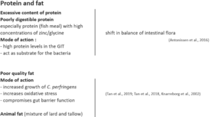
Formulating low protein diets/diets with highly digestible amino acids
Feeding low-protein diets supplemented with crystalline amino acids might be beneficial to reduce the risk of necrotic enteritis (Dahiya et al., 2007). To improve protein digestibility and therefore reduce the proliferation of C. perfringens, proteases may be added to the feed.
Avoiding/Minimizing poor quality fats / animal fats in the diet
These fats tend to increase the count of Clostridium perfringens; thus, they should be replaced by higher quality and/or vegetable fats, respectively.
Feed form
In terms of feed form, Engberg et al. (2002) found that birds fed pellets showed a reduced number of Clostridium perfringens in the caeca and the rectum than mash-fed birds. Branton and co-workers (1987) reported a lower mortality by feeding roller-milled (coarsely ground) than hammer-milled feed.
Additives
Additives can be used either to prevent the proliferation of Clostridium perfringens or to change the environmental conditions in a way that proliferation of C. perfringens is prevented.
Probiotics
These live microbial supplements can be used to help to establish, maintain or re-establish the intestinal microflora.
Mode of action:
compete with pathogenic bacteria for substrates and attachment sites
produce antimicrobial substances inhibiting the growth of pathogenic bacteria (Gillor et al., 2008)
bind and neutralize enterotoxins (Mathipa and Thantsha, 2017)
promote immune function of the host (Yang et al., 2012)
Prebiotics
These feed ingredients serve as substrates to promote beneficial bacteria in the intestine.
Mode of action:
D-mannose or fructose, starches non-digestible by birds, selectively stimulate the growth and the activity of the “good” gut flora
Fructooligosaccharides decrease C.perfringens and E. coli in the gut and increase the diversity of Lactobacillus Spp. (Kim et al., 2011)
Galactooligosaccharides, in combination with a B. lactis based probiotic, have been reported to selectively promote the proliferation of Bifidobacterium ssp. (Jung et al., 2008).
Organic acids
Organic acids are often used in animal diets to improve intestinal health.
Mode of action:
decreased pH promotes beneficial bacteria
caprylic acid suppresses C. perfringens, but also Salmonella Spp. by inhibiting their utilization of glucose (Skrivanova et al., 2006)
lauric, citric, oleic and linoleic acid as well as medium-chain fatty acids (C8-C14) impede the growth of C. perfringens
Phytomolecules
Phytomolecules, also known as secondary plant compounds, have been used against pathogens for centuries. In general, two subgroups of these substances are known as effective against Clostridium perfringens:
Tannins
Many studies have shown the efficacy of tannins against different pathogens such as helminths, Eimeria, viruses, and bacteria
Extracts from the chestnut and quebracho trees are effective not only against C. perfringens, but also its toxins (Elizando et al., 2010)
Activity of tannins against Eimeria (Cejas et al., 2011) and Salmonella Sp., two predisposing factors for NE.
Essential Oils
Their hydrophobic characteristic enables them to interact with the lipids of the membrane of C. perfringens.
They can incorporate into the bacterial membrane and disrupt its integrity.
This increases the permeability of the cell membrane for ions and other small molecules such as ATP, leading to the decrease of the electrochemical gradient above the cell membrane and the loss of the cell’s energy equivalents.
Besides their direct effect on Clostridium Spp., a lot of phytomolecules improve gut health and help to prevent a proliferation of Clostridium ssp. and therefore necrotic enteritis.
Mycotoxin/bacterial toxin binders
These binders have two modes of action:
Binding mycotoxins, damage of the intestinal epithelium can be reduced or even prevented, so that the preconditions for Clostridium proliferation are not generated.
Binding toxins produced by Clostridium perfringens can reduce the occurrence or severity of lesions:
Alpha-toxin (phospholipase C) hydrolyses membrane phospholipids and damages erythrocytes, leucocytes, myocytes, and endothelial cells and causes their lysis (Songer, 1996). This leads to necrosis and tissue damage.
Binding NetB toxin, the key virulence factor, could reduce the severity of necrotic enteritis.
Conclusion
The ever-growing trend of reduced antibiotic and ionophore use is contributing to an increased incidence of necrotic enteritis in poultry production.
The subclinical form of necrotic enteritis generally goes unnoticed, resulting in poor feed efficiency and is a major cause of financial losses to poultry producers.
Maintaining optimum gut health is key to preventing the occurrence of necrotic enteritis. In the era of antibiotic-free poultry production, alternatives acting against this pathogenic bacterium and also against its predisposing factors must be considered to control this devastating disease.
References
Annett, C.B., J. R. Viste, M. Chirino-Trejo, H. L. Classen, D. M. Middleton, and E. Simko. “Necrotic enteritis: effect of barley, wheat and corn diets on proliferation of Clostridium perfringens type A.” Avian Pathology 31 (2002): 599– 602. https://doi.org/10.1080/0307945021000024544
Antonissen G, F. Van Immerseel, F. Pasmans, R. Ducatelle, F. Haesebrouck, L. Timbermont, M. Verlinden, G.P.J. Janssens, V. Eeckhaut, M. Eeckhout, S. De Saeger, S. Hessenberger, A. Martel, and S. Croubels. “The mycotoxin deoxynivalenol predisposes for the development of Clostridium perfringens-Induced necrotic enteritis in broiler chickens. PLoS ONE 9 no. 9 (2014): e108775. https://doi.org/10.1371/journal.pone.0108775
Antonissen, G., V. Eeckhaut, K. Van Driessche, L. Onrust , F. Haesebrouck, R. Ducatelle, R.J. Moore, and F. Van Immerseel. “Microbial Shifts Associated With Necrotic enteritis.” Avian Pathol. 45 no. 3 (2016): 308-312. https://doi.org/10.1080/03079457.2016.1152625
Branton, S.L., F.N. Reece, and W.M. Hagler. “Influence of a wheat diet on mortality of broiler chickens associated with necrotic enteritis.” Poultry Sci. 66 (1987): 1326-1330. https://doi.org/10.3382/ps.0661326
Cejas, E., S. Pinto, F. Prosdócimo, M. Batalle, H. Barrios, G. Tellez, and M. De Franceschi. “Evaluation of quebracho red wood (Schinopsis lorentzii) polyphenols vegetable extract for the reduction of coccidiosis in broiler chicks.” International Journal of Poultry Science 10 no. 5 (2011): 344–349. https://doi.org/10.3923/ijps.2011.344.349
Collier, C.T., C.L. Hofacre, A.M. Payne, D.B. Anderson, P. Kaiser, R.I. Mackie, and H.R. Gaskins. “Coccidia-induced mucogenesis promotes the onset of necrotic enteritis by supporting Clostridium perfringens growth.” Veterinary Immunology and Immunopathology 122 (2008):104–115.
Dahiya, J.P., D. Hoehler, A.G. Van Kessel, and M.D. Drew. “Effect of different dietary methionine sources on intestinal microbial populations in broiler chickens.” Poultry Science 86 (2007):2358–2366
Dahiya, J.P., D. Hoehler, D.C. Wilkie, A.G. van Kessel, and M.D. Drew. “Dietary glycine concentration affects intestinal Clostridium perfringens and Lactobacilli populations in broiler chickens.” Poultry Science 84 no.12 (2005):1875-85. https://doi.org/10.1093/ps/84.12.1875
Diaz Carrasco, J.M., L.M. Redondo, E.A. Redondo, J.E. Dominguez, A.P. Chacana, and M.E. Fernandez Miyakawa. “Use of plant extracts as an effective manner to control Clostridium perfringens induced necrotic enteritis in poultry.” BioMed Research International (2016): Article ID 3278359. https://dx.doi.org/10.1155/2016/3278359
Ducatelle, R. and F. van Immerseel. “Necrotic enteritis: emerging problem in broilers.” WATTAgNet.com – Poultry Health and Disease (April 9, 2010).
Elizondo, A.M., E.C. Mercado, B.C. Rabinovitz, and M.E. Fernandez-Miyakawa. “Effect of tannins on the in vitro growth of Clostridium perfringens.” Veterinary Microbiology 145 no. 3-4 (2010): 308–314. https://doi.org/10.1016/j.vetmic.2010.04.003
Engberg, R.M., M.S. Hedemann, and B.B. Jensen. “The influence of grinding and pelleting of feed on the microbial composition and activity in the digestive tract of broiler chickens.” · British Poultry Science 43 no. 4 (2002):569-579. https://doi.org/10.1080/0007166022000004480
Fischetti, V.A. “Bacteriophage endolysins: A novel anti-infective to control Gram-positive pathogens.” J Med Microbiol. 300 no. 6 (2010): 357–362. https://doi.org/10.1016/j.ijmm.2010.04.002
Gillor, O., A. Etzion and M.A. Riley. “The dual role of bacteriocins as anti- and probiotics.” Appl Microbiol Biotechnol. 81 no. 4 (2008): 591–606. https://doi.org/10.1007/s00253-008-1726-5
Hassan, J. O., and R. Curtiss III. “Virulent Salmonella typhimurium induced lymphocyte depletion and immunosuppression in chickens.” Infect. Immun. 62 (1994):2027–2036 https://doi.org/10.1128/IAI.62.5.2027-2036.1994
Hofacre, C.L., J.A. Smith, and G.F. Mathis. “Invited Review. An optimist’s view on limiting necrotic enteritis and maintaining broiler gut health and performance in today’s marketing, food safety, and regulatory climate.” Poultry Science 97 (2018):1929–1933. https://dx.doi.org/10.3382/ps/pey082
Jung, S.J., R. Houde, B. Baurhoo, X. Zhao, and B. H. Lee. “Effects of galacto-oligosaccharides and a bifidobacteria lactis-based probiotic strain on the growth performance and fecal microflora of broiler chickens.” Poultry Science 87 (2008):1694–1699. https://doi.org/10.3382/ps.2007-00489
Kaldhusdal and Skjerve. “Association between cereal contents in the diet and incidence of necrotic enteritis in broiler chickens in Norway.” Preventive Veterinary Medicine 28 (1996):1-16. https://doi.org/10.1016/0167-5877(96)01021-5
Keyburn, A. L., S. A. Sheedy, M. E. Ford, M. M. Williamson, M. M. Awad, J. I. Rood, and R. J. Moore. “Alpha-toxin of Clostridium perfringens is not an essential virulence factor in necrotic enteritis in chickens.” Infect. Immun. 74 (2006): 6496–6500. https://doi.org/10.1128/IAI.00806-06
Keyburn, A.L., J.D. Boyce, P. Vaz, T.L. Bannam, M.E. Ford, D. Parker, A. Di Rubbo, J.I. Rood, and R.J. Moore. “NetB, a new toxin that is associated with avian necrotic enteritis caused by Clostridium perfringens.” PLoS Pathog 4 no. 2, e26 (2008): 0001-0011. https://doi.org/10.1371/journal.ppat.0040026
Kim, G.-B., Y. M. Seo , C. H. Kim , and I. K. Paik. “Effect of dietary prebiotic supplementation on the performance, intestinal microflora, and immune response of broilers.” Poultry Science 90 (2011):75–82. https://doi.org/10.3382/ps.2010-00732
Knap, I., B. Lund, A. B. Kehlet, C. Hofacre, and G. Mathis. “Bacillus licheniformis prevents necrotic enteritis in broiler chickens.” Avian Diseases 54 no. 2 (2010):931-935. https://doi.org/10.1637/9106-101509-ResNote.1
Knarreborg, A., M.A. Simon, R.M. Engberg, B.B. Jensen, and G.W. Tannock. “Effects of Dietary Fat Source and Subtherapeutic Levels of Antibioticon the Bacterial Community in the Ileum of Broiler Chickensat Various Ages.” Applied and Environmental Microbiology 68 no. 12 (2002): 5918-5924. https://doi.org/0.1128/AEM.68.12.5918–5924.2002
Kocher, A. and M. Choct. “Improving broiler chicken performance. The efficacy of organic acids, prebiotics and enzymes in controlling necrotic enteritis.” Australian Government-Rural Industries Research and Development Corporation. Publ. no. 08/149 (2008).
Kondo, F. “In vitro lecithinase activity and sensitivity to 22 antimicrobial agents of Clostridium perfringens isolated from necrotic enteritis of broiler chickens.” Research in veterinary Science 45 (1988): 337-340. https://doi.org/10.1016/S0034-5288(18)30961-5
Kubena, L.F., J.A. Byrd, C.R. Young, and D.E. Corrier. “Effects of tannic acid on cecal volatile fatty acids and susceptibility to Salmonella typhimurium colonization in broiler chicks.” Poultry Science 80, no. 9 (2001): 1293–1298. https://doi.org/10.1093/ps/80.9.1293
Los, F.C.O., T.M. Randis, R.V. Aroian, and A.J. Ratner. “Role of pore-forming toxins in bacterial infectious diseases.” Microbiology and Molecular Biology Reviews 77 (2013): 173-207 https://doi.org/10.1128/MMBR.00052-12
M’Sadeq S.A., Shubiao Wu, Robert A. Swick, Mingan Choct. “Towards the control of necrotic enteritis in broiler chickens with in-feed antibiotics phasing-out worldwide.” Animal Nutrition 1 (2015): 1-11. https://dx.doi.org/10.1016/j.aninu.2015.02.004
Mathipa, M.G. and M.S. Thantsha. “Probiotic engineering: towards development of robust probiotic strains with enhanced functional properties and for targeted control of enteric pathogens.” Gut Pathog. 9 no. 28 (2017). https://doi.org/10.1186/s13099-017-0178-9
McDevitt, R.M., J.D. Brooker, T. Acamovic, and N.H.C. Sparks. “Necrotic enteritis, a continuing challenge for the poultry industry.” World’s Poultry Science Journal 62; World’s Poultry Science Association (June 2006). https://doi.org/10.1079/WPS200593
Miller, R.W., J. Skinner, A. Sulakvelidze, G.F. Mathis, and C.L. Hofacre. “Bacteriophage therapy for control of necrotic enteritis of broiler chickens experimentally infected with Clostridium perfringens.” Avian Diseases 54 no. 1 (2010): 33-40. https://doi.org/10.1637/8953-060509-Reg.1
Mitsch, P., K. Zitterl-Eglseer, B. Köhler, C. Gabler, R. Losa, and I. Zimpernik. “The Effect of Two Different Blends of Essential Oil Components on the Proliferation of Clostridium perfringens in the Intestines of Broiler Chickens.” Poultry Science 83 (2004):669–675. https://doi.org/10.1093/ps/83.4.669
Olkowski, A.A., C. Wojnarowicz, M. Chirino-Trejo, B. Laarveld, and G. Sawicki. “Sub-clinical necrotic enteritis in broiler chickens: Novel etiological consideration based on ultra-structural and molecular changes in the intestinal tissue.” Veterinary Science 85 (2008): 543–553. https://doi.org/10.1016/j.rvsc.2008.02.007
Pan, D. and Z. Yu. “Intestinal microbiome of poultry and its interaction with host and diet.” Gut Microbes 5 no. 1 (2014): 108–119. https://dx.doi.org/10.4161/gmic.26945
Rougière, N. and B. Carré. “Comparison of gastrointestinal transit times between chickens from D + and D- genetic lines selected for divergent digestion efficiency.” Animal 4 no. 11 (2010): 1861-1872. https://doi.org/10.1017/S1751731110001266
Santos, F.B.O., B.W. Sheldon, A.A. Santos Jr., and P.R. Ferket. ”Influence of housing system, grain type, and particle size on Salmonella colonization and shedding of broilers fed triticale or corn-soybean meal diets.” Poultry Science 87 (2008): 405-420. https://dx.doi.org/10.3382/ps.2006-00417
Schiavone, A. , K. Guo, S. Tassone, L .Gasco, E. Hernandez, R. Denti, and I. Zoccarato. “Effects of a Natural Extract of Chestnut Wood on Digestibility, Performance Traits, and Nitrogen Balance of Broiler Chicks.” Poult Sci. 87 no. 3 (2008): 521-527. https://doi.org/10.3382/ps.2007-00113
Shivaramaiah, S., R. E. Wolfenden, J. R. Barta, M. J. Morgan, A. D. Wolfenden, B. M. Hargis, and G. Téllez. „The role of an early Salmonella typhimurium infection as a predisposing factor for necrotic enteritis in a laboratory challenge model.” Avian Diseases 55 (2011): 319-323. https://doi.org/10.1637/9604-112910-ResNote.1
Singh, Y., V. Ravindran, T.J. Wester, A.L. Molan, and G. Ravindran. “ Influence of feeding coarse corn on performance, nutrient utilization, digestive tract measurements, carcass characteristics, and cecal microflora counts of broilers.” Poultry Science 93 (2014): 607–616. https://dx.doi.org/10.3382/ps.2013-03542
Skrivanova, E., M. Marounek, V. Benda, and P. Brezina. “Susceptibility of Escherichia coli, Salmonella sp. and Clostridium perfringens to organic acids and monolaurin.” Veterinarni Medicina 51 no. 3 (2006): 81–88. https://doi.org/10.17221/5524-VETMED
Stanley D., Wu S.-B., Rodgers N., Swick R.A., and Moore R.J. “Differential Responses of Cecal Microbiota to Fishmeal, Eimeria and Clostridium perfringens in a Necrotic Enteritis Challenge Model in Chickens.” PLoS ONE 9 no. 8 (2014): e104739. https://doi.org/10.1371/journal.pone.0104739
Tan, L., D. Rong, Y. Yang, and B. Zhang. “Effect of Oxidized Soybean Oils on Oxidative Status and Intestinal Barrier Function in Broiler Chickens.” Brazilian Journal of Poultry Science 18 no. 2 (2018): 333-342. http://dx.doi.org/10.1590/1806-9061-2017-0610
Tan, L., D. Rong, Y. Yang, and B. Zhang. “The Effect of Oxidized Fish Oils on Growth Performance, Oxidative Status, and Intestinal Barrier Function in Broiler Chickens.” J. Appl. Poult. Res. 28 (2019): 31-41. http://dx.doi.org/10.3382/japr/pfy013
Timbermont L., A. Lanckriet, J. Dewulf, N. Nollet, K. Schwarzer, F. Haesebrouck, R. Ducatelle, and F. Van Immerseel. “Control of Clostridium perfringens-induced necrotic enteritis in broilers by target-released butyric acid, fatty acids and essential oils.” Avian Pathol. 39 no. 2 (2010): 117-21. https://doi.org/10.1080/03079451003610586
Tsiouris, V. “Poultry management: a useful tool for the control of necrotic enteritis in poultry.” Avian Pathol. 45 no. 3 (2016):323-325. https://doi.org/10.1080/03079457.2016.1154502
Van der Most, P.J., B. de Jong, H.K. Parmentier and S. Verhulst. “Trade-off between growth and immune function: a meta-analysis of selection experiments.” Functional Ecology 25 (2011): 74-80. https://doi.org/0.1111/j.1365-2435.2010.01800.x
Van der Sluis, W. “Clostridial enteritis is an often underestimated problem.” Worlds Poult. Sci. J. 16 (2000):42–43.
Van Immerseel, F., J. De Buck, F. Pasmans, G. Huyghebaert, F. Haesebrouck, and R. Ducatelle. “Clostridium perfringens in poultry: an emerging threat of animal and public health.” Avian Pathology 33 (2004): 537-549. https://doi.org/10.1080/03079450400013162
Van Immerseel, F., J.I. Rood, R.J. Moore, and R.W. Titball. “Rethinking our understanding of the pathogenesis of necrotic enteritis in chickens.” Trends in Microbiology 17 no. 1 (2008):32-36. https://doi.org/10.1016/j.tim.2008.09.005
Wade, B., A.L. Keyburn, T. Seemann, J.I. Rood, and R.J. Moore. “Binding of Clostridium perfringens to collagen correlates with the ability to cause necrotic enteritis in chickens.” Veterinary Microbiology 180 no. 3–4 (2015): 299-303. https://doi.org/10.1016/j.vetmic.2015.09.019
Williams, R.B. “Intercurrent coccidiosis and necrotic enteritis of chickens: rational, integrated disease management by maintenance of gut integrity.” Avian Pathology 34 no. 3 (2005):159-180. https://doi.org/10.1080/03079450500112195
Yang , C.M., G.T. Cao, P.R. Ferket, T.T. Liu, L. Zhou, L. Zhang, Y.P. Xiao, and A. G. Chen. “ Effects of probiotic, Clostridium butyricum, on growth performance, immune function, and cecal microflora in broiler chickens.” Poultry Science 91 (2012): 2121–2129. https://dx.doi.org/10.3382/ps.2011-02131
Fewer pathogens with egg immunoglobulins
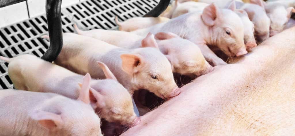
For newborn pigs there are often a host of different challenges – think of crushing or contamination of the farrowing pen. For the last problem, solutions exist. A dietary approach can help to relieve pathogenic pressure through sow manure.
The main objective of a piglet producer is to maximise the number of healthy weaned piglets per animal per year. Nowadays, it is not difficult to find production systems delivering more than 30 piglets weaned/sow/year. Combining strategies on management, feeding, and health of both piglets and sows, is crucial for increasing sow’s productivity. A unique environment that can determine the success of a piglet farm is the farrowing unit. It is important to reduce as much as possible losses during this period. Pre-weaning mortality must always be monitored and targets must be set. In European conditions, it ranges between 8-10%.
One important driver in reducing pre-weaning mortality is understanding the fragility of newborn piglets. At birth, the resources of a piglet are very scarce: low energy reserves and practically no immune defence against existing pathogens in their new environment. Problems are prone to happen and will be mostly caused by pathogens present in the environment, in the feed, in the water and most important, in the faeces of the sow. The main contamination source for newborn piglets is their mother’s manure. And this first contamination can be quite severe causing diarrhoea and increasing piglet mortality.
Together with crushing, diarrhoea definitely causes a high percentage of total losses during the first days of life. In most of the cases, the disease is caused not only by one agent but by a combination of enteric infections from different pathogens or at least different strains of a pathogenic species. E. coli and clostridia are two of the most important diarrhoea causing pathogens during the first weeks after birth.
Pathogens during the first days E. coli is well known as one of the main responsible pathogens for pre-weaning diarrhoea. And although it belongs to the normal intestinal flora of pigs, part of the different E. coli strains are pathogenic. E. coli cause about 80% of diarrhoeas in piglets and 50% of losses in piglet production. The factors making E. coli pathogenic, the so-called virulence factors include e.g. fimbria to attach to the intestinal wall and the capacity to produce toxins.
The Clostridium species are another important pathogen class. During the suckling phase, piglets are quite susceptible to Clostridium perfringens type C. This bacteria causes necrotic enteritis in piglets and the clinical symptoms appear during the first days of life. This disease provokes serious disturbances in the organism with a mortality up to 100%. It causes significant decrease in daily gain and in weaning weight.
Strategy to protect the piglets In order to maximise the sow’s performance – measured in piglets weaned per year – it is crucial to provide the best possible conditions to the piglets. Therefore the reduction of the pathogenic pressure in the farrowing unit ranks first. Cleaning of the pen is a way to get rid of germs like E. coli and Clostridium species, the most important pathogens during the first days. This should be completed by an effective gut health management in sow and piglets. For this purpose natural ingredients can be used. Supplying natural and active immune cells, the so called antibodies, has been proven to be quite efficient in supporting gut health. Applied to piglets, immunoglobulins from the egg bind to pathogens within the intestinal tract. They show efficiency in supporting piglets’ performance, decreasing the incidence of diarrhoea, mortality and increasing daily gain.
The idea was to check if these immunoglobulins from the egg could also bind pathogens in the sow’s gut and generate harmless complexes. That way pathogenic pressure for the piglets could be reduced. Thus a trial was conducted in Japan to check this thesis.
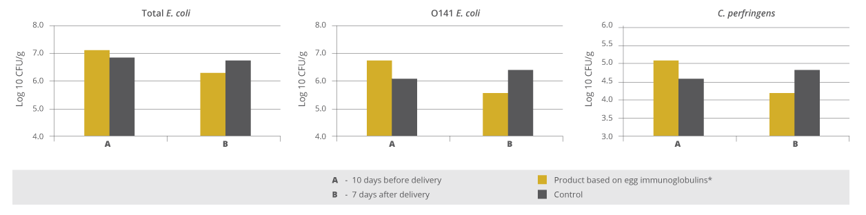
Trial In the trial two groups contained eight sows each. The sows of the control group received standard lactation feed, the trial group was also fed standard feed with a supplement containing egg immunoglobulins (Globigen Sow, EW Nutrition, at a dosage of 5 g/sow twice daily) on top during the last ten days before and the first seven days after delivery. The faeces of the sows were obtained by rectal stimulation (in order to get no contamination from the environment) on day 10 before and day 7 after delivery. The amount of colony forming units (CFU) of total E. coli, E. coli O141 and Clostridium perfringens were determined.
Results are shown in Figure 1. At the beginning of the trial, before the application of the immunoglobulin supplement, both groups showed nearly the same level of the evaluated pathogens with a slight disadvantage for the supplement group. After 17 days of applying the product based on egg immunoglobulins, a reduction of the colony forming units of total E. coli, E. coli O141 and of Clostridium perfringens could be seen. The sows of the supplement-fed group showed a lower level of pathogens in their excrements than the sows of the control group.
Conclusion It is important for swine producers to understand what adversely influences the results on the farm. One consideration is to improve farrowing unit conditions of the piglets, aiming to reduce pre-weaning mortality. The results of the trial showed that a supplement based on egg immunoglobulins supplied on top of standard sow diets substantially reduced the amount of pathogenic colonies in sow manure. The reduction on pathogenic pressure and therefore the incidence of diarrhoea may be an alternative for increasing the profitability of piglet producers by increasing the number of healthier piglets weaned/sow/year.
*References are available on request.
By Dr Inge Heinzl.
Published on PigProgress | 20th July, 2018.
Using egg immunoglobulins to enhance piglet survival
The number of healthy piglets weaned is the most important factor for the calculation of profit in piglet production.
Losses in the farrowing unit normally occur during the first seven days of life as piglets are born with very little protection in the form of immunity. The intake of immunoglobulins from colostrum is therefore of vital importance. Besides cleanliness and special feeding, piglets can be additionally supported by two strategies that mimick the effect of colostrum:
– a direct one, meaning the feeding of immunoglobulins (IgY from eggs) to piglets that would support the immune system in the gut or
– an indirect one, meaning a supply of IgY to the sow to keep the pathogenic pressure in the farrowing unit as low as possible.
Piglets are born with no immune protection and very low energy reserves It is well known that piglets are physiologically immature at birth. Their energy reserves are very low with only 1 – 2% body fat comprising mainly of structural and subcutaneous fat. Therefore, in the first hours of life they rely on the glucose supply from glycogen from the liver as their main energy source. However, this will only cover their needs for a few hours.
Due to the construction of the sow’s placenta, a transfer of immunoglobulins (antibodies) within the uterus is not possible. This means that piglets are born with practically no immune protection and depend on the immediate intake of immunoglobulins from colostrum. The immunoglobulins can be absorbed in the gastrointestinal tract and immediately transferred into the bloodstream – but also only for a short time. The absorption ability of the piglets starts to decrease soon after birth and ends after 24 to 36 hours.
Strategy 1: Making the farrowing unit as safe as possible The piglets’ environment should be warm to prevent hypoglycaemia. Piglets looking for heat close to the sow can also get crushed. Since the temperature needs of the sow and piglets are different, a piglet nest with a special heat lamp is recommended. Furthermore, the farrowing unit should be clean. Due to their low immune status, piglets are susceptible to common pathogens such as E. coli, Clostridium perfringens, and rotavirus that can all lead to diarrhoea.
Most pathogens can be traced to those found in the sow’s faeces. To keep this amount as low as possible, different measures can be taken:
– A vaccination increases the immune defences of the sow. The antibodies fight against the pathogens so that less “functioning” pathogens are excreted.
– Feeding of probiotics increases the number of good bacteria like Lactobacilli and Bifidobacteria competing with the pathogens for binding sites and nutrients.
– Administration of egg immunoglobulins, which bind to the pathogens within the gastrointestinal tract and make them harmless. These pathogen-immunoglobulin-complexes can be ingested by the piglets without any danger.
Strategy 2: Supporting the piglets with immunoglobulins The aim here is to strengthen the local immunity in the gastrointestinal tract by increasing the amount of immunoglobulins (Ig). As already mentioned, the intake of sow colostrum is of vital importance. With the vaccination of the sow, the content of antibodies in the colostrum can even be enhanced.
An additional measure would be to orally supply the piglets with egg immunoglobulins (IgY). Both classes of immunoglobulins (IgG from mammals, and IgY from birds) can bind to pathogens in the gut, preventing them from binding to the intestinal wall and reducing the incidence of diarrhoea. The difference is in the degree of effectiveness and specificity.
Conclusion To maximize the number of piglets weaned, it is necessary to support their immune system during the first days of life. Besides good hygiene management, the administration of egg antibodies to the sow will also help reduce the amount of shed pathogens keeping the pathogenic pressure low. The application of egg antibodies directly to the piglets supports their immune system by binding the pathogens in the gut, minimizing the risk of diarrhoea.


 Figures 1 a-c: Reduction of bacterial count due to the addition of Formycine
 Figure 2: Preventive effect of Formycine Gold Px concerning necrotic enteritis gut lesions
 Figure 3a and 3b: Performance-maintaining effect of Formycine Gold Px
 Figure 4: MBC of Acidomix AFG against different pathogens (%)
 Figure 5: MIC of Acidomix AFG against different pathogens (%)

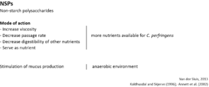

 To prevent a “feeding” of Clostridium perfringens, high content of water-soluble but indigestible NSPs such as wheat, wheat by-products, and barley should be avoided or at least minimized. Additionally, xylanases should be included in the feed formulation to reduce the deleterious effects of NSPs and improve feed energy utilization. Instead of these cereals, maize could be included in the diet. It is considered a perfect ingredient in broiler diets due to its high energy content and high nutrient availability.