Mycotoxins in poultry – External signs can give a hint

Part 3: Bone disorders and foot pad lesions

By Dr. Inge Heinzl, Editor, and Marisabel Caballero, Global Technical Manager Poultry


Bone health is essential for animals and humans. Besides giving structural support, allowing movement, and protecting vital organs, the bones release hormones that are crucial for mineral homeostasis and acid balance and serve as reservoirs of energy and minerals (Guntur & Rosen, 2012; Rath, N.C. & Durairaj, 2022; Suchacki et al., 2017).

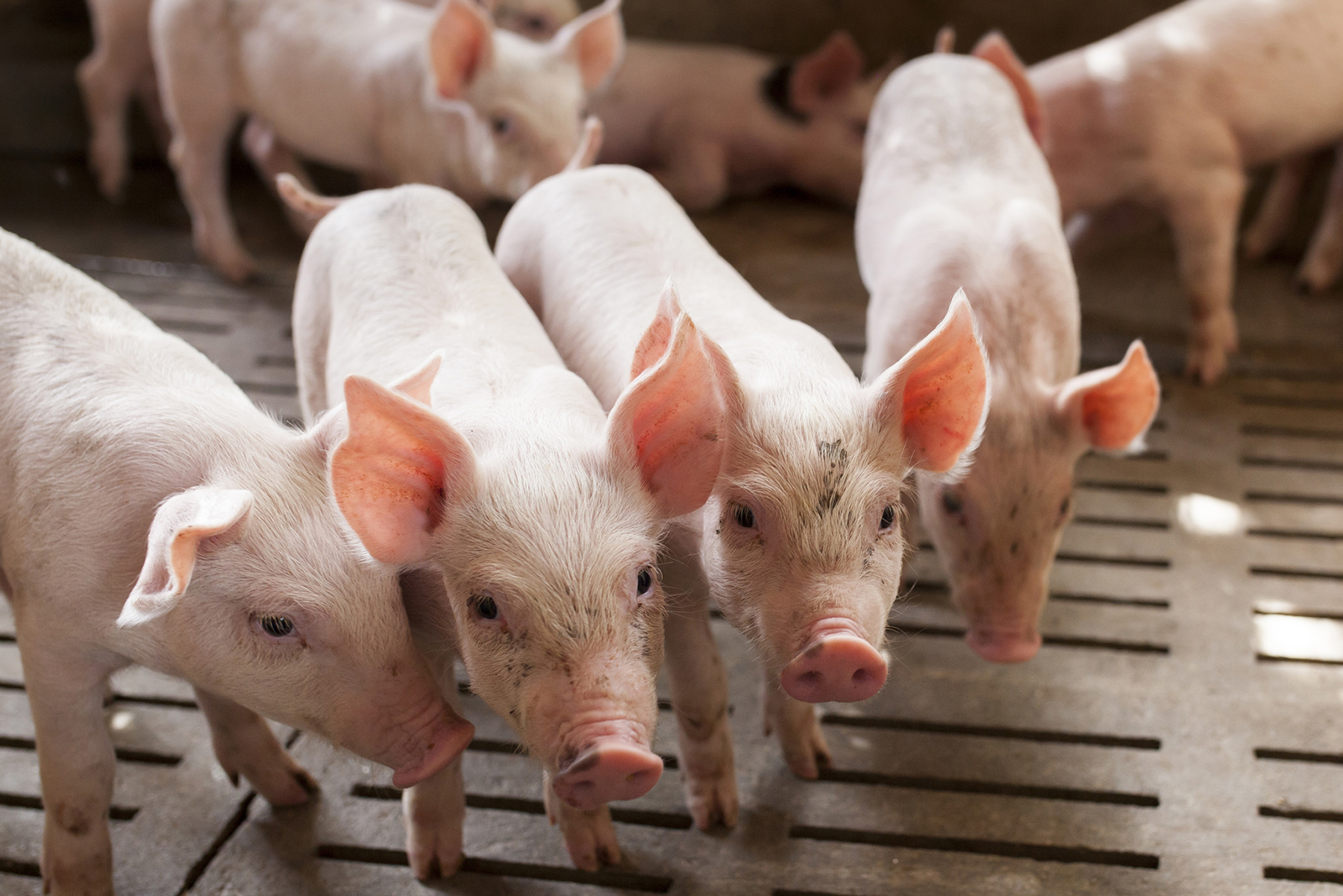
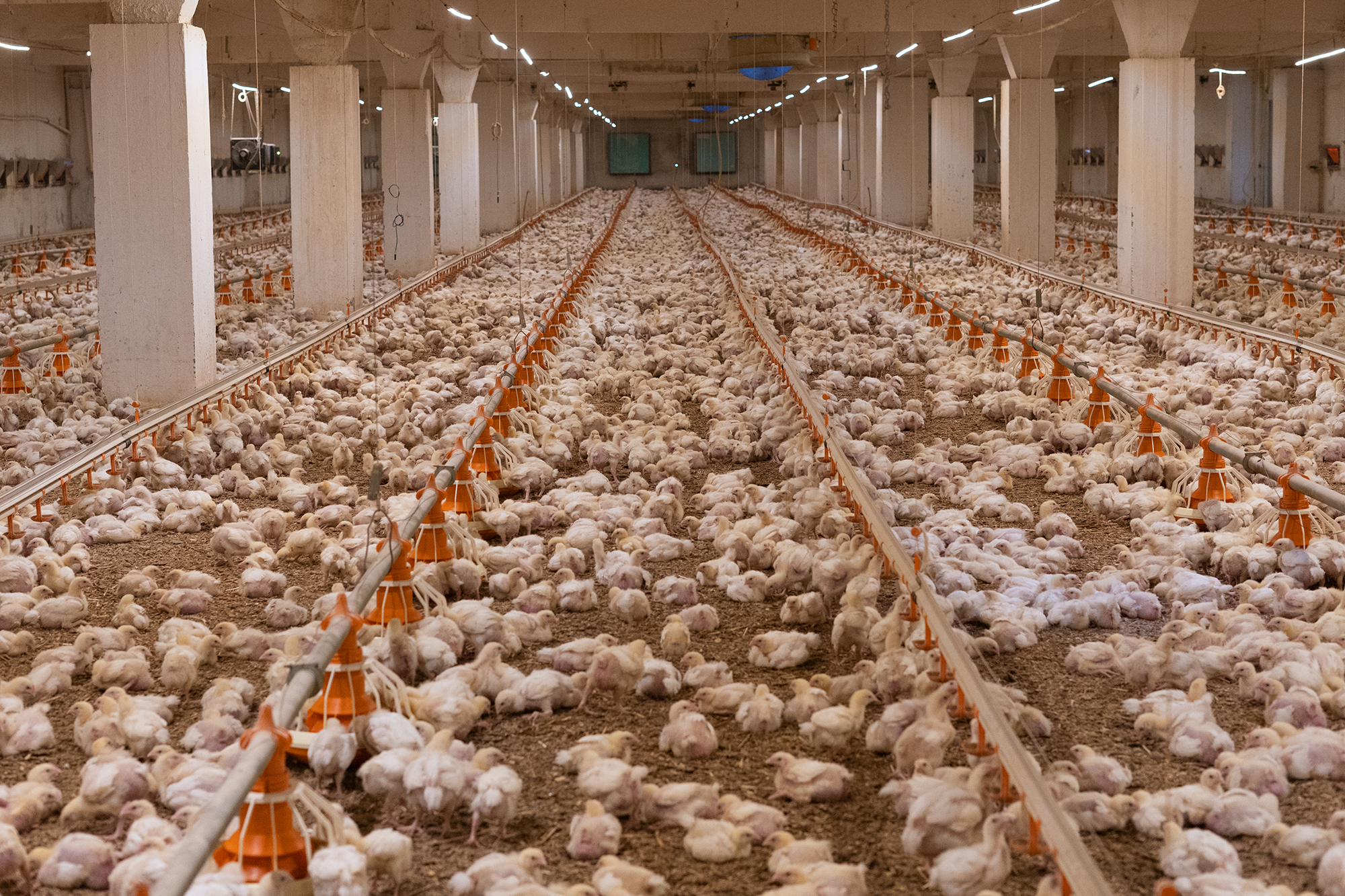
Bone disorders and foot pad lesions are considerable challenges in poultry production, especially for fast-growing birds with high final weights. Due to pain, the animals do not move, and dominant, healthy birds may restrict lame birds’ access to feed and water. In consequence, these birds are often culled. Moreover, processing these birds is problematic, and often, they must be discarded or downgraded.

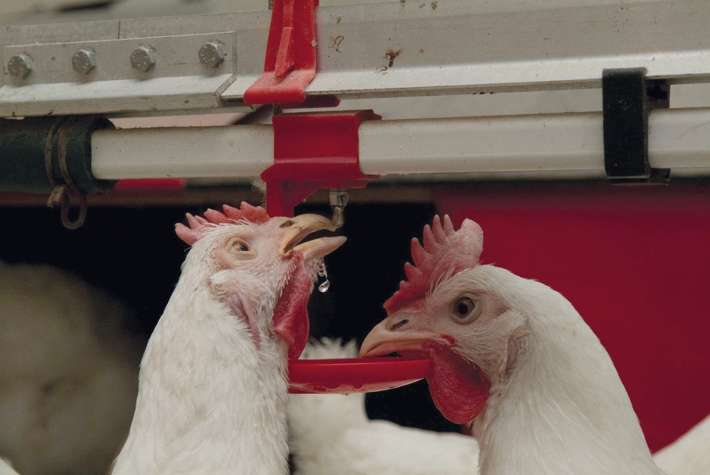
Foot pad lesions, another common issue in poultry production, can also have significant economic implications. On the one hand, pain restricts birds from eating and drinking and reduces weight gain. On the other hand, for many producers, chicken feet constitute a substantial part of the economic value of the bird; therefore, discarding them represents a significant financial loss. Additionally, to push poultry production in the right direction concerning animal health and welfare, a foot pad scoring system at the processing plant is in place in European countries.

Mycotoxins affect bones in different ways

Mycotoxins, depending on their target organs, can have diverse effects on the skeleton of birds. For example, mycotoxins that target the liver can disrupt calcium metabolism, which in turn affects the mineralization of the bones (rickets) and the impairment of chondrocytes can slow down bone growth (e.g., tibial dyschondroplasia). When the kidneys are impacted, urate clearance decreases, plasma uric acid consequently increases, and urate crystals form in the synovial fluid and tendon sheaths of various joints, particularly the hock joints. These examples highlight the complex and varied ways mycotoxins can impact poultry bone health.

Inadequate bone mineralization and strength – Rickets and layer cage fatigue

Sufficient bone mineralization is essential for the stability of the skeleton. Calcium (Ca), Vitamin D, and Phosphorous (P) deficiency leads to inadequate mineralization, weakens the bone, and can cause soft and bent bones or, in the case of layers, cage fatigue – a collapse of the spinal bone- and paralysis. Inadequate bone mineralization can be caused in different ways, among them:

  1. Decrease in the availability of the nutrients necessary for mineralization. This can occur if the digestibility of these nutrients deteriorates
  2. Impact on the Ca/P ratio—A ratio of 1 – 2:1 is vital for adequate bone development (Loughrill et al., 2016). Mycotoxins can alter absorption and transporters for one or both elements, altering their ratio.
  3. Impact on the Vitamin D receptor, affecting its expression or the transporters for Ca and P.

Aflatoxins can impair bone mineralization by different modes of action. An important one is the impairment of the digestibility of Ca and P: Kermanshahi et al. (2007) fed broilers diets with high levels of aflatoxins (0.8 to 1.2 mg AFB1/kg feed) for three weeks, which resulted in a significant reduction of Ca and P digestibility. Other researchers, however, did not find an effect on Ca and P digestibility with lower aflatoxin levels:  Bai et al. (2014) feeding diets contaminated with 96 (starter) and 157 µg Aflatoxins (grower) per kg of feed to broilers and Han et al. (2008) saw no impact on cherry valley ducks with levels of 20 and 40 µg AFB1/kg diet.

Indirectly, a decrease in the availability of Ca and P due to aflatoxin-contaminated feed can be shown by blood or tibia levels of these minerals, as demonstrated by  Zhao et al. (2010): They conducted a trial with broilers, resulting in blood serum levels of Ca and P levels significantly (P<0.05) dropped with feed contaminated with 2 mg/kg of AFB1. Another trial conducted by Bai et al. (2014) showed decreased Ca in the tibia and reduced tibial break strength.

To get more information about the effect of mycotoxins on bone mineralization and the utilization of Ca, P, and Vit. D in animal organisms, Costanzo et al. (2015) challenged osteosarcoma cells with 5 and 50 ppb of aflatoxin B1. They asserted a significant down-modulation of the expression of the Vitamin D receptor. Furthermore, they assumed an interference of AFB1 with the actions of vitamin D on calcium-binding gene expression in the kidney and intestine.  Paneru et al. (2024) could confirm this downregulation of the Vit D receptor and additionally of the Ca and P transporters in broilers with levels of ≥75 ppb AFB1. They also saw a significant reduction in tibial bone ash content at AFB1 levels >230 ppb, a decreased trabecular bone mineral content and density at AFB1 520 ppb, and a reduced bone volume and tissue volume of the cortical bone of the femur at the level of 230 ppb (see Figure 1). They concluded that AFB1 levels of already 230 ppb contribute to bone health issues in broilers.

Figure
Figure 1: Increasing doses of AFB1 (<2 ppb – 560 ppb) deteriorate bone quality (Paneru, 2024): Cross-sectional images of femoral metaphysis with increasing AFB1 levels (left to right). The outer cortical bone is shown in light grey, and the inner trabecular bone in blue. Higher levels of AFB1 (T4 and T5) show a disruption of the trabecular bone pattern (less dense blue pattern with thinner and more fragmented bone strands and with wide spaces between the trabecular bone) (shown in white).

All experiments strongly suggest that aflatoxins harm bone homeostasis. Additional liver damage, oxidative stress, and impaired cellular processes can exacerbate bone health issues.

Trichothecenes also negatively impact bone mineralization. Depending on the mycotoxin, they may affect the gut, decreasing the absorption of Ca and P and probably provoking an imbalance in the Ca/P ratio.

For instance, when T-2 toxin was fed to Yangzhou goslings at 0.4, 0.6, and 0.8 mg/kg of diet, it decreased the Ca levels (halved at 0.8 mg/kg) and increased the P levels in the blood serum, so the Ca/P ratio decreased from the adequate ratio of 1 – 2 to 0.85, 0.66, and 0.59 (P<0.05) (Gu et al., 2023). The alterations of the Ca and P levels, the resulting decreasing Ca/P ratio, and an additional increase in alkaline phosphatase (ALP) suggest that T-2 toxin negatively impacts Ca absorption, increases ALP, and, therefore, disturbs calcification and bone development.

Other studies show that serum P levels decreased in broilers fed DON-contaminated feed with levels of only 2.5 mg/kg (Keçi et al., 2019). One reason for the lower P level is probably the lower dry matter intake, affecting Ca and P intake. Ca serum level is not typically reduced, which can be explained by the fact that Ca plays many critical physiological roles (e.g., nerve communication, blood coagulation, hormonal regulation), so the body keeps the blood levels by reducing bone mineralization. Another explanation is delivered by Li et al. (2020): After their trial with broilers, they stated that dietary P deficiency is more critical for bone development than Ca deficiency or Ca & P deficiency. The results of the trial conducted by Keçi et al. with DON (see above) were reduced bone mineralization, affected bone density, ash content, and ash density in the femur and tibiotarsus with a stronger impact on the tibiotarsus than on the femur.

In line with trichothecenes effects in Ca and P absorption, Ledoux et al. (1992) suppose that diarrhea caused by intake of fumonisins leads to malabsorption or maldigestion of vitamin D, calcium and phosphorus, having birds with rickets as a secondary effect.

Ochratoxin A (OTA) impairs kidney function, negatively affects vitamin D metabolism, reduces Ca absorption, and contributes to deteriorated bone strength (Devegowda and Ravikiran, 2009). Indications from Huff et al. (1980) show decreased tibia strength after feeding chickens OTA levels of 2, 4, and 8 µ/g, and Duff et al. (1987) report similar results also in turkey poults.

A further mycotoxin possibly contributing to leg weakness is cyclopiazonic acid produced by Aspergillus and Penicillium. This mycotoxin is known for leading to eggs with thin or visibly racked shells, indicating an impairment of calcium metabolism (Devegowda and Ravikiran, 2009). Tran et al. (2023) also showed this fact with multiple mycotoxins.

The co-occurrence of different mycotoxins in the feed – the standard in praxis – increases the risk of leg issues. A trial with broiler chickens conducted by Raju and Devegowda (2000) showed a bone ash-decreasing effect of AFB1 (300 µg/kg), OTA (2 mg/kg), and T-2 toxin (3 mg/kg), fed individually but an incomparable higher effect when fed in combination.

Impairment of bone growth – tibial dyschondroplasia (TD)

In TD, the development of long bones is impaired, and abnormal cartilage development occurs. It is frequent in broilers, with a higher incidence in males than females. It happens when the bone grows, as the soft cartilage tissue is not adequately replaced by hard bone tissue. Some mycotoxins have been related to this condition: According to Sokolović et al. (2008), actively dividing cells such as bone marrow are susceptible to T-2 toxin, including the tibial growth plates, which regulate chondrocyte formation, maturation, and turnover.

T-2 toxin: In a study with primary cultures of chicken tibial growth plate chondrocytes (GPCs) and three different concentrations of T-2 toxin (5, 50, and 500 nM), He et al. (2011) found that T-2 toxin decreased cell viability, alkaline phosphatase activity, and glutathione content (P < 0.05). Additionally, it increased the level of reactive oxygen species and malondialdehyde in a dose-dependent way, which could be partly recompensated by adding an antioxidant (N-acetyl-cysteine). They concluded that T-2 toxin inhibits the proliferation and differentiation of GPCs and contributes, therefore, to the development of TD, altering cellular homeostasis. Antioxidants may help to reduce these effects.

Gu et al. (2023) investigated the closely bodyweight-related shank length and the tibia development in Yangzhou goslings fed feed with six different levels (0 to 2.0 mg/kg) of T-2 toxin for 21 days. They determined a clear dose-dependent slowed tibial length and weight growth (p<0.05), as well as abnormal morphological structures in the tibial growth plate. As tibial growth and shank length are closely related to weight gain (Gu et al., 2023; Gao et al., 2010; Ukwu et al., 2014; Yu et al., 2022), their slowdown indicates lower growth performance.

Fumonisin B1 is also a potential cause of this kind of leg issue. Feeding 100 and 200 mg/kg to day-old turkey poults for 21 days led to the development of TD (Weibking et al., 1993). Possible explanations are the reduced viability of chondrocytes, as found by Chu et al. (1995) after 48 h of exposure, or the toxicity of FB1 to splenocytes and chondrocytes, which was shown in different primary cell cultures from chicken (Wu et al., 1995).

Bacterial chondronecrosis with osteomyelitis lameness (BCO) can be triggered by DON and FUM

BCO presents a highly critical health and welfare issue in broiler production worldwide, and it is estimated that 1-2 % of condemnations in birds at the marketing age result from this disease. What is the reason? Today’s fast-growing broilers are susceptible to stress. This enables pathogenic bacteria to compromise epithelial barriers, translocate from the gastrointestinal tract or the pulmonary system into the bloodstream, and colonize osteochondrotic microfractures in the growth plate of the long bone. This can lead to bone necrosis and subsequent lameness.

In their experiment with DON and FUM in broilers, Alharbi et al. (2024) showed that these mycotoxins reduce the gut’s barrier strength and trigger immunosuppressive effects. They used contaminations of 0.76, 1.04, 0.94, and 0.93 mg DON/kg of feed and 2.40, 3.40, 3.20, and 3.50 mg FUM/kg diet in the starter, grower, finisher, and withdrawal phases, respectively. The team observed lameness on day 35; the mycotoxin groups always showed a significantly (P<0.05) higher incidence of cumulative lameness.

The increase in uric acid leads to gout

In general, mycotoxins, which damage the kidneys and, therefore, impact the renal excretion of uric acid, are potentially a factor for gout appearance.

One of these mycotoxins is T-2 toxin. With the trial mentioned before (Yangzhou goslings, 21 days of exposure), Gu et al. (2023) showed that the highest dosage of the toxin (2.0 mg/kg) significantly increased uric acid in the blood (P<0.05), possibly leading to the deposit of uric acid crystals in the joints and to gout.

Huff et al. (1975) applied Ochratoxin to chicks at 0, 0.5, 1.0, 2.0, 4.0, and 8.0 µg/g of feed during the first three weeks of life. They found ochratoxin A as a severe nephrotoxin in young broilers as it caused damage to the kidneys with doses of 1.0 µg/g and higher. At 4.0 and 8.0 µg/g doses, uric acid increased by 38 and 48%, respectively (see Figure 2). Page et al. (1980) also reported increased uric acid after feeding 0.5 or 1.0 mg/kg of Ochratoxin A to adult white Leghorn chickens.

FigureFigure 2: Effect of Ochratoxin A on plasma uric acid (mg/100 ml) (according to Huff et al., 1975)

Foot pad lesions – a further hint of mycotoxicosis

Foot pad lesions often result from wet litter, originating from diarrhea due to harmed gut integrity. Frequently, mycotoxins impact the intestinal tract and create ideal conditions for the proliferation of diarrhea-causing microorganisms and, therefore, secondary infections. Some also negatively impact the immune defense system, allowing pathogens to settle down or aggravate existing bacterial or viral parasitic diseases. In general, mycotoxins affect the physical (intestinal cell proliferation, cell viability, cell apoptosis), chemical (mucins, AMPs), immunological, and microbial barriers of the gut, as reported by Gao et al. (2020). Here are some examples of the adverse effects of mycotoxins leading to intestinal disorders and diarrhea:

  • Mycotoxins can modulate intestinal epithelial integrity and the renewal and repair of epithelial cells, negatively impacting the intestinal barrier’s intrinsic components; for instance, DON can significantly reduce the transepithelial electrical resistance (TEER)(Grenier and Applegate, 2013). A higher permeability of the epithelium and a decreased absorption of dietary proteins can lead to higher protein in the digesta in the small intestine, which serves as a nutrient for pathogens including perfringens (Antonissen et al., 2014; Antonissen et al., 2015).
  • The application of Ochratoxin A (3 mg/kg) increased the number of S. typhimurium in the duodenum and ceca of White Leghorn chickens (Fukata et al., 1996). Another trial with broiler chicks at a concentration of 2 mg/kg aggravated the symptoms due to an infection by S. gallinarum (Gupta et al., 2005).
  • In a trial by Grenier et al., 2016, feed contaminated with DON (1.5 mg/kg), Fumonisin B (20 mg/kg), or both mycotoxins aggravated lesions caused by coccidia.
  • DON impacts the mucus layer composition by downregulating the expression of the gene coding for MUC2, as shown in a trial with human goblet cells (Pinton et al., 2015). The mucus layer prevents pathogenic bacteria in the intestinal lumen from contacting the intestinal epithelium (McGuckin et al., 2011).
  • Furthermore, DON and other mycotoxins decrease the populations of lactic acid-producing bacteria, indicating a shift in the microbial balance (Antonissen et al., 2016).
  • FB1 causes intestinal disturbances such as diarrhea, although it is poorly absorbed in the intestine. According to Bouhet and Oswald (2007), the main toxicological effect ascertained in vivo and in vitro is the accumulation of sphingoid bases associated with the depletion of complex sphingolipids. This negative impact on the sphingolipid biosynthesis pathway could explain other adverse effects, such as reduced intestinal epithelial cell viability and proliferation, modification of cytokine production, and impairment of intestinal physical barrier function.
  • T-2 toxin can disrupt the immune response, enhance the proliferation of coli in the gut, and increase its efflux (Zhang et al., 2022).

All these mycotoxins can cause foot pad lesions by impacting gut integrity or damaging the gut mucosa. They promote pathogenic organisms and, thus, provoke diarrhea and wet litter.

Mitigating the negative impact of mycotoxins on bones and feet is crucial for performance

Healthy bones and feet are essential for animal welfare and performance. Mycotoxins can be obstructive. Consequently, the first step to protecting your animals is to monitor their feed. If the analyses show the occurrence of mycotoxins at risky levels, proactive measures must be taken to mitigate the issues and ensure the health and productivity of your poultry.

References

Alharbi, Khawla, Nnamdi Ekesi, Amer Hasan, Andi Asnayanti, Jundi Liu, Raj Murugesan, Shelby Ramirez, Samuel Rochell, Michael T. Kidd, and Adnan Alrubaye. “Deoxynivalenol and Fumonisin Predispose Broilers to Bacterial Chondronecrosis with Osteomyelitis Lameness.” Poultry Science 103, no. 5 (May 2024): 103598. https://doi.org/10.1016/j.psj.2024.103598.

Antonissen, Gunther, Filip Van Immerseel, Frank Pasmans, Richard Ducatelle, Freddy Haesebrouck, Leen Timbermont, Marc Verlinden, et al. “The Mycotoxin Deoxynivalenol Predisposes for the Development of Clostridium Perfringens-Induced Necrotic Enteritis in Broiler Chickens.” PLoS ONE 9, no. 9 (September 30, 2014). https://doi.org/10.1371/journal.pone.0108775.

Antonissen, Gunther, Filip Van Immerseel, Frank Pasmans, Richard Ducatelle, Geert P. Janssens, Siegrid De Baere, Konstantinos C. Mountzouris, et al. “Mycotoxins Deoxynivalenol and Fumonisins Alter the Extrinsic Component of Intestinal Barrier in Broiler Chickens.” Journal of Agricultural and Food Chemistry 63, no. 50 (December 10, 2015): 10846–55. https://doi.org/10.1021/acs.jafc.5b04119.

Antonissen, Gunther, Venessa Eeckhaut, Karolien Van Driessche, Lonneke Onrust, Freddy Haesebrouck, Richard Ducatelle, Robert J Moore, and Filip Van Immerseel. “Microbial Shifts Associated with Necrotic Enteritis.” Avian Pathology 45, no. 3 (May 3, 2016): 308–12. https://doi.org/10.1080/03079457.2016.1152625.

Bai, Shiping, Leilei Wang, Yuheng Luo, Xumei Ding, Jun Yang, Jie Bai, Keying Zhang, and Jianping Wang. “Effects of Corn Naturally Contaminated with Aflatoxins on Performance, Calcium and Phosphorus Metabolism, and Bone Mineralization of Broiler Chicks.” The Journal of Poultry Science 51, no. 2 (2014): 157–64. https://doi.org/10.2141/jpsa.0130053.

Bouhet, Sandrine, and Isabelle P. Oswald. “The Intestine as a Possible Target for Fumonisin Toxicity.” Molecular Nutrition &amp; Food Research 51, no. 8 (August 2007): 925–31. https://doi.org/10.1002/mnfr.200600266.

Chi, M.S., C.J. Mirocha, H.J. Kurtz, G. Weaver, F. Bates, W. Shimoda, and H.R. Burmeister. “Acute Toxicity of T-2 Toxin in Broiler Chicks and Laying Hens ,.” Poultry Science 56, no. 1 (January 1977): 103–16. https://doi.org/10.3382/ps.0560103.

Chu, Qili, Weidong Wu, Mark E. Cook, and Eugene B. Smalley. “Induction of Tibial Dyschondroplasia and Suppression of Cell-Mediated Immunity in Chickens by Fusarium Oxysporum Grown on Sterile Corn.” Avian Diseases 39, no. 1 (January 1995): 100. https://doi.org/10.2307/1591988.

Costanzo, Paola, Antonello Santini, Luigi Fattore, Ettore Novellino, and Alberto Ritieni. “Toxicity of Aflatoxin B1 towards the Vitamin D Receptor (VDR).” Food and Chemical Toxicology 76 (February 2015): 77–79. https://doi.org/10.1016/j.fct.2014.11.025.

Costanzo, Paola, Antonello Santini, Luigi Fattore, Ettore Novellino, and Alberto Ritieni. “Toxicity of Aflatoxin B1 towards the Vitamin D Receptor (VDR).” Food and Chemical Toxicology 76 (February 2015): 77–79. https://doi.org/10.1016/j.fct.2014.11.025.

Debouck, C., E. Haubruge, P. Bollaerts, D. van Bignoot, Y. Brostaux, A. Werry, and M. Rooze. “Skeletal Deformities Induced by the Intraperitoneal Administration of Deoxynivalenol (Vomitoxin) in Mice.” International Orthopaedics 25, no. 3 (March 24, 2001): 194–98. https://doi.org/10.1007/s002640100235.

Devegowda, G., and D. Ravikiran. “Mycotoxins and Skeletal Problems in Poultry.” World Mycotoxin Journal 2, no. 3 (August 1, 2009): 331–37. https://doi.org/10.3920/wmj2008.1085.

Duff, S.R.I., R.B. Burns, and P. Dwivedi. “Skeletal Changes in Broiler Chicks and Turkey Poults Fed Diets Containing Ochratoxin a.” Research in Veterinary Science 43, no. 3 (November 1987): 301–7. https://doi.org/10.1016/s0034-5288(18)30798-7.

Fukata, T., K. Sasai, E. Baba, and A. Arakawa. “Effect of Ochratoxin A on Salmonella Typhimurium-Challenged Layer Chickens.” Avian Diseases 40, no. 4 (October 1996): 924. https://doi.org/10.2307/1592318.

Gao, Y., Z.‐Q. Du, C.‐G. Feng, X.‐M. Deng, N. Li, Y. Da, and X.‐X. Hu. “Identification of Quantitative Trait Loci for Shank Length and Growth at Different Development Stages in Chicken.” Animal Genetics 41, no. 1 (January 6, 2010): 101–4. https://doi.org/10.1111/j.1365-2052.2009.01962.x.

Grenier, Bertrand, Ilse Dohnal, Revathi Shanmugasundaram, Susan Eicher, Ramesh Selvaraj, Gerd Schatzmayr, and Todd Applegate. “Susceptibility of Broiler Chickens to Coccidiosis When Fed Subclinical Doses of Deoxynivalenol and Fumonisins—Special Emphasis on the Immunological Response and the Mycotoxin Interaction.” Toxins 8, no. 8 (July 27, 2016): 231. https://doi.org/10.3390/toxins8080231.

Gu, Wang, Qiang Bao, Kaiqi Weng, Jinlu Liu, Shuwen Luo, Jianzhou Chen, Zheng Li, et al. “Effects of T-2 Toxin on Growth Performance, Feather Quality, Tibia Development and Blood Parameters in Yangzhou Goslings.” Poultry Science 102, no. 2 (February 2023): 102382. https://doi.org/10.1016/j.psj.2022.102382.

Guntur, Anyonya R., and Clifford J. Rosen. “Bone as an Endocrine Organ.” Endocrine Practice 18, no. 5 (September 2012): 758–62. https://doi.org/10.4158/ep12141.ra.

Gupta, S., N. Jindal, R.S. Khokhar, A.K. Gupta, D.R. Ledoux, and G.E. Rottinghaus. “Effect of Ochratoxin A on Broiler Chicks Challenged withSalmonella Gallinarum.” British Poultry Science 46, no. 4 (August 2005): 443–50. https://doi.org/10.1080/00071660500190850.

Han, Xin-Yan, Qi-Chun Huang, Wei-Fen Li, Jun-Fang Jiang, and Zi-Rong Xu. “Changes in Growth Performance, Digestive Enzyme Activities and Nutrient Digestibility of Cherry Valley Ducks in Response to Aflatoxin B1 Levels.” Livestock Science 119, no. 1–3 (December 2008): 216–20. https://doi.org/10.1016/j.livsci.2008.04.006.

He, Shao‐jun, Jia‐fa Hou, Yu‐yi Dai, Zhen‐lei Zhou, and Yi‐feng Deng. “N‐acetyl‐cysteine Protects Chicken Growth Plate Chondrocytes from T‐2 Toxin‐induced Oxidative Stress.” Journal of Applied Toxicology 32, no. 12 (July 28, 2011): 980–85. https://doi.org/10.1002/jat.1697.

Hou, Hai-Feng, Jin-Ping Li, Guo-Yong Ding, Wen-Jing Ye, Peng Jiao, and Qun-Wei Li. “The Cytotoxic Effect and Injury Mechanism of Deoxynivalenol on Articular Chondrocytes in Human Embryo.” Zhonghua Yu Fang Yi Xue Za Zhi 45, no. 7 (July 2011): 629–32.

Huff, W. E., R. D. Wyatt, and P. B. Hamilton. “Nephrotoxicity of Dietary Ochratoxin A in Broiler Chikens1.” Applied Microbiology 30, no. 1 (1975): 48–51. https://doi.org/10.1128/aem.30.1.48-51.1975.

Huff, William E., John A. Doerr, Pat B. Hamilton, Donald D. Hamann, Robert E. Peterson, and Alex Ciegler. “Evaluation of Bone Strength during Aflatoxicosis and Ochratoxicosis.” Applied and Environmental Microbiology 40, no. 1 (July 1980): 102–7. https://doi.org/10.1128/aem.40.1.102-107.1980.

Kermanshahi, H., M.R. Akbari, M. Maleki, and M. Behgar. “Effect of Prolonged Low Level Inclusion of Aflatoxin B1 into Diet on Performance, Nutrient Digestibility, Histopathology and Blood Enzymes of Broiler Chickens.” J of Anim and Vet Adv 6, no. 5 (2007): 686–92.

Keçi, Marsel, Annegret Lucke, Peter Paulsen, Qendrim Zebeli, Josef Böhm, and Barbara U. Metzler-Zebeli. “Deoxynivalenol in the Diet Impairs Bone Mineralization in Broiler Chickens.” Toxins 11, no. 6 (June 18, 2019): 352. https://doi.org/10.3390/toxins11060352.

Ledoux, David R., Tom P. Brown, Tandice S. Weibking, and George E. Rottinghaus. “Fumonisin Toxicity in Broiler Chicks.” Journal of Veterinary Diagnostic Investigation 4, no. 3 (July 1992): 330–33. https://doi.org/10.1177/104063879200400317.

Li, Tingting, Guanzhong Xing, Yuxin Shao, Liyang Zhang, Sufen Li, Lin Lu, Zongping Liu, Xiudong Liao, and Xugang Luo. “Dietary Calcium or Phosphorus Deficiency Impairs the Bone Development by Regulating Related Calcium or Phosphorus Metabolic Utilization Parameters of Broilers.” Poultry Science 99, no. 6 (June 2020): 3207–14. https://doi.org/10.1016/j.psj.2020.01.028.

Loughrill, Emma, David Wray, Tatiana Christides, and Nazanin Zand. “Calcium to Phosphorus Ratio, Essential Elements and Vitamin D Content of Infant Foods in the UK: Possible Implications for Bone Health.” Maternal &amp; Child Nutrition 13, no. 3 (September 9, 2016). https://doi.org/10.1111/mcn.12368.

McGuckin, Michael A., Sara K. Lindén, Philip Sutton, and Timothy H. Florin. “Mucin Dynamics and Enteric Pathogens.” Nature Reviews Microbiology 9, no. 4 (March 16, 2011): 265–78. https://doi.org/10.1038/nrmicro2538.

Morishita, Y., K. Nagasawa, Naoko Nakano, and Kimiko Shiromizu. “Bacterial Overgrowth in the Jejunum of ICR Mice and Wistar Rats Orally Administered with a Single Lethal Dose of Fusarenon‐x, a Trichothecene Mycotoxin.” Journal of Applied Bacteriology 66, no. 4 (April 1989): 263–70. https://doi.org/10.1111/j.1365-2672.1989.tb02478.x.

Paneru, Deependra, Milan Kumar Sharma, Hanyi Shi, Jinquan Wang, and Woo Kyun Kim. “Aflatoxin B1 Impairs Bone Mineralization in Broiler Chickens.” Toxins 16, no. 2 (February 2, 2024): 78. https://doi.org/10.3390/toxins16020078.

Pegram, R.A., and R.D. Wyatt. “Avian Gout Caused by Oosporein, a Mycotoxin Produced by Chaetomium Trilaterale.” Poultry Science 60, no. 11 (November 1981): 2429–40. https://doi.org/10.3382/ps.0602429.

Persico, Marco, Raffaele Sessa, Elena Cesaro, Irene Dini, Paola Costanzo, Alberto Ritieni, Caterina Fattorusso, and Michela Grosso. “A Multidisciplinary Approach Disclosing Unexplored Aflatoxin B1 Roles in Severe Impairment of Vitamin D Mechanisms of Action.” Cell Biology and Toxicology 39, no. 4 (September 6, 2022): 1275–95. https://doi.org/10.1007/s10565-022-09752-y.

Pinton, Philippe, Fabien Graziani, Ange Pujol, Cendrine Nicoletti, Océane Paris, Pauline Ernouf, Eric Di Pasquale, Josette Perrier, Isabelle P. Oswald, and Marc Maresca. “Deoxynivalenol Inhibits the Expression by Goblet Cells of Intestinal Mucins through a PKR and MAP Kinase Dependent Repression of the Resistin‐like Molecule β.” Molecular Nutrition &amp; Food Research 59, no. 6 (April 27, 2015): 1076–87. https://doi.org/10.1002/mnfr.201500005.

Raju, M.V.L.N., and G. Devegowda. “Influence of Esterified-Glucomannan on Performance and Organ Morphology, Serum Biochemistry and Haematology in Broilers Exposed to Individual and Combined Mycotoxicosis (Aflatoxin, Ochratoxin and T-2 Toxin).” British Poultry Science 41, no. 5 (December 2000): 640–50. https://doi.org/10.1080/713654986.

Rath, Narayan C., and Vijay Durairaj. “Avian Bone Physiology and Poultry Bone Disorders.” Sturkie’s Avian Physiology, 2022, 549–63. https://doi.org/10.1016/b978-0-12-819770-7.00037-2.

Siller, W.G. “Renal Pathology of the Fowl — a Review.” Avian Pathology 10, no. 3 (July 1981): 187–262. https://doi.org/10.1080/03079458108418474.

Suchacki, Karla J, Fiona Roberts, Andrea Lovdel, Colin Farquharson, Nik M Morton, Vicky E MacRae, and William P Cawthorn. “Skeletal Energy Homeostasis: A Paradigm of Endocrine Discovery.” Journal of Endocrinology 234, no. 1 (July 2017). https://doi.org/10.1530/joe-17-0147.

Tran, Si-Trung, Y. Ruangpanit, K. Rassmidatta, K. Pongmanee, K. Palanisamy, and M. Caballero. “The World Mycotoxin Forum, 14th Conference.” In WMF Meets Belgium – Abstracts of Lectures and Posters, 120–21. Antwerp: Conference Secretariat Bastiaanse Communication, 2023.

Ukwu, H.O, V.M.O. Okoro, and R.J. Nosike. “Statistical Modelling of Body Weight and Linear Body Measurements in Nigerian Indigenous Chicken.” IOSR Journal of Agriculture and Veterinary Science (IOSR-JAVS), Ver. V, 7, no. 1 (2014): 27–30.

Wright, G.C., Walter F.O. Marasas, and Leon Sokoloff. “Effect of Fusarochromanone and T-2 Toxin on Articular Chondrocytes in Monolayer Culture in Monolayer Culture.” Toxicological Sciences 9, no. 3 (1987): 595–97. https://doi.org/10.1093/toxsci/9.3.595.

Wu, Weidong, Mark E. Cook, Qili Chu, and Eugene B. Smalley. “Tibial Dyschondroplasia of Chickens Induced by Fusarochromanone, a Mycotoxin.” Avian Diseases 37, no. 2 (April 1993): 302. https://doi.org/10.2307/1591653.

Wu, Weidong, Tianxing Liu, and Ronald F. Vesonder. “Comparative Cytotoxicity of Fumonisin B1 and Moniliformin in Chicken Primary Cell Cultures.” Mycopathologia 132, no. 2 (November 1995): 111–16. https://doi.org/10.1007/bf01103783.

Yu, Jun, Yu Wan, Haiming Yang, and Zhiyue Wang. “Age- and Sex-Related Changes in Body Weight, Muscle, and Tibia in Growing Chinese Domestic Geese (Anser Domesticus).” Agriculture 12, no. 4 (March 25, 2022): 463. https://doi.org/10.3390/agriculture12040463.

Zhang, Jie, Xuerun Liu, Ying Su, and Tushuai Li. “An Update on T2-Toxins: Metabolism, Immunotoxicity Mechanism and Human Assessment Exposure of Intestinal Microbiota.” Heliyon 8, no. 8 (August 2022). https://doi.org/10.1016/j.heliyon.2022.e10012.

Zhao, J., R.B. Shirley, J.D. Dibner, F. Uraizee, M. Officer, M. Kitchell, M. Vazquez-Anon, and C.D. Knight. “Comparison of Hydrated Sodium Calcium Aluminosilicate and Yeast Cell Wall on Counteracting Aflatoxicosis in Broiler Chicks.” Poultry Science 89, no. 10 (October 2010): 2147–56. https://doi.org/10.3382/ps.2009-00608.


Mycotoxins in poultry – External signs can give a hint

Part 1: Impact on Feathering

By Dr. Inge Heinzl, Editor, and Marisabel Caballero, Global Technical Manager, EW Nutrition


Mycotoxins are known to decrease health and performance in poultry production. Their modes of action, such as reducing protein synthesis and promoting oxidative stress and apoptosis, lead to cell destruction and lower cell replacement, affecting several organs and tissues.

When different stress factors collude, such as high temperatures and humidity, poor ventilation, high stocking density, and management events, the effects of in-feed mycotoxins can reach a higher level, which may include external signs.

The most common and recognized external sign of mycotoxicosis is mouth lesions caused by trichothecenes, which are highly associated with the presence of T-2 in the feed. However, other signs may appear, such as paleness of combs, shanks, and feet, as well as leg problems, ruffled feathers and poor feather coverage, feed passage, and abnormal feces.

In a series of articles, we want to report on external signs facilitating a differential diagnosis of mycotoxin contamination. This is necessarily followed by feed or raw material mycotoxin analysis and strategies to avoid or mitigate the effects of mycotoxin contamination in poultry production. In the first article, we will cover feathers.

A healthy plumage is crucial for growth and reproduction

Feathering is a crucial aspect of poultry health and productivity. Feathers are essential for thermoregulation, locomotion, adequate skin protection, and reproductive success, protecting hens from injury during mating. Inadequate feathering can lead to lower feed efficiency (Leeson and Walsh, 2004) as well as loss in fertility and chick production (Fisher, 2016). Mycotoxins in poultry feed can compromise feather quality in poultry production animals. This first article delves into the relationship between mycotoxins and poor feathering, exploring different mycotoxins and their mechanisms of action.

In which way do mycotoxins compromise feathering?

On the one hand, chronic mycotoxin exposure impairs the digestive process, hindering the absorption and utilization of vital nutrients essential for feather growth. This disruption can lead to malnutrition, directly impacting the quality and health of feathers. On the other hand, mycotoxins also interfere with metabolic processes critical for feather development, such as keratin synthesis (Wyatt et al., 1975;  Nguansangiam, 2004). Enzymatic pathways involved in synthesizing keratin, the protein building block of feathers, are particularly vulnerable to mycotoxin-induced disruptions. The presence of mycotoxins in feed has been associated with the manifestation of sparse feathering and the sticking out of feathers at an unnatural angle (Emous and Krimpen, 2019). In the case of multiple mycotoxins occurring in the feed, even at singularly unimportant concentrations, a negative impact on feathering is possible. Different mycotoxins have different target organs and consequences for the animal, so their ways of compromising feathering also vary. As feathering needs protein availability, all mycotoxins affecting the protein metabolism or the absorption of nutrients also impact the feathering process. Let us look at the most prominent mycotoxins.

1.   T-2 toxin

Due to climate change, T-2 toxins are on the rise. In the US, more than 50% of the tested samples contained T-2 toxin; in Europe, we found it in 31%, and in China, in 82% of the samples (EW Nutrition, 2024). The highest level was found in Europe, with 850 ppb.

Adverse effects of T-2 toxin in goslings were shown by Gu et al. (2023), who exposed the animals to 6 different levels of T-2 toxin, from 0.2 to 2.0 mg T-2 toxin/kg of feed. The goslings showed a sparse covering with short, dry, rough, curly, and gloss-free feathers on their back with dosages ≥0.8 mg/kg. When zooming on, T-2 can cause necroses of the layer of regenerative cells in the feather base, implying malformation or absence of new feathers, as well as structural damage to existing feathers on the base of the ramus and barb ridges (Hoerr et al. (1982), Leeson et al. (1995)).

The effects in feather regenerative cells are dose-dependent, as confirmed by Hoerr et al. (1982), who applied different doses of T-2 toxin (1.5, 2, 2.5, and 3 mg/kg body weight/day) to 7-day-old broilers for 14 days. Delayed feather development, especially at high dosages, was noticed, as well as malformations and opaque bands in the feathers, the latter probably caused by a segmental reduction in diameter.

Manafi et al. (2015) noticed feather malformations when broiler chickens were challenged with 0.5 ppm T-2 toxin in the feed in combination with an inoculation of 2.4×10⁸ cfu Mycoplasma gallisepticum. When the chickens were challenged only with T-2 toxin, the feathers were ruffled, showing that a coincidence of stress factors even aggravates the symptoms.

2.   Aflatoxins

Aflatoxins, produced by certain Aspergillus species, are among the most notorious mycotoxins. Looking at test results of the last year, Aflatoxin shows incidences between 25 (USA) over 40-65% (Europe, LATAM, MEA, and SEAP) up to 84-88% (China and South Asia) with average levels up to 42 ppb in South Asia (EW Nutrition, 2023). However, more information about the concrete impact of aflatoxins on feathering is needed. They may indirectly affect feathering because they impact digestion and the utilization of nutrients or trace minerals such as zinc, which is essential for the feather construction process. Damage to the liver impacts protein metabolism, and keratin is also necessary for feather production.

In other studies, Muhammad et al. (2017) fed 5 mg AFB1/kg to Arbor Acres broilers, and the birds showed ruffled feathers. A significantly lower feather shine was noticed by Saleemi et al. (2020) when they gave the animals 300 μg AFB1/kg of feed, and the birds of Zafar et al. (2017) showed ruffled, broken, dull, and dirty feathers after six weeks of feeding an aflatoxin-contaminated diet.

3.   Ochratoxin

Ochratoxins, commonly produced by Aspergillus and Penicillium fungi, also pose a significant threat to poultry. When looking at the mycotoxin report, this mycotoxin was found in 16% (Europe) to 70% (SEAP) of the samples (EW Nutrition, 2023). Ochratoxins primarily affect feathering by compromising the structural integrity of feathers and causing delayed feathering in broilers (Leeson, 2021).

Several trials have shown the negative impact of ochratoxin on feather quality. Hassan et al. (2010) fed OTA to laying hens and saw a dose-dependent (dosages from 0 to 10 mg/kg feed) occurrence of ruffled and broken feathers in the OTA group, whereas the plumage of the control group was shiny and well-formed. Hameed et al. (2012) also realized dull feathers when feeding 0.4 and 0.8 mg OTA per kg of feed. A further dose-dependent decrease in feather quality was described by Khan et al. (2023) in broiler chicks. He injected them with dosages from 0.1 to 1.7 mg/kg body weight on day 5 of age and saw a deterioration of feather appearance (rippled feathers) in the groups with the higher dosages of 1.3 and 1.7 mg/kg. Abidin et al. (2016) observed a similar dose-dependent deterioration of the feather quality in white Leghorn cockerels when feeding 1 or 2mg OTA/kg feed.

Combinations of aflatoxins and ochratoxins were also tested. Khan et al. (2017) fed moldy feed naturally containing 56 µg OTA and 136 µg AFB1 per kg to layer hens and saw a deterioration of feather quality with increasing feeding time. Qubih (2017) noticed ruffled feathers when feeding a diet naturally contaminated with 800 ppb of OTA and 100 ppb of AFB1.

4.   Scirpenol mycotoxins

Parkhurst et al. (1992) examined the effects of different scirpenol mycotoxins. After feeding graded levels of fusarium mycotoxins to broiler chicks until three weeks of age, they discovered that the impact of scirpenols stretched across the entire feathered body parts and that the degree of feather alteration is dose-dependent. The main alteration was a frayed or even missing web on the medial side of the outer end of the feather due to poor development of the barbs, barbules, and barbicels, and the tip of the feathers became square instead of rounded—the thinner and weaker shafts of the feathers inclined to show an accentuated medial curve.

Figure Feathering Affected By Scirpenol MycotoxinsParkhurst et al. (1992)

Figure 1: Feathering affected by scirpenol mycotoxins

In their trial, Parkhurst and Hamilton realized that 15-monoacetoxyscirpenol (15-MAS) caused the most severe alterations of feathers, and they determined a minimum effective dose (MED) of 0.5 µg/g diet. The MEDs for 4,15-diacetoxyscirpenol (4,15-DAS) and 3,4,15-triacetoxyscirpenol (TAS) were higher, 2 µg/g and > 8 µg/g, respectively.

How can we enable adequate feathering in poultry?

Adequate feathering of poultry is necessary for the animal’s health and welfare and to ensure fertility and productivity. The occurrence of mycotoxins in the feed – and the probability is high! – can cause poor feathering or the development of malformed feathers.

To best equip broilers, layers, and breeders, their feed must contain all nutrients essential for healthy growth and appropriate feathering. As the risk of contamination of the feed materials is very high (see EW Nutrition’s mycotoxin report 2023), it is of crucial importance to have an efficient mycotoxin risk management in place, which includes sampling, analysis of samples, and the use of mycotoxin binders. EW Nutrition offers MasterRisk, an online tool where farmers and feed millers can feed the results of their feed analysis concerning mycotoxins and get a risk management recommendation.

In the next part of the series, we will report on beak lesions and skin paleness, two other external signs of mycotoxin contamination.

References:

Abidin, Zain ul, Muhammad Zargham Khan, Aisha Khatoon, Muhammad Kashif Saleemi, and Ahrar Khan. “Protective Effects Ofl-Carnitine upon Toxicopathological Alterations Induced by Ochratoxin A in White Leghorn Cockerels.” Toxin Reviews 35, no. 3–4 (August 22, 2016): 157–64. https://doi.org/10.1080/15569543.2016.1219374.

Emous, R. A., and M. M. Krimpen. “Effects of Nutritional Interventions on Feathering of Poultry – a Review.” Poultry Feathers and Skin: The Poultry Integument in Health and Welfare, 2019, 133–50. https://doi.org/10.1079/9781786395115.0133.

Fisher, Colin. “Feathering in Broiler Breeder Females – Aviagen.” https://aviagen.com/, 2016. http://en.aviagen.com/assets/Tech_Center/Broiler_Breeder_Tech_Articles/English/Feathering-in-Broiler-Breeeder-Females-EN-2016.pdf.

Gu, Wang, Qiang Bao, Kaiqi Weng, Jinlu Liu, Shuwen Luo, Jianzhou Chen, Zheng Li, et al. “Effects of T-2 Toxin on Growth Performance, Feather Quality, Tibia Development and Blood Parameters in Yangzhou Goslings.” Poultry Science 102, no. 2 (February 2023): 102382. https://doi.org/10.1016/j.psj.2022.102382.

Hameed, Muhammad  Raza, Muhammad Khan, Ahrar Khan, and Ijaz Javed. “Ochratoxin Induced Pathological Alterations in Broiler Chicks: Effect of Dose and Duration.” Pakistan Veterinary Journal Pakistan Veterinary Journal 8318, no. 2 (December 2012): 2074–7764.

Hassan, Zahoor-Ul, M. Zargham Khan, Ahrar Khan, and Ijaz Javed. “Pathological Responses of White Leghorn Breeder Hens Kept on Ochratoxin A Contaminated Feed.” Pakistan Veterinary Journal 30, no. 2 (2010): 118–23.

Hoerr, F. J., W. W. Carlton, and B. Yagen. “Mycotoxicosis Caused by a Single Dose of T-2 Toxin or Diacetoxyscirpenol in Broiler Chickens.” Veterinary Pathology 18, no. 5 (September 1981): 652–64. https://doi.org/10.1177/030098588101800510.

Hoerr, F.J., W.W. Carlton, B. Yagen, and A.Z. Joffe. “Mycotoxicosis Produced in Broiler Chickens by Multiple Doses of Either T‐2 Toxin or Diacetoxyscirpenol.” Avian Pathology 11, no. 3 (January 1982): 369–83. https://doi.org/10.1080/03079458208436112.

Khan, Ahrar, Muhammad Mustjab Aalim, M. Zargham Khan, M. Kashif Saleemi, Cheng He, M. Noman Naseem, and Aisha Khatoon. “Does Distillery Yeast Sludge Ameliorate Moldy Feed Toxic Effects in White Leghorn Hens?” Toxin Reviews, January 25, 2017, 1–8. https://doi.org/10.1080/15569543.2017.1278707.

Khan, Shahzad Akbar, Eiko N. Itano, Anum Urooj, and Kashif Awan. “Ochratoxin-a Induced Pathological Changes in Broiler Chicks.” Pure and Applied Biology 12, no. 4 (December 10, 2023): 1608–16. https://doi.org/10.19045/bspab.2023.120162.

Leeson, S., and T. Walsh. “Feathering in Commercial Poultry II. Factors Influencing Feather Growth and Feather Loss.” World’s Poultry Science Journal 60, no. 1 (March 1, 2004): 52–63. https://doi.org/10.1079/wps20045.

Leeson, Steve. “Effects of Nutrition on Feathering.” Poultry World, May 22, 2021. https://www.poultryworld.net/specials/effects-of-nutrition-on-feathering/.

Leeson, Steven, Gonzalo J. Diaz Gonzalez, and John D. Summers. Poultry metabolic disorders and Mycotoxins. Guelph, Ontario, Canada: University Books, 1995.

Manafi, M., N. Pirany, M. Noor Ali, M. Hedayati, S. Khalaji, and M. Yari. “Experimental Pathology of T-2 Toxicosis and Mycoplasma Infection on Performance and Hepatic Functions of Broiler Chickens.” Poultry Science 94, no. 7 (July 2015): 1483–92. https://doi.org/10.3382/ps/pev115.

Muhammad, Ishfaq, Xiaoqi Sun, He Wang, Wei Li, Xinghe Wang, Ping Cheng, Sihong Li, Xiuying Zhang, and Sattar Hamid. “Curcumin Successfully Inhibited the Computationally Identified CYP2A6 Enzyme-Mediated Bioactivation of Aflatoxin B1 in Arbor Acres Broiler.” Frontiers in Pharmacology 8 (March 21, 2017). https://doi.org/10.3389/fphar.2017.00143.

Nguansangiam, Sudarat, Subhkij Angsubhakorn, Sutatip Bhamarapravati, and Apichart Suksamrarn. The Southeast Asian J of Tropical Medicine 34, no. 4 (2004): 899–905.

Parkhurst, Carmen R., Pat B. HamiltonON, and Adedamola A. AdemoyeroERO. “Abnormal Feathering of Chicks Caused by Scirpenol Mycotoxins Differing in Degree of Acetylation.” Poultry Science 71, no. 5 (May 1992): 833–37. https://doi.org/10.3382/ps.0710833.

Qubih, T. S. “Relationship between Mycotoxicosis and Calcium during Preproduction Period in Layers.” Iraqi Journal of Veterinary Sciences 26, no. 1 (June 28, 2012): 11–14. https://doi.org/10.33899/ijvs.2012.46888.

Saleemi, M. Kashif, Kamran Ashraf, S. Tehseen Gul, M. Noman Naseem, M. Sohail Sajid, Mashkoor Mohsin, Cheng He, Muhammad Zubair, and Ahrar Khan. “Toxicopathological Effects of Feeding Aflatoxins B1 in Broilers and Its Amelioration with Indigenous Mycotoxin Binder.” Ecotoxicology and Environmental Safety 187 (January 2020): 109712. https://doi.org/10.1016/j.ecoenv.2019.109712.

Wyatt, R.D., P.B. Hamilton, and H.R. Burmeister. “Altered Feathering of Chicks Caused by T-2 Toxin.” Poultry Science 54, no. 4 (July 1975): 1042–45. https://doi.org/10.3382/ps.0541042.

Zafar, Roheena, Farhat Ali Khan, and Muhammad Zahoor. “In Vivo Amelioration of Aflatoxin B1 in Broiler Chicks by Magnetic Carbon Nanocomposite.” Pesquisa Veterinária Brasileira 37, no. 11 (November 2017): 1213–19. https://doi.org/10.1590/s0100-736×2017001100005.


Organic acids can play a crucial role in zinc oxide replacement

Dr. Inge Heinzl, Editor EW Nutrition &
Juan Antonio Mesonero Escuredo, GTM Swine/GPM Organic Acids EW Nutrition

The use of high levels of Zinc Oxide (ZnO) in the EU before 2022 was one of the most common methods to prevent postweaning diarrhea (PWD) in pig production. Pharmacologically high levels of ZnO (2000-3000 ppm) increase growth and reduce the incidence of enteric bacterial diseases such as post-weaning diarrhea (PWD)( Carlson et al., 1999; Hill et al., 2000; Hill et al., 2001; Poulsen & Larsen, 1995; De Mille et al., 2019).

However, ZnO showed adverse effects, such as the accumulation of heavy metal in the environment, the risk for antimicrobial resistance (AMR), and problems of mineral toxicity and adverse growth effects when feeding it longer than 28 days (Jensen et al., 2018; Cavaco et al., 2011; Vahjen, 2015; Romeo et al., 2014; Burrough et al., 2019). To replace ZnO in pig production, let us first look at its positive effects to know what we must compensate for.

ZnO has a multifactorial mode of action

ZnO shows several beneficial characteristics that positively influence gut health, the immune system, digestion, and, therefore, also overall health and growth performance.

FigureFigure 1. Beneficial effects and ZnO mode of action in postweaning piglets

1.   ZnO acts as an antimicrobial

Concerning the antimicrobial effects of ZnO, different possible modes of action are discussed:

  • ZnO in high dosages generates reactive oxygen species (ROS) that can damage the bacterial cell walls (Pasquet et al., 2014)
  • The death of the bacterial cell due to direct contact of the metallic Zn to the cell (Shearier et al., 2016)
  • Intrinsic antimicrobial properties of the ZnO2+ ions after dissociation. The uptake of zinc into cells is regulated by homeostasis. A concentration of the ZnO2+ ions higher than the optimal level of 10-7 to 10-5 M (depending on the microbial strain) allows the invasion of Zn2+ ions into the cell, and the zinc starts to be cytotoxic (Sugarman, 1983; Borovanský et al., 1989).

ZnO shows activity against, e.g., Staphylococcus aureus, Pseudomonas aeruginosa, E. coli, Streptococcus pyogenes, and other enterobacteria (Ann et al., 2014; Vahjen et al., 2016). However, Roselli et al. (2003) did not see a viability-decreasing effect of ZnO on ETEC.

2.   ZnO modulates the immune system

Besides fighting pathogenic organisms as described in the previous chapter and supporting the immune system, ZnO is an essential trace element and has a vital role in the immune system. ZnO improves the innate immune response, increasing phagocytosis and oxidative bursts from macrophages and neutrophils. It also ameliorates the adaptative immune response by increasing the number of T lymphocytes (T cells) in general and regulatory T lymphocytes (T-regs) in particular. These cells control the immune response and inflammation (Kloubert et al., 2018). Macrophage capacity for phagocytosis (Ercan and Bor, 1991) and to kill parasites (Wirth et al., 1989), and also the killing activity of natural killer cells depends on Zn (Rolles et al., 2018). By reducing bacterial adhesion and blocking bacterial invasion, ZnO disburdens the immune system (Roselli et al., 2003).

ZnO reduces the expression of several proinflammatory cytokines induced by ETEC (Roselli et al., 2003). Several studies have also shown a modulation effect on intestinal inflammation, decreasing levels of IFN-γ, TNF-α, IL-1ß and IL-6, all pro-inflammatory, in piglets supplemented with ZnO (Zhu et al., 2017; Grilli et al., 2015).

3.   ZnO improves digestion and promotes growth

Besides protecting young piglets against diarrhea, the goal is to make them grow optimally. For this target, an efficient digestion and a high absorption of nutrients is essential. Stimulating diverse pancreatic enzymes such as amylase, carboxypeptidase A, trypsin, chymotrypsin, and lipase increases digestibility (Hedemann et al., 2006; Pieper et al., 2015). However, Pieper et al. (2015) also showed that a long-term supply of very high dietary zinc triggers oxidative stress in the pancreas of piglets.

By stimulating the secretion of ghrelin at the stomach level and thereby promoting the release of insulin-like growth factor (IGF-1) and cholecystokinin (CCK), ZnO enhances muscle protein synthesis, cell proliferation, and feed intake (Yin et al., 2009; MacDonald et al., 2000)).

The result of improved digestion is increased body weight and average daily gain, which can be seen, e.g., in a study by Zhu et al. (2017).

4.   ZnO protects the intestinal morphology

ZnO prevents the decrease of the trans-endothelial electrical resistance (TEER), usually occurring in the case of inflammation, by downregulating TNF-α and IFN-γ. TNF-α, as well as IFN-γ, increase the permeability of the epithelial tight junctions and, therefore, the intestinal barrier (Al-Sadi et al., 2009).

The enterotrophic and anti-apoptotic effect of ZnO is reflected by a higher number of proliferating and PCNA-positive cells and an increased mucosa surface in the ileum (higher villi, higher villi/crypt ratio)(Grilli et al., 2015). Zhu et al. (2017) also saw an increase in villus height in the duodenum and ileum and a decrease in crypt depth in the duodenum due to the application of 3000 mg of ZnO/kg. Additionally, they could notice a significant (P<0.05) upregulation of the mRNA expression of the zonula occludens-1 and occluding in the mucosa of the jejunum of weaned piglets.

In a trial conducted by Roselli et al. (2003), the supplementation of 0.2 mmol/L ZnO prevented the disruption of the membrane integrity when human Caco-2 enterocytes were challenged with ETEC.

5.   ZnO acts antioxidant

The antioxidant effect of ZnO was shown in a study conducted by Zhu et al., 2017. They could demonstrate that the concentration of malondialdehyde (MDA), a marker for lipid peroxidation, decreased on day 14 or 28, and the total concentration of superoxide dismutase (SOD), comprising enzymes that transform harmful superoxide anions into hydrogen peroxide, increased on day 14 (P<0.05). Additionally, Zn is an essential ion for the catalytic action of these enzymes.

Which positive effects of ZnO can be covered by organic acids (OAs)?

1.   OAs act antimicrobial

OAs, on the one hand, lower the pH in the gastrointestinal tract. Some pathogenic bacteria are susceptible to low pH. At a pH<5, the proliferation of, e.g., Salmonella, E. coli, and Clostridium is minimized. The good thing is that some beneficial bacteria, such as lactobacilli or bifidobacteria, survive as they are acid-tolerant. The lactobacilli, on their side, can produce hydrogen peroxide, which inhibits, e.g., Staphylococcus aureus or Pseudomonas spp. (Juven and Pierson, 1996).

Besides this more indirect mode of action, a more direct one is also possible: Owing to their lipophilic character, the undissociated form of OAs can pass the bacterial membrane (Partanen and Mroz, 1999). The lower the external pH, the more undissociated acid is available for invading the microbial cells. Inside the cell, the pH is higher than outside, and the OA dissociates. The release of hydrogen ions leads to a decrease in the internal pH of the cell and to a depressed cell metabolism. To get back to “normal conditions”, the cell expels protons. However, this is an energy-consuming process; longer exposure to OAs leads to cell death. The anion remaining in the cell, when removing the protons, disturbs the cell’s metabolic processes and participates in killing the bacterium.

These theoretical effects could be shown in a practical trial by Ahmed et al. (2014). He fed citric acid (0.5 %) and a blend of acidifiers composed of formic, propionic, lactic, and phosphoric acid + SiO2 (0.4 %) and saw a reduction in fecal counts of Salmonella and E. coli for both groups.

2.   OAs modulate the immune system

The immune system is essential in the pig’s life, especially around weaning. Organic acids have been shown to support or stimulate the immune system. Citric acid (0.5%), as well as the blend of acidifiers mentioned before (Ahmed et al., 2014), significantly increased the level of serum IgG. IgG is part of the humoral immune system. They mark foreign substances to be eliminated by other defense systems.

Ren et al. (2019) could demonstrate a decrease in plasma tumor necrosis factor-α that regulates the activity of diverse immune cells. He also found lower interferon-γ and interleukin (Il)-1ß values in the OA group than in the control group after the challenge with ETEC. This trial shows that inflammatory response can be mitigated through the addition of organic acids.

3.   OAs improve digestion and promote growth

In piglets, the acidity in the stomach is responsible for the activation and stimulation of certain enzymes. Additionally, it keeps the feed in the stomach for a longer time. Both effects lead to better digestion of the feed.

In the stomach, the conversion of pepsinogen to pepsin, which is responsible for protein digestion, is catalyzed under acid conditions (Sanny et al., 1975)group. Pepsin works optimally at two pH levels: pH 2 and pH 3.5 (Taylor, 1959). With increasing pH, the activity decreases; at pH 6, it stops. Therefore, a high pH can lead to poor digestion and undigested protein arriving in the intestine.

These final products of pepsin protein digestion are needed in the lower parts of the GIT to stimulate the secretion of pancreatic proteolytic enzymes. If they do not arrive, the enzymes are not activated, and the inadequate protein digestion continues. Additionally, gastric acid is the primary stimulant for bicarbonate secretion in the pancreas, neutralizing gastric acid and providing an optimal pH environment for the digestive enzymes working in the duodenum.

As already mentioned, the pH in the stomach influences the transport of digesta. The amount of digesta being transferred from the stomach to the small intestine is related to the acidity of the chyme leaving the stomach and arriving in the small intestine. Emptying of the stomach can only take place when the duodenal chyme can be neutralized by pancreatic or other secretions (Pohl et al., 2008); so, acid-sensitive receptors provide feedback regulation and a higher pH in the stomach leads to a faster transport of the digesta and a worse feed digestion.

4.   OAs protect the intestinal morphology

Maintaining an intact gut mucosa with a high surface area is crucial for optimal nutrient absorption. Research suggests organic acids play a significant role in improving mucosal health:

Butyric acid promotes epithelial cell proliferation, as demonstrated in an in vitro pig hindgut mucosa study (Sakata et al., 1995). Fumaric acid, serving as an energy source, may locally enhance small intestinal mucosal growth, aiding in post-weaning epithelial cells’ recovery and increasing absorptive surface and digestive capacity (Blank et al., 1999). Sodium butyrate supplementation at low doses influences gastric morphology and function, thickening the stomach mucosa and enhancing mucosal maturation and differentiation (Mazzoni et al., 2008).

Studies show that organic acids affect gut morphology, with a mixture of short-chain and mid-chain fatty acids leading to longer villi (Ferrara et al., 2016) and Na-butyrate supplementation increasing crypt depth and villi length in the distal jejunum and ileum (Kotunia et al., 2004). However, the villi length and mucosa thickness in the duodenum were reduced. Dietary sodium butyrate has been linked to increased microvilli length and cecal crypt depth in pigs (Gálfi and Bokori, 1990).

5.   OAs show antioxidant activity

The last characteristic, the antioxidant effect, cannot be provided at the same level as with ZnO; however, Zhang et al. (2019) attest to OAs a certain antioxidant activity. Oxalic, citric, acetic, malic, and succinic acids, which were extracted from Camellia oleifera, also showed good antioxidant activity in a trial conducted by Zhang et al. (2020).

Organic acids are an excellent tool to compensate for the ban on ZnO

The article shows that organic acids have similar positive effects as zinc oxide. They act antimicrobial, modulate the immune system, maintain the gut morphology, fight pathogenic microbes, and also act – slightly – antioxidant. Additionally, they have a significant advantage: they are not harmful to the environment. Organic acids used in the proper pH range and combination are good tools for replacing zinc oxide.

References on request


Salmonella in pigs: a threat for humans and a challenge for pig producers

By Dr. Inge Heinzl, Editor, EW Nutrition

Salmonellosis is third among foodborne diseases leading to death (Ferrari, 2019). More than 91,000 human cases of Salmonellosis are reported by the EU each year, generating overall costs of up to €3 billion a year (EFSA, 2023), 10-20% of which are attributed to pork consumption (Soumet, 2022). The annual costs arising from the resulting human health losses in 2010 were about €90 million (FCC Consortium, 2010). Take the example of Ireland, where a high prevalence of Salmonella in lymph nodes still shows a severe issue pre-slaughter and a big challenge for slaughterhouses to stick to the process hygiene requirements (Deane, 2022).

Several governments already have monitoring programs in place, and the farms are categorized according to the salmonella contamination of their pigs. In some countries, e.g., Denmark, an economic penalty of 2% of the carcass value must be paid if the farm has level 2 (intermediate seroprevalence) and 4-8% if the level is 3. Other countries, e.g., Germany, the UK, Ireland, or the Netherlands, use quality assurance schemes. The farmers can only sell their carcasses under this label if their farm has a certain level.

Let’s take a quick look at the genus of Salmonella

Salmonellas are rod-shaped gram-negative bacteria of the family of enterobacteria that use flagella for their movement. They were named after the American vet Daniel Elmer Salmon. The genus of Salmonella consists of two species (S. bongori and S. enterica with seven subspecies) with in total more than 2500 serovars (see Figure 1). The effects of the different serovars can range from asymptomatic carriage to severe invasive systemic disease (Gal-Mor, 2014). All Salmonella serovars generally can cause disease in humans; the rosa-marked ones already showed infections.

Figure Genus Salmonella For ART PIGFigure 1: the genus of Salmonella with Salmonella serovars relevant for pigs (according to Bonardi, 2017: Salmonella in the pork production chain and its impact on human health in the European Union)

Within the group of Salmonella, some serovars can only reside in one or few species, e.g., S. enterica spp. enterica Serovar Dublin (S. Dublin) in bovines (Waldron, 2018) or S. Cholerasuis in pigs (Chiu, 2004). An infection in humans with these pathogens is often invasive and life-threatening (WHO, 2018). On the contrary, serovars like S. Typhimurium and S. Enteritidis are not host-specific and can cause disease in various species.

The serotypes S. Typhi and S. Paratyphi A, B, or C are highly adapted to humans and only for them pathogenic; they are responsible for the occurrence of typhus.

Serovars occurring in pigs and relevant for humans are, for example, S. Typhimurium (Hendriksen, 2004), S. Serotype 4,[5],12:I (Hauser et al., 2010), S. Cholerasuis (Chiu, 2004), S. Derby (Gonzalez-Santamarina, 2021), S. Agona (Brenner Michael, 2006) and S. Rissen (Elbediwi, 2021).

Transmission of Salmonella mostly happens via contaminated food

The way of transmission to humans depends on the serovar:
Human-specific and, therefore, only in humans and higher primates residing serovars S. Typhi and Paratyphi A, B, or C (typhoidal) are excreted via feces or urine. Therefore, any food or water contaminated with the feces or urine of infected people can transmit this disease (Government of South Australia, 2023). Typhoid and paratyphoid Salmonellosis occur endemic in developing countries with the lack of clean water and, therefore, inadequate hygiene (Gal-Mor, 2014).

Serovars which can cause disease in humans and animals (non-typhoidal), can be transmitted by
– animal products such as milk, eggs, meat
– contact with infected persons/animals (pigs, cows, pets, reptiles…) or
– other feces- or urine-contaminated products such as sprouts, vegetables, fruits….

Farm animals take salmonellas from their fellows, contaminated feed or water, rodents, or pests.

Symptoms of Salmonellosis can be severe

In the case of typhoid or paratyphoid Salmonellosis, the onset of illness is gradual. People can suffer from sustained high fever, unwellness, severe headache, and decreased appetite, but also from an enlarged spleen irritating the abdomen and dry cough.

A study conducted in Thailand with children suffering from enteric fever caused by the typhoid serovars S. Typhi and Paratyphi showed a sudden onset of fever and gastrointestinal issues (diarrhea), rose spots, bronchitis, and pneumonia (Thisyakorn et al., 1987)

The non-typhoid Salmonellosis is typically characterized by an acute onset of fever, nausea, abdominal pain with diarrhea, and sometimes vomiting (WHO, 2018). However, 5% of the persons – children with underlying conditions, e.g., babies, or people who have AIDS, malignancies, inflammatory bowel disease, gastrointestinal illness caused by non-typhoid serovars, and hemolytic anemia, or receiving an immunosuppressive therapy can be susceptible to bacteremia. Additionally, serovars like S. Cholerasuis or S. Dublin are apt to develop bacteremia by entering the bloodstream with little or no involvement of the gut (Chiu, 1999). In these cases, consequences can be septic arthritis, pneumonia, peritonitis, cutaneous abscess, mycotic aneurysm, and sometimes death (Chen et al., 2007; Chiu, 2004, Wang et al., 1996).

In pigs, S. Cholerasuis causes high fever, purple discolorations of the skin, and thereinafter diarrhea. The mortality rate in pigs suffering from this type of Salmonellosis is high. Barrows orally challenged with S. Typhimurium showed elevated rectal temperature by 12h, remaining elevated until the end of the study. Feed intake decreased with a peak at 48h after the challenge and remained up to 120h after the challenge. Daily gain reduced during the following two weeks after infection. A higher plasma cortisol level and a lower IGF-I level could also be noticed. All these effects indicate significant changes in the endocrine stress and the somatotropic axis, also without significant alterations in the systemic pro-inflammatory mediators (Balaji et al., 2000)

To protect humans, Salmonella in pork must be restraint

There are three main steps to keep the contamination of pork as low as possible:

  1. Keeping Salmonella out of the pig farm
  2. Minimizing spreading if Salmonella is already on the farm
  3. Minimizing contamination in the slaughterhouse

1. How to keep Salmonella out of the pig farm?

To answer this question, we must look at how the pathogen can be transported to the farm. According to the Code of Practice for the Prevention and Control of Salmonella on Pig Farms (Ministry of Agriculture, Fisheries and Food and the Scottish Executive Rural Affairs Department), there are several possibilities to infiltrate the pathogen into the farm:

  • Diseased pigs or pigs which are ill but don’t show any symptoms
  • Feeding stuff or bedding contaminated with dung
  • Pets, rodents, wild birds, or animals
  • Farm personnel or visitors
  • Equipment or vehicles

Caution with purchased animals!

To minimize/prevent the entry of Salmonella into the livestock, bought-in animals must come from reputable breeding farms with a salmonella monitoring system in place. As possible carrier animals are more likely to excrete Salmonella when stressed; they should be kept in isolation after purchasing. Additionally, the animals must go through a disinfectant foot bath before entering the farm.

Keep rodents, wild animals, and vermin in check!

Generally, the production site must be kept clean and as unattractive as possible for all these animals. Rests of feed must be removed, and dead animals and afterbirths must be promptly and carefully disposed of. A well-planned baiting and trapping policy should be in place to effectively control rodents.

Only selected people should enter the hog houses

In any case, the number of persons entering the hog house must be kept as low as possible. Farmworkers should be trained in the principles of hygiene. They should wear adequate clothing (waterproof boots and protective overalls) that can be easily cleaned/laundered and disinfected. The clothes/shoes should always be used only at this site. Thorough hand washing and the disinfection of the boots when entering and leaving the pig unit are a must.

If visits are necessary, the visitors should take the same measures as the farm workers. And, of course, they should not have had contact with another pig farm during the last 48 hours.

Keep pens, farm equipment, and vehicles clean!

Farm equipment should not be shared with other farms. If this cannot be avoided, it must be cleaned and disinfected before re-entering the farm. Also, the vehicles for the transport of the animals must be cleaned and disinfected as soon as possible after usage, as contaminated transporters always pose the risk of infection.

Feed should be Salmonella-free!

To get high feed quality, the feed should be purchased from feed mills/sources with a well-functioning bacterial control to guarantee the absence of Salmonella. It is essential that birds, domestic and wild animals cannot enter the feed stores.
It is also advised to keep dry feed dry as possibly contaminating Salmonella can multiply in such humid conditions. Additionally, all feed bins and delivery pipes for dry and wet feed must be consciously cleaned, and the damp feed pipes also disinfected.
The change from pellets to mash could be helpful as the pellets facilitate Salmonella colonization by stimulating the secretion of mucins (Hedemann et al., 2005).

For sanitation of the feed, we offer organic acids (Acidomix product range) or mixtures of organic acids and formaldehyde in countries where formaldehyde products are allowed (Formycine) to decrease the pathogenic load of the feed materials. In vitro trials show the effectiveness of the products:


For the in vitro trial with Formycine, autoclaved feed samples were inoculated with Salmonella enteritidis serovar Typhimurium DSM 19587 strain to reach a Salmonella contamination of 106 CFU/g of feed. After incubating at room temperature for three hours, Formycine Liquido was added to the contaminated feed samples at 0, 500, 1000, and 2000 ppm. The control and inoculated feed samples were further incubated at room temperature, and Salmonella counts (CFU/g) were carried out at 24, 48, 72 hours and on day 15. The limit of Salmonella detection was set at 100 CFU/g (102). Results are shown in figure 2.

Figure FormycineFig. 2: Effect of treatment time and different inclusion levels of Formycine Liquido on the Salmonella count in feed

As important as uncontaminated feed is clean water for drinking. It can be achieved by taking the water from a main or a bacteriologically controlled water borehole. Regular cleaning/disinfection of the tanks, pipes, and drinkers is essential.

Bedding should be Salmonella-free

Straw material containing feces of other animals (rodents, pets) always carries the risk of Salmonella contamination. Also, wet or moldy bedding is not recommended because it is an additional challenge for the animal. To optimize the quality of bedding, the straw should be bought from reliable and as few as possible sources. The material must be stored dry and as far as practicable from the pig buildings (Ministry of Agriculture, Fisheries and Food & Scottish Executive Rural Affairs Department, 2000).

Vaccination is a beneficial measure

For the control of Salmonella in swine herds, vaccination is an effective tool. De Ridder et al. (2013) showed that an attenuated vaccine reduced the transmission of Salmonella Typhimurium in pigs. The vaccination with an attenuated S. Typhimurium strain, followed by a booster vaccination with inactivated S. Cholerasuis, showed better effects than an inactivated S. Cholerasuis vaccine alone (Alborali et al., 2017). Bearson et al. (2017) could delimitate transmission through less shedding and protect the animals against systemic disease.
To achieve the best effects, the producer must understand the diversity of Salmonella serovars to choose the most promising vaccination strategy (FSIS, 2023).

2. How to minimize the spreading of Salmonella on the farm?

If there are already cases of Salmonella on the farm, infected animals must be separated from the rest of the herd. Small batch sizes are beneficial, as well as not mixing different litters after weaning. If feasible, separate units for different production phases with an all-in/all-out system could break the reinfection cycle and help reduce Salmonella contamination on the farm. And also in this case, vaccination is helpful.

Salmonella doesn’t like acid conditions

An effective tool is acidifying the feed with organic acids, as Salmonella doesn’t like acid conditions. A trial was conducted with Acidomix AFG and Acidomix AFL to show their effects against Salmonella. For the test, 105 CFU/g of Salmonella enterica ser. Typhimurium was added to feed containing 1000 ppm, 2000 ppm, and 3000 ppm of Acidomix AFG or AFL. The stomach and intestine were simulated in vitro by adjusting the pH with HCl and NaHCO3 as follows:
Stomach              2.8
Intestine              6.8-7.0

After the respective incubation, the microorganisms were recovered from feed and plated on an appropriate medium for CFU counting. The results are shown in figures 3 and 4.

Combi
Combi

Figures 3 + 4: Effects of different concentrations of Acidomix AFG and Acidomix AFL against Salmonella enterica ser. Typhimurium in feed

Phytomolecules can support pigs against Salmonella

Plant compounds or phytomolecules can also be used against Salmonella in pigs. Some examples of phytomolecules to be used are Piperine, Allicin, Eugenol, and Carvacrol. Eugenol, e.g., increases the permeability of the Salmonella membrane, disrupts the cytoplasmic membrane, and inhibits the production of bacterial virulence factors (Keita et al., 2022; Mak et al., 2019). Thymol and Carvacrol interact with the cell membrane by H bonding, also resulting in a higher permeability.

An already published in vitro trial conducted with our product Ventar D also showed excellent effects against Salmonella while sparing the beneficial gut flora. A further trial once more demonstrated the susceptibility of Salmonella to Ventar D. It showed that Ventar D controls Salmonella by suppressing their motility and, at higher concentrations, inactivating the cells (see figures 5 + 6):

Figure Motility TestFigure 5: S. enterica motility test: on the left side – control; on the right side – motility medium containing.750 µg/mL of Ventar
Figure Disk DiffusionFig 6 . Disk diffusion assay employing S. enterica. upper left side – disk containing 10 µL of Ventar; upper right – 5 µL; lower left – control; lower right – 1µL.

In addition to the direct Salmonella-reducing effect, essential oils / secondary plant compounds / phytomolecules improve digestive enzyme activity and digestion, leading to increased nutrient absorption and better feed conversion (Windisch et al., 2008).

3. How can the farmer keep Salmonella contamination low in the slaughterhouse?

In general, the slaughterhouse personnel is responsible for adequate hygiene management to prevent contamination of carcasses and meat. However, also the farmer can make his contribution to maintain the risk of contamination in the slaughterhouse as low as possible. A study by Vieira-Pinto (2006) revealed that one Salmonella-positive pig can contaminate several other carcasses.

According to a trial conducted by Hurd et al. (2002), infection and, therefore, “contamination” of other pigs can rapidly occur, meaning that cross-contamination is a topic during transport to the slaughterhouse and in the lairages when the pigs come together with animals from other farms. The stress to which the pigs are exposed influences physiological and biochemical processes. The microbiome and animal’s immunity are affected, leading to higher excretion of Salmonella during transport and in the lairages. So, the animals should not be stressed during loading and unloading or transportation. The trailer poses a further risk of infection if it was not cleaned and disinfected before. So, reliable people who treat the animals well and keep their trailers clean should be chosen for transportation.

Pig producers are obliged to keep Salmonella in check – phytomolecules can help

At least in the EU, pig producers have the big duty to keep Salmonella low in their herds; otherwise, they will have financial losses. They are not only responsible for their farm, but also the slaughterhouses count on them. Besides the standard strict hygiene management and vaccination, farmers can use products provided by the industry to sanitize feed but also to support their animals directly with phytomolecules acting against pathogens and supporting gut health.

All these measures together should be a solution to the immense challenge of Salmonella, to protect people and prevent economic losses.

References:

Alborali, Giovanni Loris, Jessica Ruggeri, Michele Pesciaroli, Nicola Martinelli, Barbara Chirullo, Serena Ammendola, Andrea Battistoni, Maria Cristina Ossiprandi, Attilio Corradi, and Paolo Pasquali. “Prime-Boost Vaccination with Attenuated Salmonella Typhimurium Δznuabc and Inactivated Salmonella Choleraesuis Is Protective against Salmonella Choleraesuis Challenge Infection in Piglets.” BMC Veterinary Research 13, no. 1 (2017): 284. https://doi.org/10.1186/s12917-017-1202-5.

Balaji, R, K J Wright, C M Hill, S S Dritz, E L Knoppel, and J E Minton. “Acute Phase Responses of Pigs Challenged Orally with Salmonella Typhimurium.” Journal of Animal Science 78, no. 7 (2000): 1885. https://doi.org/10.2527/2000.7871885x.

Bearson, Bradley L, Shawn M. Bearson, Brian W Brunelle, Darrell O Bayles, In Soo Lee, and Jalusa D Kich. “Salmonella Diva Vaccine Reduces Disease, Colonization, and Shedding Due to Virulent S. Typhimurium Infection in Swine.” Journal of Medical Microbiology 66, no. 5 (2017): 651–61. https://doi.org/10.1099/jmm.0.000482.

Brenner Michael, G, M Cardoso, and S Schwarz. “Molecular Analysis of Salmonella Enterica Subsp. Enterica Serovar Agona Isolated from Slaughter Pigs.” Veterinary Microbiology 112, no. 1 (2006): 43–52. https://doi.org/10.1016/j.vetmic.2005.10.011.

Chen, P.-L., C.-M. Chang, C.-J. Wu, N.-Y. Ko, N.-Y. Lee, H.-C. Lee, H.-I. Shih, C.-C. Lee, R.-R. Wang, and W.-C. Ko. “Extraintestinal Focal Infections in Adults with Non-typhoid Salmonella Bacteraemia: Predisposing Factors and Clinical Outcome.” Journal of Internal Medicine 261, no. 1 (2007): 91–100. https://doi.org/10.1111/j.1365-2796.2006.01748.x.

Chiu, Cheng-Hsun, Lin-Hui Su, and Chishih Chu. “Salmonella EntericaSerotype Choleraesuis: Epidemiology, Pathogenesis, Clinical Disease, and Treatment.” Clinical Microbiology Reviews 17, no. 2 (2004): 311–22. https://doi.org/10.1128/cmr.17.2.311-322.2004.

De Ridder, L., D. Maes, J. Dewulf, F. Pasmans, F. Boyen, F. Haesebrouck, E. Méroc, P. Butaye, and Y. Van der Stede. “Evaluation of Three Intervention Strategies to Reduce the Transmission of Salmonella Typhimurium in Pigs.” The Veterinary Journal 197, no. 3 (2013): 613–18. https://doi.org/10.1016/j.tvjl.2013.03.026.

Deane, Annette, Declan Murphy, Finola C. Leonard, William Byrne, Tracey Clegg, Gillian Madigan, Margaret Griffin, John Egan, and Deirdre M. Prendergast. “Prevalence of Salmonella spp. in Slaughter Pigs and Carcasses in Irish Abattoirs and Their Antimicrobial Resistance.” Irish Veterinary Journal 75, no. 1 (2022). https://doi.org/10.1186/s13620-022-00211-y.

Edel, W., M. Schothorst, P. A. Guinée, and E. H. Kampelmacher. “Effect of Feeding Pellets on the Prevention and Sanitation of Salmonella Infections in Fattening Pigs1.” Zentralblatt für Veterinärmedizin Reihe B 17, no. 7 (2010): 730–38. https://doi.org/10.1111/j.1439-0450.1970.tb01571.x.

EFSA. “Salmonella.” European Food Safety Authority. Accessed August 7, 2023. https://www.efsa.europa.eu/en/topics/topic/salmonella.

Elbediwi, Mohammed, Daiwei Shi, Silpak Biswas, Xuebin Xu, and Min Yue. “Changing Patterns of Salmonella Enterica Serovar Rissen from Humans, Food Animals, and Animal-Derived Foods in China, 1995–2019.” Frontiers in Microbiology 12 (2021). https://doi.org/10.3389/fmicb.2021.702909.

Elnekave, Ehud, Samuel Hong, Alison E Mather, Dave Boxrud, Angela J Taylor, Victoria Lappi, Timothy J Johnson, et al. “Salmonella Enterica Serotype 4,[5],12:I:- In Swine in the United States Midwest: An Emerging Multidrug-Resistant Clade.” Clinical Infectious Diseases 66, no. 6 (2018): 877–85. https://doi.org/10.1093/cid/cix909.

FCC Consortium. “Final Report – Food Safety.” European Commission, 2010. https://food.ec.europa.eu/system/files/2016-10/biosafety_food-borne-disease_salmonella_fattening-pigs_slaughthouse-analysis-costs.pdf.

Ferrari, Rafaela G., Denes K. Rosario, Adelino Cunha-Neto, Sérgio B. Mano, Eduardo E. Figueiredo, and Carlos A. Conte-Junior. “Worldwide Epidemiology of Salmonella serovars in Animal-Based Foods: A Meta-Analysis.” Applied and Environmental Microbiology 85, no. 14 (2019). https://doi.org/10.1128/aem.00591-19.

“FSIS Guideline to Control Salmonella in Swine Slaughter and Pork Processing Establishments.” FSIS Guideline to Control Salmonella in Swine Slaughter and Pork Processing Establishments | Food Safety and Inspection Service. Accessed August 14, 2023. https://www.fsis.usda.gov/guidelines/2023-0003.

Gal-Mor, Ohad, Erin C. Boyle, and Guntram A. Grassl. “Same Species, Different Diseases: How and Why Typhoidal and Non-Typhoidal Salmonella Enterica Serovars Differ.” Frontiers in Microbiology 5 (2014). https://doi.org/10.3389/fmicb.2014.00391.

González-Santamarina, Belén, Silvia García-Soto, Helmut Hotzel, Diana Meemken, Reinhard Fries, and Herbert Tomaso. “Salmonella Derby: A Comparative Genomic Analysis of Strains from Germany.” Frontiers in Microbiology 12 (2021). https://doi.org/10.3389/fmicb.2021.591929.

Government of South Australia. Typhoid and paratyphoid – including symptoms, treatment, and prevention, April 3, 2022. https://www.sahealth.sa.gov.au/wps/wcm/connect/public+content/sa+health+internet/conditions/infectious+diseases/typhoid+and+paratyphoid/typhoid+and+paratyphoid+-+including+symptoms+treatment+and+prevention.

Hauser, Elisabeth, Erhard Tietze, Reiner Helmuth, Ernst Junker, Kathrin Blank, Rita Prager, Wolfgang Rabsch, Bernd Appel, Angelika Fruth, and Burkhard Malorny. “Pork Contaminated with            Salmonella            Enterica            Serovar 4,[5],12:I:−, an Emerging Health Risk for Humans.” Applied and Environmental Microbiology 76, no. 14 (2010): 4601–10. https://doi.org/10.1128/aem.02991-09.

Health and Wellbeing; address=11 Hindmarsh Square, Adelaide scheme=AGLSTERMS.AglsAgent; corporateName=Department for. “Sa Health.” Typhoid and paratyphoid – including symptoms, treatment, and prevention, April 3, 2022. https://www.sahealth.sa.gov.au/wps/wcm/connect/public+content/sa+health+internet/conditions/infectious+diseases/typhoid+and+paratyphoid/typhoid+and+paratyphoid+-+including+symptoms+treatment+and+prevention.

Hedemann, M. S., L. L. Mikkelsen, P. J. Naughton, and B. B. Jensen. “Effect of Feed Particle Size and Feed Processing on Morphological Characteristics in the Small and Large Intestine of Pigs and on Adhesion of Salmonella Enterica Serovar Typhimurium DT12 in the Ileum in Vitro1.” Journal of Animal Science 83, no. 7 (2005): 1554–62. https://doi.org/10.2527/2005.8371554x.

Hendriksen, Susan W.M., Karin Orsel, Jaap A. Wagenaar, Angelika Miko, and Engeline van Duijkeren. “Animal-to-Human Transmission ofSalmonellaTyphimurium DT104A Variant.” Emerging Infectious Diseases 10, no. 12 (2004): 2225–27. https://doi.org/10.3201/eid1012.040286.

Keita, Kadiatou, Charles Darkoh, and Florence Okafor. “Secondary Plant Metabolites as Potent Drug Candidates against Antimicrobial-Resistant Pathogens.” SN Applied Sciences 4, no. 8 (2022). https://doi.org/10.1007/s42452-022-05084-y.

Ministry of Agriculture, Fisheries and Food, and Scottish Executive Rural Affairs Department. “Salmonella on Pig Farms – Code of Practice for the Prevention and Control Of.” ReadkonG.com, 2000. https://www.readkong.com/page/code-of-practice-for-the-prevention-and-control-of-5160969.

Morrow, W.E. Morgan, and Julie Funk. Ms. Salmonella as a Foodborne Pathogen in Pork. North Carolina State University Animal Science, n.d.

Soumet, C., A. Kerouanton, A. Bridier, N. Rose, M. Denis, I. Attig, N. Haddache, and C. Fablet. Report, Salmonella excretion level in pig farms and impact of quaternary ammonium compounds based disinfectants on Escherichia coli antibiotic resistance § (2022).

Thisyakorn, Usa. “Typhoid and Paratyphoid Fever in 192 Hospitalized Children in Thailand.” Archives of Pediatrics &amp; Adolescent Medicine 141, no. 8 (1987): 862. https://doi.org/10.1001/archpedi.1987.04460080048025.

Ung, Aymeric, Amrish Y. Baidjoe, Dieter Van Cauteren, Nizar Fawal, Laetitia Fabre, Caroline Guerrisi, Kostas Danis, et al. “Disentangling a Complex Nationwide Salmonella Dublin Outbreak Associated with Raw-Milk Cheese Consumption, France, 2015 to 2016.” Eurosurveillance 24, no. 3 (2019). https://doi.org/10.2807/1560-7917.es.2019.24.3.1700703.

Vieira-Pinto, M, R Tenreiro, and C Martins. “Unveiling Contamination Sources and Dissemination Routes of Salmonella Sp. in Pigs at a Portuguese Slaughterhouse through Macrorestriction Profiling by Pulsed-Field Gel Electrophoresis.” International Journal of Food Microbiology 110, no. 1 (2006): 77–84. https://doi.org/10.1016/j.ijfoodmicro.2006.01.046.

Waldron, P. “Keeping Cows and Humans Safe from Salmonella Dublin.” Cornell University College of Veterinary Medicine, December 25, 2018. https://www.vet.cornell.edu/news/20181218/keeping-cows-and-humans-safe-salmonella-dublin.

Wang, J.-H., Y.-C. Liu, M.-Y. Yen, J.-H. Wang, Y.-S. Chen, S.-R. Wann, and D.-L. Cheng. “Mycotic Aneurysm Due to Non-Typhi Salmonella: Report of 16 Cases.” Clinical Infectious Diseases 23, no. 4 (1996): 743–47. https://doi.org/10.1093/clinids/23.4.743.

WHO. “Salmonella (Non-Typhoidal).” World Health Organization, February 20, 2018. https://www.who.int/news-room/fact-sheets/detail/salmonella-(non-typhoidal).

Windisch, W., K. Schedle, C. Plitzner, and A. Kroismayr. “Use of Phytogenic Products as Feed Additives for Swine and Poultry1.” Journal of Animal Science 86, no. suppl_14 (2008). https://doi.org/10.2527/jas.2007-0459.

Windisch, W., K. Schedle, C. Plitzner, and A. Kroismayr. “Use of Phytogenic Products as Feed Additives for Swine and Poultry1.” Journal of Animal Science 86, no. suppl_14 (2008). https://doi.org/10.2527/jas.2007-0459.


Masked mycotoxins – particularly dangerous for dairy cows

By Si-Trung Tran, SEAP Regional Technical Manager, EW Nutrition

Marisabel Caballero, Global Technical Manager Poultry, EW Nutrition, and
Inge Heinzl, Editor, EW Nutrition

Mycotoxins are secondary metabolites of fungi, commonly found as contaminants in agricultural products. In some cases, these compounds are used in medicine or industry, such as penicillin and patulin. In most cases, however, they are considered xenobiotics that are toxic to animals and humans, causing the disease collectively known as mycotoxicosis. The adverse effects of mycotoxins on human and animal health have been documented in many publications. Aflatoxins (AFs) and deoxynivalenol (DON, vomitoxin) are amongst the most critical mycotoxins affecting milk production and -quality.

Aflatoxins do not only affect cows

Aflatoxins (AFs) are highly oxygenated, heterocyclic difuranocoumarin compounds produced by Aspergillus flavus and Aspergillus parasiticus. They colonize crops, including many staple foods and feed ingredients. Within a group of over 20 AFs and derivatives, aflatoxin B1 (AFB1), B2, G1, and G2 are the most important naturally occurring compounds.

Among the aflatoxins, AFB1 is the most widespread and most toxic to humans and animals. Concern about mycotoxin contamination in dairy products began in the 1960s with the first reported cases of contamination by aflatoxin M1 (AFM1), a metabolite of AFB1 formed in the liver of animals and excreted in the milk.

There is ample evidence that lactating cows exhibit a significant reduction in feed efficiency and milk yield within a few days of consuming aflatoxin-contaminated feed. At the cellular level, aflatoxins cause degranulation of endoplasmic membranes, loss of ribosomes from the endoplasmic reticulum, loss of nuclear chromatin material, and altered nuclear shapes. The liver, as the organ mainly dealing with the decontamination of the organism, gets damaged, and performance drops. Immune cells are also affected, reducing immune competence and vaccination success (Arnold and Gaskill, 2023).

DON reduces cows’ performance

Another mycotoxin that can also reduce milk quality and affect metabolic parameters, as well as the immune function of dairy cows, is DON. DON is produced by different fungi of the Fusarium genus that infect plants. DON synthesis is associated with rainy weather from crop flowering to harvest. Whitlow and co-workers (1994) reported the association between DON and poor performance in dairy herds and showed decreased milk production in dairy cows fed 2.5 mg DON/kg. However, in cows fed 6 to 12 mg DON/kg dry matter for 10 weeks, no DON or its metabolite DOM-1 residues were detected in milk.

Masked mycotoxins hide themselves during analysis

Plants suffering from fungal infestations and thus confronted with mycotoxins convert the harmful forms of mycotoxins into less harmful or harmless ones for themselves by conjugation to sulfates, organic acids, or sugars. Conjugated mycotoxins cannot always be detected by standard analytical methods. However, in animals, these forms can be released and transformed into parent compounds by enzymes and microorganisms in the gastrointestinal tract. Thus, the feed may show a concentration of mycotoxins that is still below the limit value, but in the animal, this concentration is suddenly much higher. In dairy cows, the release of free mycotoxins from conjugates during digestion may play an important role in understanding the silent effects of mycotoxins.

Fusarium toxins, in particular, frequently occur in this “masked form”. They represent a serious health risk for animals and humans.

Aflatoxins first show up in the milk

Masked aflatoxins may also play a role in total aflatoxin contamination of feed materials. Research has harvested little information on masked aflatoxins that may be present in TMR ingredients. So far, metabolites such as Aflatoxin M2 have been identified (Righetti, 2021), which may reappear later in milk as AFM1.

DON-related symptoms without DON?

Sometimes, animals show DON-related symptoms, with low levels detected in the feed or raw materials. Besides sampling errors, this enigma could be due to conjugated or masked DON, which is structurally altered DON bound to various compounds such as glucose, fatty acids, and amino acids. These compounds escape conventional feed analysis techniques because of their modified chemical properties but can be released as their toxic precursors after acid hydrolysis.

Masked DON was first described in 1984 by Young and co-workers, who found that the DON content of yeast-fermented foods was higher than that of the contaminated wheat flour used in their production. The most plausible reason for this apparent increase was that the toxin from the wheat had been converted to a compound other than DON, which could be converted back to DON under certain conditions. Since this report, there has been much interest in conjugated or masked DON.

Silage: masked DON is a challenge for dairy producers

Silage is an essential feed for dairy cows, supporting milk production. Most silage is made from corn and other grains. The whole green plant is used, which can be infected by fungi. Since infection of corn with Fusarium spp. and subsequent DON contamination is usually a major problem in the field worldwide, a relatively high occurrence of this toxin in silage must be expected. The ensiling process may reduce the amount of Fusarium fungi, but the DON formed before ensiling is very stable.

Corn Silage

Silage samples show DON levels of concern

It is reasonable to assume that the DON biosynthesized by the fungi was metabolized by the plants to a new compound and thus masked DON. Under ensiling conditions, masked DON can be hydrolyzed, producing free DON again. Therefore, the level of free DON in the silage may not reflect the concentration measured in the plants before ensiling.

A study analyzed 50 silage samples from different farms in Ontario, Canada. Free DON was found in all samples, with levels ranging from 0.38 to 1.72 µg/g silage (unpublished data). Eighty-six percent of the samples contained DON at concentrations higher than 0.5 µg/g. Together with masked DON, it poses a potential threat to dairy cattle.

Specific hydrolysis conditions allow detection

However, in the natural ensiling process, the conditions for hydrolysis of masked DON are not optimal. The conditions that allow improved analysis of masked DON were recently described. This method detected masked DON in 32 of 50 silage samples (64%) along with free DON, increasing DON concentration by 23% in some cases (unpublished data).

Mycotoxins impact humans and animals

Aflatoxins, as well as DON, have adverse effects. In the case of DON, the impact on the animal is significant; in the case of aflatoxin, the possible long-term effects on humans are of higher relevance.

DON has more adverse effects on the animal and its performance

Unlike AFs, DON may be found in milk at low or trace concentrations. It is more associated with negative effects in the animal, altered rumen fermentation, and reduced flow of usable protein into the duodenum. For example, milk fat content was significantly reduced when cows were fed 6 µg DON/kg. However, the presence of DON also indicates that the feed probably contains other mycotoxins, such as zearalenone (ZEA) (estrogenic mycotoxin) and fusaric acid (pharmacologically active compound). All these mycotoxins may interact to cause symptoms that are different or more severe than expected, considering their individual effects. DON and related compounds also have immunosuppressive effects, resulting in increased somatic cell counts in milk. The U.S. FDA has established an action level for DON in wheat and wheat-derived products intended for cows, which is 5µg DON/g feed and the contaminated ingredient must not exceed 40% of the ration.

Aflatoxins decrease milk quality and pose a risk to humans

Aflatoxins are poorly degraded in the rumen, with aflatoxicol being the main metabolite that can be reconverted to AFB1. Most AFs are absorbed and extensively metabolized/hydrolyzed by enzymes found mainly in the liver. This results in the formation of AFM1, a part of which is conjugated to glucuronic acid and subsequently excreted in the bile. The other part enters the systemic circulation. It is either excreted in urine or milk. AFM1 appears within 12-48 hours after ingestion in cow’s milk. The excreted amount of AFM1 in milk from dairy cows usually ranges from 0.17% to 3% of the ingested AFB1. However, this carryover rate may vary from day to day and from one milking to the next in individual animals, as it is influenced by various factors, such as feeding regime, health status, individual biotransformation capacity, and, of course, by actual milk production. Carryover rates of up to 6.2% have been reported in high-yielding dairy cows producing up to 40 liters of milk per day.

In various experiments, AFM1 showed both carcinogenic and immunosuppressive effects. Accordingly, the International Agency for Research on Cancer (IARC) classified AFM1 as being in Group 2B and, thus, possibly carcinogenic in humans. The action level of 0.50 ppb and 0.05 ppb for AFM1 in milk is strictly adhered to by the U.S. Food and Drug Administration (FDA) and the European Food Safety Authority (EFSA), respectively.

Trials show the high adsorption capacity of Solis Max

A trial was conducted at an independent laboratory located in Spain. The evaluation of the performance of Solis Max was executed with the following inclusion levels:

  • 0.10% equivalent to 1.0 kg of Solis Max per ton of feed
  • 0.20% equivalent to 2.0 kg of Solis Max per ton of feed

A phosphate buffer solution at pH 7 was prepared for the trial to simulate rumen conditions. Each mycotoxin was tested separately, preparing solutions with known contamination (final concentration described in the table below). The contaminated solutions were divided into 3 parts: A positive control, 0.10% Solis Max and 0.20% Solis Max. All samples were incubated at 41°C for 1 hour, centrifuged, and the supernatant was analyzed for the mycotoxin added to determine the binding efficacy. All analyses were carried out by high-performance liquid chromatography (HPLC) with standard detectors.

Mycotoxin Contamination Level (ppb)
Aflatoxin B1 800
DON 800
Fumonisin B1 2000
ZEA 1200

Results:
The higher concentration of Solis max showed a higher adsorption rate for most mycotoxins. The high dose of Solis Max adsorbed 99% of the AFB1 contamination. In the case of DON, more than 70% was bound. For fumonisin B1 and zearalenone, Solis max showed excellent binding rates of 87.7% and 78.9%, respectively (Figure 1).

FigureFigure 1: Solis Max showed a high binding capacity for the most relevant mycotoxins

Another trial was conducted at an independent laboratory serving the food and feed industry and located in Valladolid, Spain.

All tests were carried out as duplicates and using a standard liquid chromatography/mass spectrometry (LC/MS/MS) quantification. Interpretation and data analysis were carried out with the corresponding software. The used pH was 3.0, toxin concentrations and anti-mycotoxin agent application rates were set as follows (Table 1):

TableTable 1: Trial set-up testing the binding capacity of Solis Plus 2.0 for several mycotoxins in different contamination levels

Results:

Under acidic conditions (pH3), Solis Plus 2.0 effectively adsorbs the three tested mycotoxins at low and high levels. 100% binding of aflatoxin was achieved at a level of 150ppb and 98% at 1500ppb.In the case of fumonisin, 87% adsorption could be reached at 500ppb and 86 for a challenge with 5000ppb. 43% ochratoxin was adsorbed at the contamination level of 150ppb and 52% at 1500ppb.

FigureFigure 2: The adsorption capacity of Solis Plus 2.0 for three different mycotoxins at two challenge levels

Mycotoxins – Effective risk management is of paramount importance

Although the rumen microflora may be responsible for conferring some mycotoxin resistance to ruminants compared to monogastric animals, there are still effects of mycotoxins on rumen fermentation and milk quality. In addition, masked mycotoxins in feed present an additional challenge for dairy farms because they are not readily detectable by standard analyses.

Feeding dairy cows with feed contaminated with mycotoxins can lead to a reduction in milk production. Milk quality may also deteriorate due to an adverse change in milk composition and mycotoxin residues, threatening the innocuousness of dairy products. Dairy farmers should therefore have feed tested regularly, consider masked mycotoxins, and take action. EW Nutrition’s MasterRisk tool provides a risk evaluation and corresponding recommendations for the use of products that mitigate the effects of mycotoxin contamination and, in the end, guarantee the safety of all of us.


Toxin Mitigation 101: Essentials for Animal Production

By Monish Raj, Assistant Manager-Technical Services, EW Nutrition
Inge Heinzl, Editor, EW Nutrition  

Mycotoxins, toxic secondary metabolites produced by fungi, are a constant and severe threat to animal production. They can contaminate grains used for animal feed and are highly stable, invisible, and resistant to high temperatures and normal feed manufacturing processes. Mycotoxin-producing fungi can be found during plant growth and in stored grains; the prevalence of fungi species depends on environmental conditions, though in grains, we find mainly three genera: Aspergillus, Penicillium, and Fusarium. The most critical mycotoxins for poultry production and the fungi that produce them are detailed in Fig 1.

FigureFigure 1: Fungi species and their mycotoxins of worldwide importance for poultry production (adapted from Bryden, 2012).

The effects of mycotoxins on the animal are manifold

When, usually, more than one mycotoxin enters the animal, they “cooperate” with each other, which means that they combine their effects in different ways. Also, not all mycotoxins have the same targets.

The synergistic effect: When 1+1 ≥3

Even at low concentrations, mycotoxins can display synergistic effects, which means that the toxicological consequences of two or more mycotoxins present in the same sample will be higher than the sum of the toxicological effects of the individual mycotoxins. So, disregarded mycotoxins can suddenly get important due to their additive or synergistic effect.

Table 1: Synergistic effects of mycotoxins in poultry

Synergistic interactions
DON ZEN T-2 DAS
FUM * * *
NIV * * *
AFL * *

Table 2: Additive effects of mycotoxins in poultry

Additive interactions
AFL T2 DAS MON
FUM + + + +
DON + +
OTA + +

Recognize the effects of mycotoxins in animals is not easy

The mode of action of mycotoxins in animals is complex and has many implications. Research so far could identify the main target organs and effects of high levels of individual mycotoxins. However, the impact of low contamination levels and interactions are not entirely understood, as they are subtle, and their identification requires diverse analytical methods and closer observation.

With regard to the gastrointestinal tract, mycotoxins can inhibit the absorption of nutrients vital for maintaining health, growth, productivity, and reproduction. The nutrients affected include amino acids, lipid-soluble vitamins (vitamins A, D, E, and K), and minerals, especially Ca and P (Devegowda and Murthy, 2005). As a result of improper absorption of nutrients, egg production, eggshell formation, fertility, and hatchability are also negatively influenced.

Most mycotoxins also have a negative impact on the immune system, causing a higher susceptibility to disease and compromising the success of vaccinations. Besides that, organs like kidneys, the liver, and lungs, but also reproduction, endocrine, and nervous systems get battered.

Mycotoxins have specific targets

Aflatoxins, fumonisins, and ochratoxin impair the liver and thus the physiological processes modulated and performed by it:

  • lipid and carbohydrate metabolism and storage
  • synthesis of functional proteins such as hormones, enzymes, and nutrient transporters
  • metabolism of proteins, vitamins, and minerals.

For trichothecenes, the gastrointestinal tract is the main target. There, they hamper digestion, absorption, and intestinal integrity. T-2 can even produce necrosis in the oral cavity and esophagus.

Figure Main Targets Of Important MycotoxinsFigure 2: Main target organs of important mycotoxins

How to reduce mycotoxicosis?

There are two main paths of action, depending on whether you are placed along the crop production, feed production, or animal production cycle. Essentially, you can either prevent the formation of mycotoxins on the plant on the field during harvest and storage or, if placed at a further point along the chain, mitigate their impact.

Preventing mycotoxin production means preventing mold growth

To minimize the production of mycotoxins, the development of molds must be inhibited already during the cultivation of the plants and later on throughout storage. For this purpose, different measures can be taken:

Selection of the suitable crop variety, good practices, and optimal harvesting conditions are half of the battle

Already before and during the production of the grains, actions can be taken to minimize mold growth as far as possible:

  • Choose varieties of grain that are area-specific and resistant to insects and fungal attacks.
  • Practice crop rotation
  • Harvest proper and timely
  • Avoid damage to kernels by maintaining the proper condition of harvesting equipment.

Optimal moisture of the grains and the best hygienic conditions are essential

The next step is storage. Here too, try to provide the best conditions.

  • Dry properly: grains should be stored at <13% of moisture
  • Control moisture: minimize chances of moisture to increase due to condensation, and rain-water leakage
  • Biosecurity: clean the bins and silos routinely.
  • Prevent mold growth: organic acids can help prevent mold growth and increase storage life.

Mold production does not mean that the war is lost

Even if molds and, therefore, mycotoxins occur, there is still the possibility to change tack with several actions. There are measures to improve feed and support the animal when it has already ingested the contaminated feed.

1.    Feed can sometimes be decontaminated

If a high level of mycotoxin contamination is detected, removing, replacing, or diluting contaminated raw materials is possible. However, this is not very practical, economically costly, and not always very effective, as many molds cannot be seen. Also, heat treatment does not have the desired effect, as mycotoxins are highly heat stable.

2.    Effects of mycotoxins can be mitigated

Even when mycotoxins are already present in raw materials or finished feed, you still can act. Adding products adsorbing the mycotoxins or mitigating the effects of mycotoxins in the organism has been considered a highly-effective measure to protect the animals (Galvano et al., 2001).

This type of mycotoxin mitigation happens at the animal production stage and consists of suppressing or reducing the absorption of mycotoxins in the animal. Suppose the mycotoxins get absorbed in the animal to a certain degree. In that case, mycotoxin mitigation agents help by promoting the excretion of mycotoxins, modifying their mode of action, or reducing their effects. As toxin-mitigating agents, the following are very common:

Aluminosilicates: inorganic compounds widely found in nature that are the most common agents used to mitigate the impact of mycotoxins in animals. Their layered (phyllosilicates) or porous (tectosilicates) structure helps “trap” mycotoxins and adsorbs them.

  • Bentonite / Montmorillonite: classified as phyllosilicate, originated from volcanic ash. This absorbent clay is known to bind multiple toxins in vivo. Incidentally, its name derives from the Benton Shale in the USA, where large formations were discovered 150 years ago.
    Bentonite mainly consists of smectite minerals, especially montmorillonite (a layered silicate with a larger surface area and laminar structure).
  • Zeolites: porous crystalline tectosilicates, consisting of aluminum, oxygen, and silicon. They have a framework structure with channels that fit cations and small molecules. The name “zeolite” means “boiling stone” in Greek, alluding to the steam this type of mineral can give off in the heat). The large pores of this material help to trap toxins.

Activated charcoal: the charcoal is “activated” when heated at very high temperatures together with gas. Afterward, it is submitted to chemical processes to remove impurities and expand the surface area. This porous, powdered, non-soluble organic compound is sometimes used as a binder, including in cases of treating acute poisoning with certain substances.

Yeast cell wall: derived from Saccharomyces cerevisiae. Yeast cell walls are widely used as adsorbing agents. Esterified glucomannan polymer extracted from the yeast cell wall was shown to bind to aflatoxin, ochratoxin, and T-2 toxin, individually and combined (Raju and Devegowda 2000).

Bacteria: In some studies, Lactic Acid Bacteria (LAB), particularly Lactobacillus rhamnosus, were found to have the ability to reduce mycotoxin contamination.

Which characteristics are crucial for an effective toxin-mitigating solution

If you are looking for an effective solution to mitigate the adverse effects of mycotoxins, you should keep some essential requirements:

  1. The product must be safe to use:
    1. safe for the feed-mill workers.
    2. does not have any adverse effect on the animal
    3. does not leave residues in the animal
    4. does not bind with nutrients in the feed.
  2. It must show the following effects:
    1. effectively adsorbs the toxins relevant to your operation.
    2. helps the animals to cope with the consequences of non-bound toxins.
  3. It must be practical to use:
    1. cost-effective
    2. easy to store and add to the feed.

Depending on

  • the challenge (one mycotoxin or several, aflatoxin or another mycotoxin),
  • the animals (short-cycle or long-living animals), and
  • the economical resources that can be invested,

different solutions are available on the market. The more cost-effective solutions mainly contain clay to adsorb the toxins. Higher-in-price products often additionally contain substances such as phytogenics supporting the animal to cope with the consequences of non-bound mycotoxins.

Solis – the cost-effective solution

In the case of contamination with only aflatoxin, the cost-effective solution Solis is recommended. Solis consists of well-selected superior silicates with high surface area due to its layered structure. Solis shows high adsorption of aflatoxin B1, which was proven in a trial:

FigureFigure 3: Binding capacity of Solis for Aflatoxin

Even at a low inclusion rate, Solis effectively binds the tested mycotoxin at a very high rate of nearly 100%. It is a high-efficient, cost-effective solution for aflatoxin contamination.

Solis Max 2.0: The effective mycotoxin solution for sustainable profitability

Solis Max 2.0 has a synergistic combination of ingredients that acts by chemi- and physisorption to prevent toxic fungal metabolites from damaging the animal’s gastrointestinal tract and entering the bloodstream.

Figure

Figure 4: Composition and effects of Solis Max 2.0

Solis Max 2.0 is suitable for more complex challenges and longer-living animals: in addition to the pure mycotoxin adsorption, Solis Max 2.0 also effectively supports the liver and, thus, the animal in its fight against mycotoxins.

In an in vitro trial, the adsorption capacity of Solis Max 2.0 for the most relevant mycotoxins was tested. For the test, the concentrations of Solis Max 2.0 in the test solutions equated to 1kg/t and 2kg/t of feed.

FigureFigure 5: Efficacy of Solis Max 2.0 against different mycotoxins relevant in poultry production

The test showed a high adsorption capacity: between 80% and 90% for Aflatoxin B1, T-2 Toxin (2kg/t), and Fumonisin B1. For OTA, DON, and Zearalenone, adsorption rates between 40% and 80% could be achieved at both concentrations (Figure 5). This test demonstrated that Solis Max 2.0 could be considered a valuable tool to mitigate the effects of mycotoxins in poultry.

Broiler trial shows improved performance in broilers

Protected and, therefore, healthier animals can use their resources for growing/laying eggs. A trial showed improved liver health and performance in broilers challenged with two different mycotoxins but supported with Solis Max 2.0.

For the trial, 480 Ross-308 broilers were divided into three groups of 160 birds each. Each group was placed in 8 pens of 20 birds in a single house. Nutrition and management were the same for all groups. If the birds were challenged, they received feed contaminated with 30 ppb of Aflatoxin B1 (AFB1) and 500 ppb of Ochratoxin Alpha (OTA).

Negative control: no challenge no mycotoxin-mitigating product
Challenged group: challenge no mycotoxin-mitigating product
Challenge + Solis Max 2.0 challenge Solis Max 2.0, 1kg/t

The body weight and FCR performance parameters were measured, as well as the blood parameters of alanine aminotransferase and aspartate aminotransferase, both related to liver damage when increased.

Concerning performance as well as liver health, the trial showed partly even better results for the challenged group fed with Solis Max 2.0 than for the negative, unchallenged control (Figures 6 and 7):

  • 6% higher body weight than the negative control and 18.5% higher body weight than the challenged group
  • 12 points and 49 points better FCR than the negative control and the challenged group, respectively
  • Lower levels of AST and ALT compared to the challenged group, showing a better liver health

The values for body weight, FCR, and AST, even better than the negative control, may be owed to the content of different gut and liver health-supporting phytomolecules.

FigureFigure 6: Better performance data due to the addition of Solis Max 2.0

FigureFigure 7: Healthier liver shown by lower values of AST and ALT

Effective toxin risk management: staying power is required

Mycotoxin mitigation requires many different approaches. Mycotoxin mitigation starts with sewing the appropriate plants and continues up to the post-ingestion moment. From various studies and field experience, we find that besides the right decisions about grain crops, storage management, and hygiene, the use of effective products which mitigate the adverse effects of mycotoxins is the most practical and effective way to maintain animals healthy and well-performing. According to Eskola and co-workers (2020), the worldwide contamination of crops with mycotoxins can be up to 80% due to the impact of climate change and the availability of sensitive technologies for analysis and detection. Using a proper mycotoxin mitigation program as a precautionary measure is, therefore, always recommended in animal production.

Toxin Risk ManagementFigure

EW Nutrition’s Toxin Risk Management Program supports farmers by offering a tool (MasterRisk) that helps identify and evaluate the risk and gives recommendations concerning using toxin solutions.


Feed hygiene protects animals and humans

By Vaibhav Gawande, Assistant Manager Technical Services, Dr. Inge Heinzl, Editor, and Marisabel Caballero, Global Technical Manager Poultry, EW Nutrition

The utility value of feed consists of the nutritional value and the quality. The first covers all characteristics concerning the essential nutrients and is important for feed formulation and the adequate supply of the animals.

Feed quality comprises all characteristics of a feed influenced by treatment, storage, conservation, hygiene, and its content of specific substances. For many factors, guidance and threshold values are available which should be met to guarantee animal health and welfare, as well as to protect public health, since some undesirable substances can be transferred to animal products such as meat, eggs, and milk.

In this article, we will focus on feed hygiene. We will talk about the consequences of low feed quality, how to understand it, its causes, and possible solutions.

What are the effects of deficient feed hygiene?

The consequences of deficient feed hygiene can be divided into two parts, impurities and spoilage.

Impurities comprise:

  • the presence of soil, sand, or dust
  • contamination with or residues of heavy metals, PCB, dioxins, pesticides, fertilizers, disinfectants, toxic plants, or banned feed ingredients

In the case of spoilage, we see:

  • degradation of organic components by the action of molds and bacteria
  • growth of pathogens such as E. coli, salmonella, etc.
  • accumulation of toxins such as mycotoxins or bacterial toxins (Hoffmann, 2021)

Bad feed hygiene can also negatively impact the feed’s nutritional value by leading to a loss of energy as well as decreasing the bioavailability of vitamins A, D3, E, K, and B1.

But, how can all signs of deficient feed hygiene be recognized? Soil, sand, and probably dust can be seen in well-taken samples and impurities can be analyzed. But is it possible to spot spoilage? In this case, agglutinated particles, rancid odor, moisture, and discoloration are indicators. Sometimes, also the temperature of the feed or ingredient increases. However, spoilage is not always obvious and an analysis of the feed can give more information about the spoilage-related organisms present. It also helps to decide if the feed is safe for the animals or not. In the case of obvious alterations, the feed should not be consumed by any animal.

Different organisms decrease feed quality and impact health

Several organisms can be responsible for a decrease in feed quality. Besides the visible pests such as rats, mice, or beetles, which can easily be noticed and combatted, there are organisms whose mastering is much more difficult. In the following part, the different harmful organisms and substances are described and solutions are presented.

Enteropathogens can cause diarrhea and production losses

In poultry, different bacteria responsible for high production losses can be transferred via the feed. The most relevant of them are Clostridium perfringens, Escherichia coli, and some strains of Salmonella.

Clostridium perfringens, the cause of necrotic enteritis

Clostridium perfringens is a Gram-positive, anaerobic bacterium that is extremely resistant to environmental influences and can survive in soil, feed, and litter for several years and even reproduce. Clostridium perfringens causes necrotic enteritis mainly in 2-16 weeks old chickens and turkeys, being more critical in 3-6 weeks old chicks.

There is a clinical and a subclinical form of necrotic enteritis. The clinical form can be detected very well due to clear symptoms and mortality rates up to 50%. The subclinical form, while harder to detect, also raises production costs due to a significant decrease in performance. The best prophylaxis against clostridia is the maintenance of gut health, including feed hygiene.

Clostridia can be found in animal by-products, as can be seen in table 1.

Sr. No. Sample details Clostridium perfringens contamination Total number of samples Positivity %
Positive Negative
1 Meat and bone meal 39 52 91 42.86
2 Soya meal 0 3 3 0
3 Rape seed meal 0 1 1 0
4 Fish meal 21 17 38 55.26
5 Layer Feed 21 71 93 22.58
6 Dry fish 5 8 13 38.46
7 De-oiled rice bran 0 2 2 0
8 Maize 0 2 2 0
9 Bone meal 13 16 29 44.83

Table 1: Isolation of Clostridium perfringens from various poultry feed ingredients in Tamil Nadu, India (Udhayavel et al., 2017)

Salmonella is harmful to animals and humans

Salmonella is a gram-negative enterobacterium and can occur in feed. There are only two species – S. enterica and S. bongori (Lin-Hui and Cheng-Hsun, 2007), but almost 2700 serotypes. The most known poultry-specific Salmonella serotypes are S. pullorum affecting chicks and S. gallinarum affecting adult birds. The other two well-known serotypes, S. enteritidis and S. typhimurium are the most economically important ones because they can also infect humans.

Salmonella enteritidis, in particular, can be transferred via table eggs to humans. The egg content can be infected vertically as a result of a colonization of the reproductive tract of the hen (De Reu, 2015). The other possibility is a horizontal infection, as some can penetrate through the eggshell from a contaminated environment or poor egg handling.

Salmonella can also be transferred through meat. However, as there are more production steps where contamination can happen (breeder and broiler farm, slaughterhouse, processing plants, food storage…), traceability is more complicated. As feed can be vector, feed hygiene is crucial.

Moreover, different studies have found that the same Salmonella types found in feed are also detected – weeks later – in poultry farms and even further in the food chain, as reviewed by Ricke and collaborators (2019). Other researches even imply that Salmonella contamination of carcasses and eggs could be significantly reduced by minimizing the incidence of Salmonella in the feed (Shirota et al., 2000).

E. coli – some are pathogenic

E. coli is a gram-negative, not acid-resistant bacterium and most strains are inhabitants of the gut flora of birds, warm-blooded animals, and humans. Only some strains cause disease. To be infectious, the bacteria must have fimbriae to attach to the gut wall or the host must have an immune deficiency, perhaps due to stress. E. coli can be transmitted via contaminated feed or water as well as by fecal-contaminated dust.

Escherichia coli infections can be found in poultry of all ages and categories and nearly everywhere in the bird. E. coli affects the navel of chicks, the reproductive organs of hens, several parts of the gut, the respiratory tract, the bones and joints, and the skin and are part of the standard control.

The feed microbiome can contribute to a balanced gut microbial community. The origins of pathogenic E. coli in a flock can also be traced to feed contamination (Stanley & Bajagai, 2022). Especially in pre-starter/starter feeds, E. coli contamination can be critical as the day-old chick’s gut is starting to be colonized. Especially in this phase, maintaining a low microbial count in feed is crucial.

Molds cause feed spoilage and reduce nutritional value

Molds contaminate grains, both in the field and during storage, and can also grow in stored feed and even in feed stored or accumulated in storage facilities in animal production farms.

The contamination of feed by molds and their rapid growth can cause heating of the feed. As molds also need nutrients, their growth results in a reduction of energy and the availability of vitamins A, D3, E, K, and B1, thus decreasing the feed’s nutritional value. This heating occurs in most feeds with a moisture content higher than 15 /16%. Additionally, mold-contaminated feed tends to be dusty and has a bad taste impacting palatability and, as a consequence, feed intake and performance.

Molds produce spores that can, when inhaled, cause chronic respiratory disease or even death if the animals are exposed to contaminated feed for a longer time. Another consequence of mold contamination is the production of mycotoxins by several mold species. These mycotoxins can affect the animal in several ways, from decreasing performance to severe disease (Esmail, 2021; Government of Manitoba, 2023).

With effective feed hygiene management, we want to stop and prevent mold growth, as well as all its negative consequences.

Prevention is better than treatment

It is clear that when the feed is spoiled, it must be removed, and animal health supporting measures should take place. However, it is better to prevent the consequences of low feed hygiene on animals. Proper harvest and adequate storage of the feed are basic measures to stop mold growth. Additionally, different tools are available to protect the animals from feed bacterial load and other risk factors.

Solutions are available to support feed hygiene

There are several solutions to fight the organisms which decrease feed quality. Some directly act against the harmful substances / pathogens, and others act indirectly, meaning that they change the environment to a non-comfortable one for the organism.

Formaldehyde and propionic acid – an unbeatable team against bacteria

A combination of formaldehyde and propionic acid is perfect to sanitize feed. Formaldehyde results in bacterial DNA and protein damage, and propionic acid is active against bacteria and molds. Together, they improve the microbiological quality of the feed and reduce the risk of secondary diseases such as necrotic enteritis or dysbiosis on the farm. In addition to the pure hygienic aspect, organic acids support digestion.

An in-vitro trial was conducted to evaluate the effect of such a combination (Formycine Gold Px) against common poultry pathogens. Poultry feed was spiked with three different bacteria, achieving very high initial contamination of 1,000,000 CFU/g per pathogen. One batch of the contaminated feed served as a control (no additive). To the other contaminated batches, 1, 2, or 4 kg of Formycine per ton of feed were added. The results (means of triplicates) are shown in figures 1 a-c.

Figure A Salmonella

Figure B E

Figure C Clostridium PerfringensFigures 1 a-c: Reduction of bacterial count due to the addition of Formycine

Formycine Gold Px significantly reduced the bacterial counts in all three cases. A clear dose-response-effect can be seen and by using 2 kg of Formycine / t of feed, pathogens could not be detected anymore in the feed.

A further trial showed the positive effects of feeding Formycine Gold Px treated feed to the animals. Also here, the feed for both groups was contaminated with 1,000,000 CFU of Clostridium/g. The feed of the control group was not treated and to the treatment group, 2 kg of Formycine per t was added.

Figure Preventive EffectFigure 2: Preventive effect of Formycine Gold Px concerning necrotic enteritis gut lesions

Figure A Daily GainFigure 3a and 3b: Performance-maintaining effect of Formycine Gold Px

The trial showed that Formycine Gold Px reduced the ingestion of the pathogen, and thus could prevent the lesions caused by necrotic enteritis (Fig. 2). The consequence of this improved gut health is a better feed conversion and higher average daily gain (Fig.3a and 3b).

Products containing formaldehyde may represent a risk for humans, however, the adequate protection equipment helps to reduce/avoid exposure.

A combination of free acids and acid salts provides optimal hygienic effects

Additionally, another blend of organic acids (Acidomix AFG) shows the best effects against representatives of relevant feed-borne pathogens in poultry. In a test, 50 µl solution containing different microorganisms (reference strains of S. enterica, E. coli, C. perfringens, C. albicans, and A. niger; concentration 105 CFU/ml, respectively) were pipetted into microdilution plates together with 50 µl of increasing concentrations of a mixture of organic acids (Acidomix) After incubation, the MIC and MBC of each pathogen were calculated.

The test results show (figure 4, Minimal Bactericidal Concentration) that 0.5% of Acidomix AFG in the medium (≙ 5kg/t of feed) is sufficient to kill S. enterica, C. albicans, and A. niger and even only 2.5kg/t in the case of E. coli. If the pathogens should only be prevented to proliferate, even a lower amount of product is requested (figure 5, Minimal Inhibitory Concentration – MIC)

Figure MbcFigure 4: MBC of Acidomix AFG against different pathogens (%)

Figure MicFigure 5: MIC of Acidomix AFG against different pathogens (%)

In addition to the direct antimicrobial effect, this product decreases the pH of the feed and reduces its buffering capacity. The combination of free acids and acid salts provides prompt and long-lasting effects.

Feed hygiene: a critical path to animal performance

Feed accounts for 65-70% of broiler and 75-80% of layer production costs. Therefore, it is essential to use the available feed to the utmost. The quality of the feed is one decisive factor for the health and performance of the animals. Proper harvesting and storage are in the hands of the farmers and the feed millers. The industry offers products to control the pathogens causing diseases and the molds producing toxins and, therefore, helps farmers save feed AND protect the health and performance of their animals.

References:

Dinev, Ivan. Diseases of Poultry: A Colour Atlas. Stara Zagora: Ceva Sante Animal, 2007.

Esmail, Salah Hamed. “Moulds and Their Effect on Animal Health and Performance.” All About Feed, June 17, 2021. https://www.allaboutfeed.net/all-about/mycotoxins/moulds-and-their-effect-on-animal-health-and-performance/.

Government of Manitoba. “Spoiled Feeds, Molds, Mycotoxins and Animal Health.” Province of Manitoba – Agriculture. Accessed March 16, 2023. https://www.gov.mb.ca/agriculture/livestock/production/beef/spoiled-feeds-molds-mycotoxins-and-animal-health.html.

Hoffmann, M. “Tierwohl Und Fütterung.” LKV Sachsen: Tierwohl und Fütterung. Sächsischer Landeskontrollverband e.V., January 25, 2021. https://www.lkvsachsen.de/fuetterungsberater/blogbeitrag/artikel/tierwohl-und-fuetterung/.

Ricke, Steven C., Kurt Richardson, and Dana K. Dittoe. “Formaldehydes in Feed and Their Potential Interaction with the Poultry Gastrointestinal Tract Microbial Community–A Review.” Frontiers in Veterinary Science 6 (2019). https://doi.org/10.3389/fvets.2019.00188.

Shirota, Kazutoshi, Hiromitsu Katoh, Toshihiro Ito, and Koichi Otsuki. “Salmonella Contamination in Commercial Layer Feed in Japan.” Journal of Veterinary Medical Science 62, no. 7 (2000): 789–91. https://doi.org/10.1292/jvms.62.789.

Stanley, Dragana, and Yadav Sharma Bajagai. “Feed Safety and the Development of Poultry Intestinal Microbiota.” Animals 12, no. 20 (2022): 2890. https://doi.org/10.3390/ani12202890.

Su, Lin-Hui, and Cheng-Hsun Chiu. “Salmonella: Clinical Importance and Evolution of Nomenclature.” Chang Gung Med J 30, no. 3 (2007): 210–19.

Udhayavel, Shanmugasundaram, Gopalakrishnamurthy Thippichettypalayam Ramasamy, Vasudevan Gowthaman, Shanmugasamy Malmarugan, and Kandasamy Senthilvel. “Occurrence of Clostridium Perfringens Contamination in Poultry Feed Ingredients: Isolation, Identification and Its Antibiotic Sensitivity Pattern.” Animal Nutrition 3, no. 3 (2017): 309–12. https://doi.org/10.1016/j.aninu.2017.05.006.


Rancidity in fats and oils: Considerations for analytical testing

feed pellets

By Dr. Ajay Bhoyar, Global Technical Manager – Poultry, EW Nutrition


Rancidity testing is essential in the feed industry, as a key indicator of product quality and shelf life. It is conducted to determine the level of oxidation in samples of feed or feed ingredients and it can be performed through a number of analytical methods.

Rancidity is the process by which fats and oils in food become degraded, resulting into off-odor/flavor, taste, and texture. This process is caused by the oxidation of unsaturated fatty acids and can be accelerated by factors such as exposure to light, heat, and air. Rancidity can occur naturally over time, but it can also be accelerated by improper storage or processing of animal products. Fats are highly susceptible to degradation due to their chemical nature.

How does oxidative rancidity occur?

Oxidation occurs when an oxygen ion replaces a hydrogen ion within a fatty acid molecule and higher numbers of double bonds within the fatty acid increase the possibility of autoxidation. Oxidative rancidity results from the breakdown of unsaturated fatty acids in the presence of oxygen. Light and heat promote this reaction, which results in the generation of aldehydes and ketones – compounds which impart off-odors and flavors to food products. Pork and chicken fat demonstrate a higher degree of unsaturated fatty acids compared with beef fat and are therefore more prone for rancidity.

Oxidation: a three-step process

Fat/oil oxidation is a three-step process (Initiation, Propagation and Termination). Therefore, the oxidation products depend on the time. In the first phase, called Initiation, the formation of free radicals begins and accelerates.

Once the initial radicals have formed, the formation of other radicals proceeds rapidly in this second phase called Propagation. In this part of the process, a chain reaction of high energy molecules, which are variations of free radicals and oxygen, are formed and can react with other fatty acids. These reactions can proceed exponentially, if not controlled. Also in this phase, the rate of peroxide radical formation will reach equilibrium with the rate of decomposition to form a bell-shaped curve.

In the final phase, called Termination, the starting material has been consumed, and the peroxide radicals, as well as other radicals decompose into secondary oxidation by-products such as esters, short chain fatty acids, polymers, alcohols, ketones and aldehydes. It is these secondary oxidation by-products, which can negatively affect the growth and performance of animals.

Three Phase
Fig. 1: Oxidation: a three-phase series of reactions

Antioxidants preserve the quality of rendered products

Chemical antioxidants are used in the rendering industry to help preserve the quality of animal by-products. Synthetic antioxidants, such as BHA, BHT, and ethoxyquin, can help prevent the oxidation of these by-products, which can cause them to become rancid. These chemical antioxidants are added in small amounts to the raw materials prior to rendering or can be incorporated into the finished products to help extend their shelf life and maintain their nutritional value. It is important to note that the use of antioxidants in the rendering industry must be done in compliance with regulations and guidelines set forth by the FDA and other governing bodies.

Natural antioxidants like tocopherols, rosemary extract, ascorbyl palmitate, etc. are also used to prevent oxidation and maintain the freshness of rendered products, if the chemical antioxidants cannot be used.

Rancidity testing

Rancidity testing is the process of determining the level of rancidity in a product. Testing for level of rancidity is used widely as an indication of product quality and stability.

There are several methods used for rancidity testing, including:

Organoleptic rancidity testing

Oxidation of fats and oils leads to a change in taste, smell, and appearance. Organoleptic testing involves using the senses (sight, smell, taste) to determine the level of rancidity. Trained testers will examine the product for visual signs of spoilage, such as discoloration or the presence of crystals, and will also smell and taste the product to detect any off-flavors or odors.

Chemical & instrumental rancidity testing

Chemical testing involves using chemical methods to measure the level of rancidity. One common method is the peroxide value test, which measures the amount of peroxides (indicators of rancidity) in the product. Another method is the p-anisidine test, which measures the level of aldehydes (another indicator of rancidity) in the product.

Peroxide value

Peroxide Value (PV) testing determines the amount of peroxides in the lipid portion of a sample through an iodine titration reaction targeting peroxide formations. Peroxides are the initial indicators of lipid oxidation and react further to produce secondary products such as aldehydes. Because peroxide formation increases rapidly during the early stages of rancidification but subsequently diminishes over time, it is best to pair PV testing with p-Anisidine Value to obtain a fuller picture of product quality.

Three Phase Graph
Fig.2: Oxidation products changes with time

p-Anisidine (p-AV)

p-AV is a determination of the amount of reactive aldehydes and ketones in the lipid portion of a sample. Both compounds can produce strong objectionable flavors and odors at relatively low levels. The compound used for this analysis (p-Anisidine) reacts readily with aldehydes and ketones and the reaction product can be measured using a colorimeter. Samples that are particularly dark may not be the most applicable for this analysis as the colorimeter may not be able to adequately measure the wavelength required.

TBARS

Thiobarbituric acid reactive substances (TBARS) are a byproduct of lipid peroxidation (i.e. as degradation products of fats). This can be detected by the TBARS assay using thiobarbituric acid as a reagent. TBA Rancidity (TBAR) also measures aldehydes (primarily malondialdehyde) created during the oxidation of lipids. This analysis is primarily useful for low-fat samples, as the whole sample can be analyzed rather than just the extracted lipids.

The Instrumental testing involves using instruments to measure the level of rancidity.

Gas chromatography

One common method is the use of a gas chromatograph, which can detect the presence of volatile compounds that indicate rancidity.

Fourier-transform infrared spectrophotometer (FTIR)

FTIR method can detect changes in the chemical makeup of the product that indicate rancidity.

Free Fatty Acids (FFA)

FFA testing determines the fatty acids that have been liberated from their triglyceride structure. A titration is performed on the extracted fat from a specific sample. The FFA content is then determined through a calculation of the amount of titrant used to reach the final result. Knowing what type of fat or fat containing product is being tested is important for this analysis to ensure that the appropriate calculation is applied. As the test does not differentiate between fatty acid types, samples with high palmitic or lauric fatty acid composition should have a different calculation factor applied so as to accurately represent the free fatty acid result.

Oxidative Stability Index (OSI)

OSI indicates how resistant a sample is to oxidation. Samples are subjected to heat while air is injected – a process which accelerates oxidation reactions. The samples are monitored, and the time required for the sample to reach an inflection point is determined. This test is useful when testing the efficacy of an antioxidant added to a product. Antioxidants should inhibit free radical propagation and thus increase a samples ability to hold up under the stressing conditions imposed by the OSI analysis. The measuring instrument, the Rancimat.

Analytical testing considerations in rendering operations

It is common to perform regular analytical testing in a rendering operation as a part of quality control and quality assurance program. There are several methods for testing rancidity in rendering operations. It is important to choose the appropriate method based on the type of product and the desired level of accuracy.

The results of rancidity testing are used to monitor and control the rendering process to prevent or minimize rancidity. This may involve adjusting processing conditions, using antioxidants, or implementing other measures to reduce oxidation.

Test objective Analysis Remarks
Current state of oxidation
  1. Peroxide Value (PV)
  2. Secondary Oxidatives (p-Anisidine, TBARS)
  1. PV:< 5 meq/kg
  2. 50 ppm
Potential for future oxidation Oxidative Stability Index (OSI) Analyze the stability of oil/fats
Residual antioxidant Gas chromatography Value decreases as the antioxidant gets sacrificed

Table. 1: Analytical testing considerations for rendering

Conclusion

Rancidity is a common problem in rendered animal products. It can have detrimental effects on both the quality and safety of the product. It is caused by the oxidation of fats and oils, leading to the formation of harmful compounds such as free radicals and hydroperoxides. The best way to prevent rancidity is through proper storage, packaging, and handling techniques, as well as the use of antioxidants to slow down the oxidation process. It is important for manufacturers and consumers to be aware of the potential for rancidity in rendered animal products and take the necessary precautions to ensure the safety and quality of the product. 


Salmonella in poultry: What are the most effective natural solutions?

layer imgp1242 scaled

By Dr. Inge Heinzl, Editor, EW Nutrition

Salmonella infection in poultry is a problem for the producer because of the performance losses of his flock. At the same time, products of salmonella-contaminated animals pose a severe risk to human health. In the USA, Salmonellosis in poultry is estimated to cost $ 11.6 billion each year (Wernicki et al., 2017) and more than € 3 billion in the EU (Ehuwa, 2021). As the use of antibiotics needs to be reduced to keep them effective, Salmonella control in poultry requires new solutions. This article shows how organic acids and phy­tomolecules can help to fight this problematic disease.

Salmonellosis: what it is, how it works, and why it’s such a problem


Salmonellosis is a zoonosis, meaning that it can be easily transferred from animals to humans. The transfer can occur via different routes:

  • Direct contact with an infected animal
  • Handling or consuming contaminated animal products such as eggs or raw meat from pigs, turkeys, and chicken
  • Contact with infected vectors (insects or pets) or contaminated equipment

Frozen or raw chicken products, as well as the eggs of backyard hens, are the most frequent causes of animal-mediated Salmonella infections in humans. The following graphic shows a clear relationship between the occurrence of Salmonella in layer flocks and the event of disease in humans:

Salmonella Infection Populations Chart
(Source: Koutsoumanis et al., 2019)

The impact of Salmonella on poultry depends on the bird’s age

Within the poultry flock, there are two ways of spreading: the fecal-oral way (horizontal infection) or the infection of the progeny in the egg (vertical infection). The effects of the disease depend on the age of the birds: the younger the animals, the more severe the impact.

If the brood eggs already carry salmonellae, the hatchability dwindles. During their first month of life, infected chicks show ruffled downs and higher temperatures. Diarrhea leads to fluid losses and frequently to the chicks’ death.

Adult animals usually do not die from Salmonellosis; often, the infection remains unnoticed. During a substantial acute salmonella outbreak, the animals show weakness and diarrhea. They lose weight, resulting in decreased egg production in layers and worse growth performance in broilers. The birds need more water to compensate for the fluid losses, and their crowns and jowls appear pale.

Salmonella protects itself through an intelligent infection style

Salmonellae have developed a clever way to protect themselves. After they arrive in the gut, they attach to the epithelial cells and form small molecular “syringes” to inject divers substances into the gut cells (Type-3-injection system). These signaling substances make the gut cells bulge their membranes and enclose the bacterium. Finally, the manipulated gut cell absorbs the Salmonella, the host “allows” the bacterium to enter, and it can proliferate in the gut cells (Fischer, 2018).

When an antibiotic is attacking the bacterium, Salmonellae stop their cell division. Since many antibiotics are only effective against bacteria during cell division and growth, Salmonellae survive the attack by staying as dormant variants or persisters until the treatment stops (Fischer, 2018).

Salmonellae – a big “family”

The genus of Salmonella consists of more than 2600 serovars (Ranieri et al., 2015), of which less than 100 are relevant for humans (CDC, 2020). More than 1500 serovars belong to the Salmonella enterica subspecies that colonize the intestinal tract of warm-blooded animals. These serovars are responsible for 99 % of salmonella infections (Mendes Maciel et al., 2017). The main serovars relevant for poultry are S. Gallinarum and S. Pullorum, but also S. Enteritidis, Typhimurium, and in recent years, S. Kentucky, S. Heidelberg, S. Livingstone, and S. Mbandaka (Guillén et al., 2020).

(Source: Mkangara et al., 2020)

The zoonosis Salmonellosis must be controlled

Several Salmonella serovars are critical for animals and humans. Since more than 91,000 salmonellosis cases are reported for Europe and more than 1.35 million for the USA every year (EFSA, 2022; FDA, 2020), their spread must be prevented by all means. Governments have enacted some laws to curtail this disease. The EU, for example, implemented extended control programs for zoonotic diseases, with Salmonella set as a priority. These programs include the provision of scientific advice, targets for reducing Salmonella in poultry flocks, and restrictions on the trade of products from infected flocks.

For farmers and vets, this means the obligation to notify the occurrence of the disease to the authorities. Depending on the country, it also entails compulsory vaccination and the documentation of hygienic measures. In the EU, due to the risk of developing resistances, the EFSA recommends limiting the use of antimicrobials to individual cases, e.g., to prevent inordinate suffering of animals.

Prevention of Salmonella infection is the key

The best strategy for salmonella control is prevention based on three key points (Visscher, 2014):

  • Preventing the introduction of Salmonella into the farm/flock through effective hygiene measures
  • Preventing the spread of the pathogens within a flock/farm
  • Prophylactic measures to recover immune resistance of the animals against Salmonella infection

For this purpose, the following steps are requested/recommended:

1.    Keeping the litter dry

The use of well-absorptive material such as wood shavings, straw pellets, or straw granulates and regular removal of the used litter is recommended. The animals must be controlled for diarrhea to avoid wet droppings. The water supply must be adequate; an excessive water supply wets the litter.

2.    Providing a clean environment

To keep the poultry house clean, broken eggs and dead animals (potential sources of infection) must be removed. In general, the houses should be cleaned and disinfected before every restocking.

Clean feed and water are essential; therefore, feed should not be stored outside but be kept dry and protected from pests and rodents. The feeding of the animals should take place inside to avoid contamination by wild birds. Concerning the water for drinking, the flow rate must be high enough to provide the birds with sufficient water but not too high that the floor gets wet. The troughs must be clean from droppings.

3.    Limiting contacts

To limit the spread of Salmonella, only a restricted number of persons can have access to the flocks. They must wear clothes, and instruments should be exclusively used for the respective poultry house.

Knowing the optimal growth conditions for Salmonella facilitates control

Salmonellae are a genus in the family of Enterobacteriaceae. They are gram-negative, rod-shaped (size: approx. 2 µm), glucose-fermenting facultative anaerobes that are motile due to peritrichous flagella. Since Salmonellae do not form spores, they can be easily destroyed by heating them to 60°C for 15-20 min (Forsythe, 2001), especially in food/feed with higher water content.

Salmonella facilitates control

For the storage of food, Bell and Kyriakis (2002) found that most serovars of Salmonella will not grow at temperatures lower than 7°C and a pH lower than 4.5. Wessels et al. (2021) showed optimal growth conditions for Salmonella: temperatures between 5 and 46°C (optimum 38°C), a water activity of 0.94-0.99, and a pH of 3.8-9.5.

A high fat content in the feed or food increases the likelihood of infection with Salmonella because the fat protects the bacteria during the passage through the stomach. Doses of 10 to 100 Salmonella cells can already pose a severe risk (University of Georgia, 2015).

Natural alternatives to antibiotics: effective Salmonella control?

To reduce the incidence of Salmonella while simultaneously lowering the use of antibiotics in animal production, there are different possibilities. On the one hand, veterinary medicine offers vaccines. On the other hand, the feed industry provides additives that strengthen the immune system, improve gut health, or support the animals in another manner. Other than pro- and prebiotics, the main active ingredient categories for such additives are organic acids and phy­tomolecules.

Organic acids worsen the conditions for Salmonella

Already in ancient Egypt, the method of fermentation and the generated acids have been used for the conservation of food (Ohmomo et al., 2002). Nowadays, it is a standard tool to protect feed  (silage) and food from spoilage. Also for animals, organic acids added to the feed or the water have proven helpful against pathogens. These modes of action can be combined against Salmonella: reducing the pathogen load in the feed to limit the intake of bacteria and fighting against these pathogens in the animal.

Organic acids reduce Salmonella in feed materials

In general, the antimicrobial activity of organic acids in feed is based on lowering the pH (Pearlin et al., 2019). pH-sensitive bacteria such as Salmonella minimize their proliferation at a pH <5. Additionally, the organic acids attack bacteria directly. The acid’s undissociated and more lipophilic form penetrates the bacterial cell membrane. At the neutral pH within the cell, the acid dissociates, releases protons, and lowers the pH, leading to the impediment of metabolic processes in the cell. The cell spends a lot of energy trying to get the pH back to neutral (Mroz et al., 2006). Additionally, the anions become toxic for the cell metabolites and disrupt the membrane (Russel, 1992).

What do organic acids do in the bird?

According to Hernández and co-workers (2006) and Thompson and Hinton (1997), the addition of organic acids to the feed does not change the pH in the various digestive tract segments. Still, literature shows a clear reduction of Salmonella in the gut or litter when using propionic or/and formic acid (McHan and Emmett, 1992; Hinton and Linton, 1988; Humphrey & Lanning, 1988). A likely mode of action is described by Van Immerseel et al. (2004). He asserts that SCFAs such as propionic and formic acid as well as MCFAs can inhibit Salmonella’s penetration of the intestinal epithelium and, therefore, can control these invasive phenotypes of Salmonella (S. Typhimurium and S. Enteritidis).

Different acids show different efficacy

Depending on the acid, the efficacy against Salmonella varies (see figure 3). Formic acid shows the highest effect, followed by fumaric acid. Then, lactic, butyric, and citric acid follow, showing lower efficacy.

Efficacy of different organic acids against Salmonella
Figure 3: Efficacy of different organic acids against Salmonella in feed

Trials prove the efficacy of organic acids

An in-vitro trial was conducted at a commercial research facility in the US to test the efficacy of Acidomix AFL, a liquid mixture of propionic and formic acid, against Salmonella. The bacterial strain used in these studies was nalidixic acid-resistant Salmonella typhimurium. The bacteria were maintained in broth cultures of tryptic soy broth.

They were added to 5 g of dry feed in a 50 ml tube to a final concentration of 40,000 CFU/g. Next, Acidomix AFL was added to the desired inclusion rate, and the samples were incubated at room temperature. After 18 to 72 hours of incubation, viable bacteria were counted using the plate count method.

Results: As shown in figure 4, the trial found that at an inclusion rate of 2.0 %, Salmonella inhibition was nearly 100 %. Already at a 0.4 % inclusion rate, Salmonella could be reduced by 45-60 %, showing a clear dose dependency.

Efficacy of Acidomix AFL (liquid) on Salmonella Typhimurium in dry feed
Figure 4: Efficacy of Acidomix AFL (liquid) on Salmonella Typhimurium in dry feed

Phytomolecules combat Salmonella through complex modes of action

Plants produce phytogenic substances to protect themselves from molds, yeasts, and bacteria, among others. After several purification steps, these phy­tomolecules can be used to fight Salmonella in poultry. They work through different modes of action, from attacking the cell wall (terpenoids and phenols) to influencing the genetic material of the pathogenic cells or changing the whole morphology of the cell.

Due to the different modes of action, it was long thought that there would be no resistance development. Still, Khan et al. (2009) found some microorganisms such as multidrug-resistant E. coli, Klebsiella pneumoniae, S. aureus, Enterococcus faecalis, Pseudomonas aeruginosa, and Salmonella typhimurium can show a certain – perhaps natural – resistance to some components of herbal medicines.

Gram-negative bacteria such as Salmonella are usually less attackable by phytomolecules because the cell wall only allows small hydrophilic solutes to pass; however, phy­tomolecules are hydrophobic. However, mixing the phytomolecules with an emulsifier facilitates the invasion into the cell. Their efficacy depends on their chemical composition. It is also decisive if single substances or blends (possible positive or negative synergies) are used.

The best-clarified mode of action is the one of thymol and carvacrol, the major components of the oils of thyme and oregano. They can get into the bacterial membrane and disrupt its integrity. The permeability of the cell membrane for ions and other small molecules such as ATP increases, decreasing the electrochemical gradient above the cell membrane and the loss of energy equivalents.

Trials show the efficacy of phy­tomolecules against Salmonella

Two different phytogenic compositions were tested for their efficacy against Salmonella.

Trial 1: Blend of phy­tomolecules and organic acids shows best results in an in-vitro assay

To evaluate its potential as a tool for antibiotic reduction, a trial was conducted to test the antimicrobial properties of Activo Liquid, a mixture of selected phy­tomolecules and an organic acid designed for application in water. The laboratory test was carried out at the Veterinary Diagnosis Department of Kasetsart University in Thailand. Standardized suspensions [1×10⁴ CFU/ml] of three poultry-relevant Salmonella strains were incubated in LB medium, either without or with Activo Liquid. The tests were run at concentrations of 0.05%; 0.1%; 0.2% and 0.4%. After incubation at 37°C for 6-7 hours, serial dilutions of the cell suspensions were transferred onto LB agar plates and incubated for 18-22h at 37°C. Subsequently, colonies (CFU/ml) were determined.

Results: Activo Liquid was found to be growth-inhibiting to all Salmonella strains from a concentration of 0.1% onwards. At 0.2%, Activo Liquid already exhibited bactericidal efficacy against all tested Salmonella isolates, which was confirmed at a concentration of 0.4%.

Inhibiting effect of Activo Liquid against three different Salmonella serovars
Table 1: Inhibiting effect of Activo Liquid against three different Salmonella serovars

Trial 2: Blend of nature-identical phy­tomolecules inhibits Salmonella

On Mueller Hinton agar plates where Salmonella enterica were spread uniformly, small disks containing 0 (control, only methanol), 1, 5, and 10 µl of Ventar D were placed and incubated at 37 °C for 16 hours. The presence of clearing zones indicates antimicrobial activity.

Additionally, a motility test was performed in tubes with a motility test medium containing 0 (control) and 750 µL Ventar D. For this purpose, one colony of Salmonella enterica grown on the agar was stuck in the middle of the medium and incubated at 37 °C for 12-16 hours. Growth can be visualized through the formation of red color.

Result: Ventar D inhibited S. enterica in a dose-dependent manner. Clearing zones were visible within the lowest tested concentration. At its inhibitory concentration, Ventar D suppressed S. enterica motility (figures 5 and 6).

S. enterica motility test
Figure 5: S. enterica motility test

Disk diffusion assay employing S. enterica
Figure 6: Disk diffusion assay employing S. enterica

Let’s fight Salmonella through effective and sustainable natural tools

The zoonosis Salmonella generates high costs in the poultry industry. As Salmonellosis can be transferred to humans, it must be kept under control by all means. Antibiotics are one tool to fight Salmonella, but they have their “side effects”: they are no longer well respected by the consumer, and, even more critically, they create resistance. To help keep antibiotics effective, poultry producers seek to use effective but not resistance-creating natural solutions against Salmonella.

As shown with the reviewed trials, organic acids and phytomolecules are highly active against diverse Salmonella serovars. Accordingly, feed additives based on these active ingredients offer effective tools for controlling Salmonella in poultry while also contributing to the overarching aim of reducing antibiotic use in poultry production.


References

Bell, Chris, and Alec Kyriakides. Salmonella: A Practical Approach to the Organism and Its Control in Foods. Oxford: Blackwell Science, 2002.

Castro-Vargas, Rafael Enrique, María Paula Herrera-Sánchez, Roy Rodríguez-Hernández, and Iang Schroniltgen Rondón-Barragán. “Antibiotic Resistance in Salmonella Spp. Isolated from Poultry: A Global Overview.” October-2020 13, no. 10 (October 3, 2020): 2070–84. https://doi.org/10.14202/vetworld.2020.2070-2084.

CDC. “Serotypes and the Importance of Serotyping Salmonella.” Centers for Disease Control and Prevention, February 21, 2020. https://www.cdc.gov/salmonella/reportspubs/salmonella-atlas/serotyping-importance.html.

EFSA. “Salmonella.” European Food Safety Authority. Accessed February 1, 2022. https://www.efsa.europa.eu/en/topics/topic/salmonella.

Ehuwa, Olugbenga, Amit K. Jaiswal, and Swarna Jaiswal. “Salmonella, Food Safety and Food Handling Practices.” Foods 10, no. 5 (2021): 907. https://doi.org/10.3390/foods10050907.

FDA. “Get the Facts about Salmonella.” U.S. Food and Drug Administration, July 28, 2020. https://www.fda.gov/animal-veterinary/animal-health-literacy/get-facts-about-salmonella.

Fischer, Andreas. “Clever Infiziert – Die Tricks Der Bakterien.” HZI – Helmholtz Zentrum für Infektionsforschung, August 19, 2021. https://www.helmholtz-hzi.de/de/aktuelles/thema/clever-infiziert-die-tricks-der-bakterien/.

Forsythe, Steve J. The Microbiology of Safe Food. Hoboken, NJ: Wiley-Blackwell, 2020.

Gheisari, A.A., M. Heidari, R.K. Kermanshahi, M. Togani, and S. Saraeian. “Effect of Dietary Supplementation of Protected Organic …” WPSA, 2007. https://www.cabi.org/Uploads/animal-science/worlds-poultry-science-association/WPSA-france-2007/74.pdf.

Guillén, Silvia, María Marcén, Ignacio Álvarez, Pilar Mañas, and Guillermo Cebrián. “Stress Resistance of Emerging Poultry-Associated Salmonella Serovars.” International Journal of Food Microbiology 335 (2020): 108884. https://doi.org/10.1016/j.ijfoodmicro.2020.108884.

Hernández, F., V. García, J. Madrid, J. Orengo, P. Catalá, and M.D. Megías. “Effect of Formic Acid on Performance, Digestibility, Intestinal Histomorphology and Plasma Metabolite Levels of Broiler Chickens.” British Poultry Science 47, no. 1 (2006): 50–56. https://doi.org/10.1080/00071660500475574.

Hinton, M. “Antibacterial Activity of Short-Chain Fatty Acids.” The Veterinary Record 126 (n.d.): 416–21.

Hinton, M., and A. Linton. “Control of Salmonella Infections in Broiler Chickens by the Acid Treatment of Their Feed.” Veterinary Record 123, no. 16 (1988): 416–21. https://doi.org/10.1136/vr.123.16.416.

Humphrey, T. J., and D. G. Lanning. “The Vertical Transmission of Salmonellas and Formic Acid Treatment of Chicken Feed: A Possible Strategy for Control.” Epidemiology and Infection 100, no. 1 (1988): 43–49. https://doi.org/10.1017/s0950268800065547.

Khan, Rosina, Barira Islam, Mohd Akram, Shazi Shakil, Anis Ahmad Ahmad, S. Manazir Ali, Mashiatullah Siddiqui, and Asad Khan. “Antimicrobial Activity of Five Herbal Extracts against Multi Drug Resistant (MDR) Strains of Bacteria and Fungus of Clinical Origin.” Molecules 14, no. 2 (2009): 586–97. https://doi.org/10.3390/molecules14020586.

Koutsoumanis, Kostas, Ana Allende, Avelino Alvarez‐Ordóñez, Declan Bolton, Sara Bover‐Cid, Marianne Chemaly, Alessandra De Cesare, et al. “Salmonella Control in Poultry Flocks and Its Public Health Impact.” EFSA Journal 17, no. 2 (2019). https://doi.org/10.2903/j.efsa.2019.5596.

Maciel, Bianca Mendes, Rachel Passos Rezende, and Nammalwar Sriranganathan. “Salmonella Enterica: Latency.” Current Topics in Salmonella and Salmonellosis, 2017. https://doi.org/10.5772/67173.

McHan, Frank, and Emmett B. Shotts. “Effect of Feeding Selected Short-Chain Fatty Acids on the in Vivo Attachment of Salmonella Typhimurium in Chick Ceca.” Avian Diseases 36, no. 1 (1992): 139. https://doi.org/10.2307/1591728.

Mkangara, M. and M., R. Mwakapuja, J. Chilongola, P. Ndakidemi, E. Mbega, and M. Chacha. “Mechanisms for Salmonella Infection and Potential Management Options in Chicken.” The Journal of Animal & Plant Sciences 30, no. 2 (April 2, 2020): 259–79. https://doi.org/10.36899/japs.2020.2.0050.

Mroz, Z., S.-J. Koopmans, A. Bannink, K. Partanen, W. Krasucki, M. Øverland, and S. Radcliffe. “Chapter 4 Carboxylic Acids as Bioregulators and Gut Growth Promoters in Nonruminants.” Biology of Growing Animals, 2006, 81–133. https://doi.org/10.1016/s1877-1823(09)70091-8.

OHMOMO, Sadahiro, Osamu TANAKA, Hiroko K. KITAMOTO, and Yimin CAI. “Silage and Microbial Performance, Old Story but New Problems.” Japan Agricultural Research Quarterly: JARQ 36, no. 2 (2002): 59–71. https://doi.org/10.6090/jarq.36.59.

Ranieri, Matthew L., Chunlei Shi, Andrea I. Moreno Switt, Henk C. den Bakker, and Martin Wiedmann. “Comparison of Typing Methods with a New Procedure Based on Sequence Characterization for Salmonella Serovar Prediction.” Journal of Clinical Microbiology 51, no. 6 (2013): 1786–97. https://doi.org/10.1128/jcm.03201-12.

Russell, J.B. “Another Explanation for the Toxicity of Fermentation Acids at Low Ph: Anion Accumulation versus Uncoupling.” Journal of Applied Bacteriology 73, no. 5 (1992): 363–70. https://doi.org/10.1111/j.1365-2672.1992.tb04990.x.

Thompson, J. L., and M. Hinton. “Antibacterial Activity of Formic and Propionic Acids in the Diet of Hens on Salmonellas in the Crop.” British Poultry Science 38, no. 1 (1997): 59–65. https://doi.org/10.1080/00071669708417941.

USDA – United States Department of Agriculture – Research, Education & Economics Information System. University of Georgia, 2015. https://portal.nifa.usda.gov/web/crisprojectpages/0228031-effect-of-fat-content-on-the-survival-of-salmonella-in-food.html.

“USDA Launches New Effort to Reduce Salmonella Illnesses Linked to Poultry.” USDA, October 19, 2021. https://www.usda.gov/media/press-releases/2021/10/19/usda-launches-new-effort-reduce-salmonella-illnesses-linked-poultry.

Van Immerseel, F., J. B. Russell, M. D. Flythe, I. Gantois, L. Timbermont, F. Pasmans, F. Haesebrouck, and R. Ducatelle. “The Use of Organic Acids to Combatsalmonellain Poultry: A Mechanistic Explanation of the Efficacy.” Avian Pathology 35, no. 3 (2006): 182–88. https://doi.org/10.1080/03079450600711045.

Van Immerseel, Filip, Jeroen De Buck, Isabel De Smet, Frank Pasmans, Freddy Haesebrouck, and Richard Ducatelle. “Interactions of Butyric Acid– and Acetic Acid–Treated Salmonella with Chicken Primary Cecal Epithelial Cells in Vitro.” Avian Diseases 48, no. 2 (2004): 384–91. https://doi.org/10.1637/7094.

Visscher, C. “Über Das Futter Helfen – Den Salmonellen Das Leben Schwer Machen.” Bauernblatt Schleswig-Holstein + Hamburg 68/164, no. 51 (December 20, 2014): 66–68.

Wernicki, Andrzej, Anna Nowaczek, and Renata Urban-Chmiel. “Bacteriophage Therapy to Combat Bacterial Infections in Poultry.” Virology Journal 14, no. 1 (September 16, 2017). https://doi.org/10.1186/s12985-017-0849-7.

Wessels, Kirsten, Diane Rip, and Pieter Gouws. “Salmonella in Chicken Meat: Consumption, Outbreaks, Characteristics, Current Control Methods and the Potential of Bacteriophage Use.” Foods 10, no. 8 (2021): 1742. https://doi.org/10.3390/foods10081742.


Water Hygiene: The missing ingredient for successful ABF poultry

art79 header

By T.J. Gaydos

Water quality is a frequently overlooked part of animal production and it becomes even more important when producing animals in an antibiotic-free (ABF) system. Chickens drink almost twice as much water as they consume feed, and water hygiene is often a second-level priority. Microbes present in water can be primary or secondary pathogens or non-pathogenic. Consuming impure water can add a challenge to the immune system, negatively impacting performance. 

Water hygiene is essential

Water hygiene is essential for achieving antibiotic-free poultry production

Significant resources are spent on the correct nutrients in the diet and the correct additives for bird health. Water quality should be a priority, and a water quality monitoring program is essential for success in an ABF program. All things being equal, animals will perform better if they have access to high-quality water.

The variability of water quality in the grow-out region should determine how many water quality samples are taken. In highly variable areas, water quality should be measured at every season change on enough farms in every region to know if the solutes are changing. If the water quality is good and consistent, monitoring may be reduced significantly. Water quality should be a part of a “problem farm” work up or related to otherwise unexplained poor performance.

Water-soluble additives: Prevent biofilm

The use of water-soluble products is common in ABF production systems and their frequent use may provide a carbon source for bacteria. This, along with warm temperatures and slow water flow in enclosed water systems, makes the perfect environment for biofilm development.

It is important to frequently flush lines, give birds access to fresh water between additives, and sanitize water lines after using a product that can provide nutrients to bacteria in the line. The biofilm is a perfect location to harbor and protect pathogens from acids and mild or under-dosed disinfectants.

Designing a water quality program

Sample collection

The first step to building a water quality program is to understand the challenge on every farm. Correct sample collection is critical to achieving good results. Take a water sample from as close to the well as possible and submit for water quality analysis: pH, hardness, and minerals. This sample should also be submitted for bacterial load: total aerobic plate count (CFU) per mL and total coliforms per mL.

Monitor bacterial load

A drip sample should be collected from the end of the line for bacterial load analysis as well. This will help determine if the bacterial challenge begins at the source or is limited to the house. Additionally, a swab from the inside of the end of the water line should be taken to determine the level of biofilm. The total bacterial count should be less than 1,000 CFU/mL without fecal coliforms in a free-flowing sample, and total bacteria should be less than 10,000 CFU/mL on a biofilm swab.

Monitor water pH

Water should have a pH between 5 and 8. Water with a pH consistently lower than 5 can be damaging to equipment, while a pH over 8 reduces the efficacy of many disinfectants and can have a bitter taste to birds. Hard water can increase scaling of lines and equipment, leading to leaking seals. Scale also provides a matrix for biofilm formation, making cleaning and disinfection more difficult.

Clean and disinfect water lines

Cleaning water lines between flocks is the minimum program for ABF production. Stabilized hydrogen peroxide products are excellent for disinfecting water lines between flocks. The levels needed for proper disinfection of lines are generally too strong for birds, and the lines must be flushed prior to bird placement.

Water lines are often only cleaned in the house; it is important to periodically clean the lines that transport water from the well or water source to the poultry house as this may be a significant reservoir for bacteria. If the well is identified as a source of contamination, it is essential to seek the help of a qualified technician before adding any sanitizing product to a wellhead.

Designing a water quality program poultry farm

Continuous disinfection

Ideally, water should be continuously disinfected with a product that is approved for poultry consumption. One of the best products for continuous disinfection is chlorine dioxide, which is effective at reducing bacteria and also reducing the concentrations of some mineral components. High levels of iron in the water can create a favorable environment for E. coli and other bacteria such as C. perfringens.

In addition to disinfection, chlorine dioxide is an effective treatment to reduce soluble iron levels. High sodium and chloride levels can lead to flushing and promote the growth of some bacteria. If high levels of sodium and chloride are consistent across a grow-out region, it may be possible to decrease the levels in the feed to reduce flushing. If the levels of sodium and chloride are considerably high, reverse osmosis should be considered to improve water quality.

Bottom line: invest in high-quality water

Another effective product is stabilized hydrogen peroxide at an appropriate residual level for bird consumption. There are other options for water line sanitation that can be explored on a case-by-case basis.

There are excellent online resources [link] for poultry water quality. The important message remains, in any case, that investment in high-quality water is a critical step for success in ABF poultry production.


References

Austin, B.J., J. Payne, S.E. Watkins, M. Daniels, and B.E. Haggard. 2016. How to Collect Your Water Sample and Interpret the Results for the Poultry Analytical Package. Arkansas Water Resources Center, Fayetteville, AR, FS-2017-01: 8 pp.

Scantling, M. and Watkins, S. 2013. Identify Poultry Water System Contamination Challenges. FSA8011. University of Arkansas Division of Agriculture Research and Extension.

Watkins, S. 2008. Water: Identifying and correcting challenges. Avian Advice 10(3):10-15. University of Arkansas Cooperative Extension Service, Fayetteville, AR