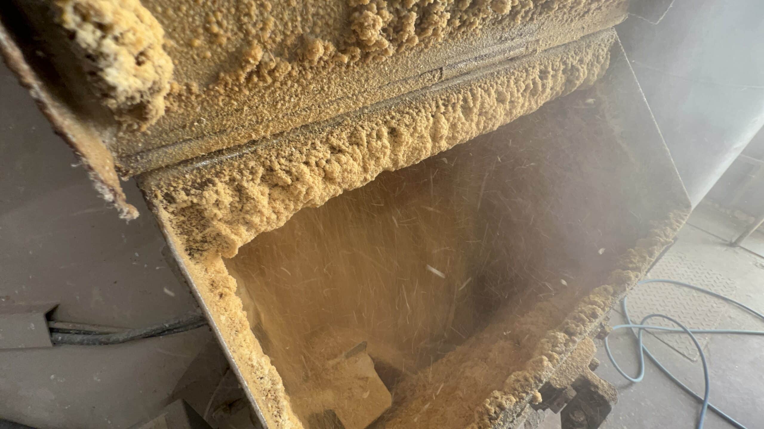
Distiller’s dried grains are produced by condensing and drying the stillage left over after starch-fermentation of corn for ethanol production. Solubles left over from the process are usually added before drying, resulting in the DDGS product that has become more and more commonplace in poultry feed formulations.
Historically, this ingredient was used in ruminant diets, as the nutrient content and quality of DDGS is considered to be somewhat variable – not only between, but also within production facilities. However, recent improvements in processing technologies and quality control systems have resulted in more consistent, higher-quality feed products.
Furthermore, newer fractionation processes continue to be implemented by some ethanol plants that are capable of fractioning out protein, fiber and oil portions of the grain, either pre- or post-fermentation, providing a wide variety of co-products which will result from the blending of these fraction streams.
Why is there more danger of oxidative damage today?
A decade ago, typical poultry diets consisted of far fewer feed ingredients than some of today’s rations. Additionally, the use of further-processed by-product meals and fats has also increased due to economic constraints associated with some raw ingredients.
This has created a diet which is highly prone to oxidative degeneration due to the higher rate of thermal, and in some cases, chemical processing of feed ingredients such as DDGS.
Currently, it is not uncommon to see DDGS inclusion rates between 5 and 12% in broiler and turkey diets, depending on bird age and feed prices.
One factor to consider when utilizing DDGS at such levels is the increase in polyunsaturated fatty acid content relative to saturated fatty acids that results from the addition of the 18:2, n-6 -rich corn oil which comprises approximately 10% of most DDGS.
Despite the fact that most modern rations have decreased the total lipid content in recent years, overall increases in the use of vegetable-based by-products invariably change the fatty acid profile of the diet to one which contains higher levels of polyunsaturated fats relative to saturated fats.
Polyunsaturated fatty acids are highly sensitive to oxidation during storage and are likely to turn rancid at high environmental temperatures. The oxidation process involves the generation of fatty acid free radicals, which then react with molecular oxygen to produce peroxide free radicals. This results in a chemical change to the fatty acid which decreases nutrient value and often produces undesirable odor.
…And that’s why risk management of oxidative damage is essential
Oxidative damage in feeds entails economic losses because of the negative impact on feed quality through the transformation of the lipid fraction of feed ingredients, decreased animal growth rate and performance, and decreased meat quality parameters, such as nutritional value and shelf-life. Therefore, lipid stability in feed is important, particularly with regard to the oxidative rancidity that occurs in high-fat ingredients, and prevention and management of oxidative stress is critical.
Respiratory health in poultry: no action is no solution
by Inge Heinzl and Ruturaj Patil, EW Nutrition
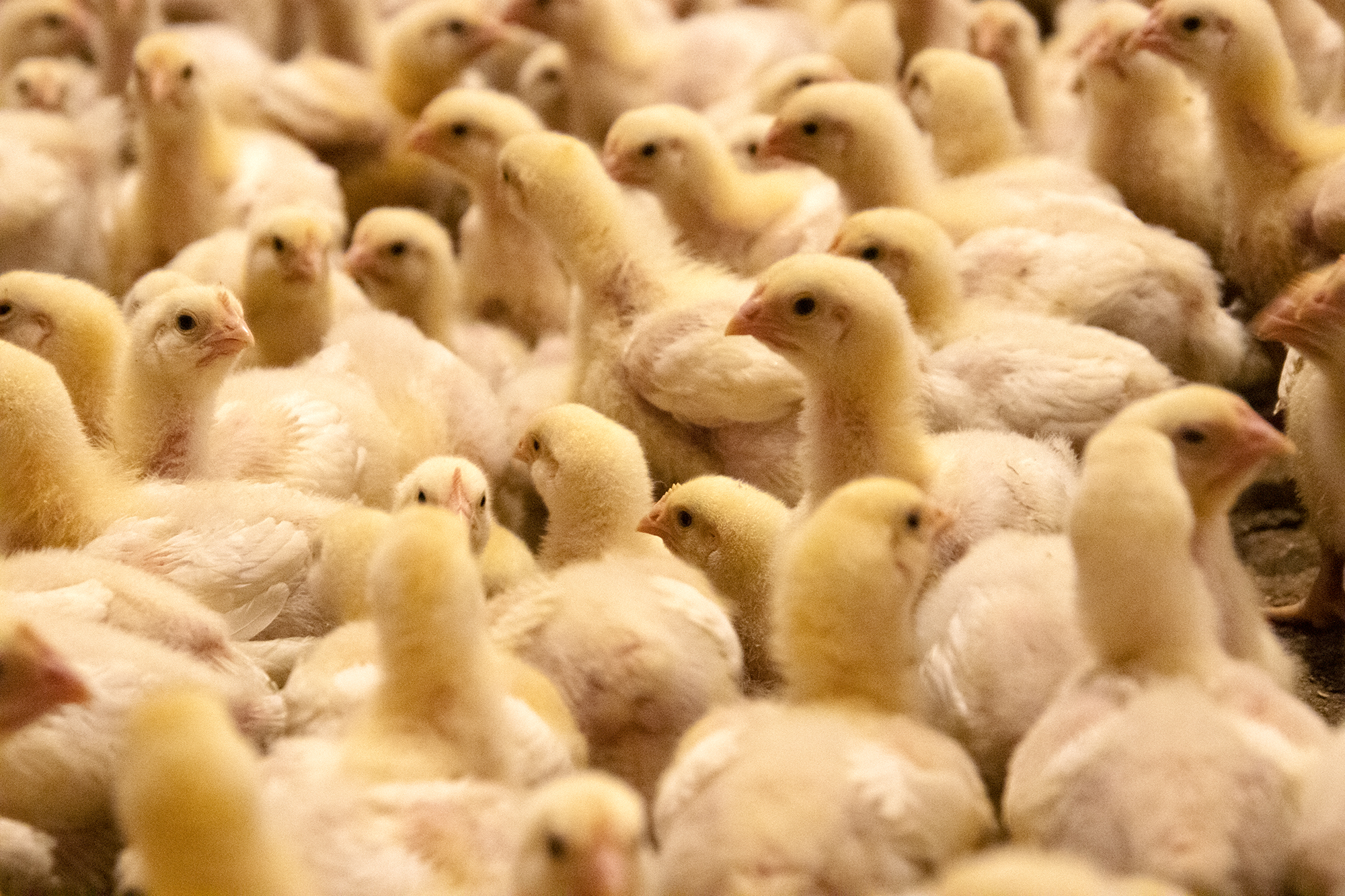
Broilers face high respiratory disease risks. In winter, they often come from lower temperatures; throughout the year, they come from improper ventilation and proximity to manure or infected birds. The confined spaces and lack of proper airflow create an environment conducive to harmful airborne particles and pathogens, significantly compromising birds’ respiratory health. In the possible presence of viruses such as ILT (Infectious Laryngotracheitis Virus), IBV (Infectious Bronchitis Virus), AIV (Avian Influenza Virus, NDV (Newcastle Disease Virus), bacteria like Mycoplasma gallisepticum, E. coli, or Chlamydia, respiratory issues are inevitable.
High efficiency takes its toll
A bird, generally a flying species, has a complex respiratory system. Instead of the diaphragm cooperating with the lung, nine additional air sacs do the job of sucking in and blowing out of the air like bellows. They increase the air volume passing through the lungs, where oxygen absorption occurs. The air sacs are situated in different parts of the birds and connected to hollow (pneumatic) bones.
The co-action of the air sacs and the lung results in a high efficiency of the bird’s respiratory system: birds can extract about 160% more oxygen from the air than mammals. However, the extended parts of the respiratory system also offer a high contact surface for pathogens. To protect themselves, the respiratory system is equipped with
– cilia in the trachea to propel entrapped particles for disposal – mucus produced by goblet cells in the trachea and cooperating with the cilia – immune cells in the lung, scavenging inhaled particles and bacteria that enter the lower respiratory tract
Additional support is recommended
To additionally support your birds against respiratory issues, stress should be kept low, and immunity to diseases should be high. If possible, decrease the stocking density. Effective litter management can help keep litter particle inhalation low. These particles irritate the respiratory system and reduce immune resistance. They often carry pathogens and possibly induce respiratory issues through several toxic mechanisms.
Another possibility is using phytogenic substances alone or combined with vaccines. Eucalyptus oil exerts antimicrobial, anti-inflammatory, mucolytic, and bronchodilator effects in the case of respiratory disease. Thyme has expectorant, mucolytic, antitussive, and antispasmodic characteristics, and mint, with its antihistamine and cooling effect, acts as a decongestant. Grippozon is such an example, based on fast-acting, concentrated phytomolecules supporting animals against respiratory challenges.
A trial with 20,000 birds showed fewer gurgling sounds and reduced post-vaccination reaction than the untreated group.
Regardless of the solution chosen, especially with the cold season coming and high stocking density a given in many parts of the world, by far the worst action is no action at all.
Feeding layers for longer laying cycles and optimized production
Conference report
At the recent EW Nutrition Poultry Academy in Jakarta Indonesia, Dr Steve Leeson, Professor Emeritus, University of Guelph, Canada, commented that “genetic progress in layer breeding has been substantial in recent decades. Since 1995, the yearly change has included +1 egg, -0.01 feed/dozen eggs, -10g final bodyweight, 0.02% mortality, and +1 week at >90% egg production. This improved persistency of commercial laying hens enables egg producers to keep flocks longer in production, provided egg shell quality can be maintained.”
He noted that “the increase in hen-housed egg production is mainly due to longer clutch length and improved uniformity of layer flocks. No doubt, there is a trend in cage layers to longer production cycles. A popular commercial goal is 500 eggs in one cycle with no moult, although this has already been surpassed in many flocks. The modern layer is capable of laying 150 eggs per clutch.”
Dr Leeson, however, stressed that “genetic progress and longer laying cycles have consequences. Long laying cycle programmes start during pullet rearing – you can’t make decisions at 72 weeks of age. Instead, you must start with your end goals, such as persistency, egg size and shell quality, in mind. You can then develop a life-cycle approach to feeding, lighting, nutrition, and general management.” Important issues to manage include:
Body weight control – early and late
Mature body weight dictates subsequent egg size. In the past, the common goal was being at, or above, management guide weight recommendations. For extended lay, a larger body weight results in too large an egg past 70 weeks of age, and so it is more difficult to maintain egg shell quality. Now the goal is to grow a slightly smaller pullet, and emphasis changes to achieving adequate early egg size from this smaller bird. This makes pre-lay nutrition for these slightly smaller pullets even more important.
The scheduling of rearing diets is more important than diet formulation. Dr Leeson’s guidelines are:
Starter diet – 19-20% CP, 2,850-2,900 kcal ME/kg from day old to target pullet body weight
Grower diet – 17-18% CP, 2,800-2,900 kcal ME/kg from target body weight to mature body size
Pre-lay diet (or layer diet?) – 16-18% CP, 2,800-2,900 ME/kg, mature body size to first egg
All nutrients are important, but energy is usually limiting for egg number, whereas protein/amino acids influence egg size (and feathering).
There is now even more emphasis on pullet growing to ensure adequate fat reserves through peak production, so birds are in a positive energy balance. The establishment of an energy reserve occurs during the rearing phase and has a significant effect on the bird’s body composition at point of lay.
Egg size control – early and late
The obvious solution to manage body weight (and egg size) is to light-stimulate a smaller pullet, or at least to not light-stimulate a heavy pullet. This achieves a balance between accepting reduced early egg size, versus limiting an increase in egg size late in the production cycle.
Egg size can be increased in smaller early-lay pullets by:
Reducing environmental temperature, if possible, to stimulate feed intake
Midnight feeding 19-29 weeks
Adequate amino acid nutrition intake, tailored to feed intake, especially methionine
Increased number of feedings/day and increased feed particle size (pellets)
Shell strength is negatively correlated with egg size. To temper egg size late in the cycle, Dr Leeson recommended:
Body weight control
Controlled day length: longer day length = increased feed intake, 14 hours maximum day length in controlled-environment houses
Warmer temperature – 26oC is ideal
Reduce number of feedings and particle size
Temper amino acid nutrition (with caution). Low crude protein/high amino acid diets limit the increase in egg size.
Midnight feeding provides about 1-hour extra light per day and therefore stimulating feed consumption in the middle of the dark period. Having access to feed during this period improves eggshell quality via the supply of calcium during the time when shell calcification takes place. The extra light period is perceived by the bird to be part of the night. The dark period after the light period must be longer than the initial dark period, as the bird perceives the start of the day is the end of the longest period of darkness. Removing midnight feeding should be done gradually – 15 minutes per week, advised Dr Leeson.
Preventing calcium depletion
Also known as cage layer fatigue, calcium depletion is becoming more common in all strains due to high sustained egg output. Calcium deficiency in the feed leads to loss of medullary or long bone (a reservoir of about 4g of calcium) and increased bone fragility. It is commonly seen at 35-40 weeks of age, with a 1-2% occurrence. If the incidence is more than 2%, seek advice for your pre-lay nutrition.
The development of the medullary bones takes about 10 days and requires additional calcium. Pre-lay rations support a smooth transition from developer feed to layer feed, with 2-2.5% calcium, while the other nutrients are similar to a layer feed. Pre-lay rations help the birds to adapt to the high calcium content of layer feed and to maintain sufficient daily feed intake.
To prevent calcium depletion, Dr Leeson suggested:
Optimise pre-lay calcium (Ca) and phosphorous (P) nutrition
Intake of 1.5g Ca, 350-450mg available P/day for at least 7 days prior to first egg
During early lay, ensure 3.5-4 g Ca and 420 mg available P/day
Consider vitamin D3 water treatment (150 IU/day, twice weekly)
Pre-lay diets provide the bird with the opportunity to deposit medullary bone. This bone deposition coincides with follicular maturation and is under the control of both estrogens and androgens. The latter hormone seems essential for medullary bone growth, and its presence is manifested in the growth and reddening of the comb and wattles. Consequently, there will be little medullary deposition, regardless of diet calcium level, if the birds are not showing comb and wattle development and this stage of maturity should be the cue for increasing the bird’s calcium intake.
Liver health
Excess energy relative to needs results in excess fat accumulation that is prone to oxidation. This is why you never see fatty liver haemorrhagic syndrome (FLHS) in poor-producing flocks. Layers normally have a very fatty liver, as 100% of egg yolk synthesis occurs in the liver.
The lower the fat content of the diet, the greater the stress/need to fat synthesis in the liver. With a low energy/low fat/carbohydrate diet FLHS is almost universal to varying degrees. One treatment is to add fat to the diet! Haemorrhage (not always FLHS) is inevitable with dietary omega-3s that are very prone to oxidation.
Dr Leeson recommended prevention/control for FLHS, which usually starts about weeks 36-40, including:
+1.0 kg choline
+0.5 kg methionine
+100 IU vitamin E
+30% does Hy-D because of impaired liver metabolism of vitamin D3 (that can also impact calcium absorption)
Add 2% dietary fat without change in diet energy level
***
EW Nutrition’s Poultry Academy took place in Jakarta and Manila in early September 2023. Dr. Steve Leeson, an expert in Poultry Nutrition & Production with nearly 50 years’ experience in the industry, was the distinguished keynote speaker.
Dr. Leeson had his Ph.D. in Poultry Nutrition in 1974 from the University of Nottingham. Over a span of 38 years, he was a Professor in the Department of Animal &Poultry Science at the University of Guelph, Canada. Since 2014, he has been Professor Emeritus at the same University. As an eminent author, he has more than 400 papers in refereed journals and 6 books on various aspects of Poultry Nutrition & Management. He also won the American Feed Manufacturer’s Association Nutrition Research Award (1981), the Canadian Society of Animal Science Fellowship Award (2001), and Novus Lifetime Achievement Award in Poultry Nutrition (2011).
Nutritional considerations for immunity and gut health
Conference report
At the recent EW Nutrition Poultry Academy in Jakarta, Indonesia, Dr Steve Leeson, Professor Emeritus, University of Guelph, Canada, opened his presentation by stating that “it is obvious that any nutrient deficiency will impact bird health, but not so obvious is that nutrition per se can positively impact immunity and health in an otherwise healthy and high-producing bird.”
Modern high-performing broilers are characterized by extremely high feed intake. This puts a lot of stress on the physiology of the entire gastrointestinal tract, but particularly so on the absorptive epithelial cells of the small intestine. Any organism requires a nutrient source for survival and reproduction. Dr Leeson asked “can we significantly reduce nutrient supply to pathogens, while sustaining bird productivity?”
He reminded the audience that no cellular function comes for free: so there is always a “cost”. A general conclusion is that 10% of nutrients can be used for immune function during disease challenge, and always get priority. Therefore, you don’t want to overstimulate the immune system, which in extreme situations leads to an inflammatory response. In his presentation, Dr Leeson considered factors determining gut health and nutritional tools which are available to support gut health.
Gut microflora
Gut pathogens impact the bird and/or the consumer. Clostridia and E. coli are the major concerns regarding bird health and productivity, whereas Salmonella and Campylobacter are major pathogens important for human health.
The chick hatches with a gut virtually devoid of microbes, so early colonizers tend to predominate quite quickly. Microbial species present on the hatching tray, during delivery and during the first few days at the farm will likely dictate early gut colonization. In some instances, the chick’s microflora may be established by the time it gets to the farm, so the probiotic faces more of a challenge to establish itself as the predominant species.
Antibiotic alternatives
Gut villi development matures at around 10-15 days of age. The broiler pre-starter diet therefore is a target for feed additives that positively impact gut structure and development.
Among the short chain fatty acids, butyric acid is considered the prime energy source for enterocytes and it is also necessary for the correct development of the gut-associated lymphoid tissue (GALT). Butyric acid can also be added indirectly via fermentation of judicious levels of soluble fiber to encourage optimal gut villi development. Dr Leeson added that he is a big believer in butyric acid, encouraging a good gut structure at 10 days, which can be worth about 50 kcal.
Exogenous enzymes should also be considered in an attempt to maximize digestion and limit the flow of nutrients to the large intestine and ceca. Protease enzymes have great potential in this regard, since they allow nutritionists to reduce dietary crude protein and hopefully reduce the supply of nitrogen that fuels proteolytic Clostridia bacteria in the large intestine and ceca.
Amino acids, particularly threonine, play a critical role in the maintenance of intestinal mucosal integrity and barrier function, especially for mucin synthesis, which protects enterocytes from adherence by pathogenic bacteria, and from attack by endogenous enzymes and acids.
Polyunsaturated fatty acids (PUFAs) – Omega-3s and especially DHA from fish oil help to reduce inflammatory response (overstimulation). Omega-3s are poorly converted to DHA by the chicken, so conventional sources such as flax are of limited application for immunity.
Blood plasma from pigs or cattle is a complex spray-dried mixture of proteins and amino acids, many of which are immunoglobulins that “temper” the immune system, much like PUFAs.
Vitamins A, D, E and C have vital roles in the normal function of the immune system and have antioxidant capacity.
Certain complex carbohydrates, such as ß-glucans, influence gut health due to their fermentation, leading to the production of short-chain fatty acids, such as butyrate.
Antioxidants – to firstly control oxidation of fats and fat-soluble vitamins in feed, and secondly to optimize birds’ cellular oxidative capacity, to prevent cell damage, therefore maintaining healthy cellular and immune function.
Betaine increases intracellular water retention, reducing “dehydration” of microvilli and increasing their volume/surface area.
Fiber – moderate levels (1-2%) of soluble (fermentable) and insoluble fiber can be beneficial to early gut development by stimulating gizzard development and endogenous enzyme production.
Phytogenics are becoming very common in combination with acidifiers (upper tract) and probiotics. Essential oils are becoming more mainstream the more we know about them.
Recommendations for optimizing gut health and immunity
Fast growth rate and high egg output are negatively correlated with immune response. Consequently, nutrient-dense diets are not optimal for immunity. With bacteria, it’s a numbers game – but these numbers quickly multiply. The first 7 days are important, therefore probiotics must be established early. Consider the role of targeted feed additives, such as butyrate, phytogenics, antioxidants, PUFAs etc.
Also, maximize feed particle size – the limit is usually pellet quality. Mitigate nutrient transition at any diet change. Review the supply of trace minerals, as there is a trend to lower levels of organic minerals. With all the factors that weigh into production performance, any support that can be rallied through nutrition needs to be considered.
***
EW Nutrition’s Poultry Academy took place in Jakarta and Manila in early September 2023. Dr. Steve Leeson, an expert in Poultry Nutrition & Production with nearly 50 years’ experience in the industry, was the distinguished keynote speaker.
Dr. Leeson had his Ph.D. in Poultry Nutrition in 1974 from the University of Nottingham. Over a span of 38 years, he was a Professor in the Department of Animal &Poultry Science at the University of Guelph, Canada. Since 2014, he has been Professor Emeritus at the same University. As an eminent author, he has more than 400 papers in refereed journals and 6 books on various aspects of Poultry Nutrition & Management. He also won the American Feed Manufacturer’s Association Nutrition Research Award (1981), the Canadian Society of Animal Science Fellowship Award (2001), and Novus Lifetime Achievement Award in Poultry Nutrition (2011).
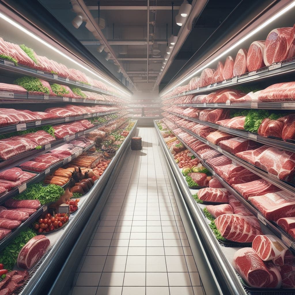
Meat labels explained
Certified Organic: (US, others) To be labeled as “Certified Organic” in the US, meat and poultry must come from animals that are raised in accordance with organic farming standards. These standards typically include restrictions on the use of synthetic pesticides, herbicides, antibiotics, and genetically modified organisms (GMOs). The animals are typically raised with organic feed and have access to the outdoors.
Chemical-free: (US) A product that contains no artificial ingredients or chemical preservatives.
Free-range or Free-roaming: (International) Poultry that has been allowed access to the outside.
Free-Range or Pasture-Raised: (US, others) These terms suggest that the animals had access to the outdoors or were raised on pasture, which can offer better living conditions than confined, industrial operations.
Fresh poultry: (US) Poultry that has never been below 26°F.
Frozen poultry: (US) Poultry that has been held at 0°F or lower.
Grain-Fed: (International) This label implies that the animals were primarily fed grains or other non-grass-based diets, which is common in many commercial meat production systems.
Grass-Fed: (International) “Grass-Fed” typically means that the animals were primarily fed a diet of grass or forage throughout their lives, although some supplemental grains may be allowed. This label does not necessarily imply organic or non-GMO practices.
Halal: (International) Halal meat is prepared following Islamic dietary laws. This includes specific slaughter methods and requirements for the handling and preparation of the meat.
Kosher: (International) Kosher meat is prepared according to Jewish dietary laws and involves specific slaughtering practices and inspections.
Mechanically separated meat: (US) A paste-like meat product produced by forcing bones, with attached edible meat, under high pressure through a sieve or similar device to separate the bone from the edible meat tissue.
Natural: (US) A product containing no artificial ingredient or added color and is only minimally processed (a process which does not fundamentally alter the raw product). The label must explain the use of the term natural (such as “no added colorings or artificial ingredients; minimally processed”).
No antibiotics: (US) The terms “no antibiotics added” may be used on labels for meat or poultry products if sufficient documentation is provided by the producer to the USDA demonstrating that the animals were raised without antibiotics. If an animal becomes sick and requires antibiotics, it cannot be sold as “no antibiotics added.”
No hormones (beef): (US) The term “no hormones administered” may be approved for use on the label of beef products if sufficient documentation is provided to USDA by the producer showing no hormones have been used in raising the animals.
No hormones (pork or poultry): (US) Federal regulations prohibit the use of hormones in raising hogs and poultry.
Non-GMO: (International) A “Non-GMO” label indicates that the animals were not fed genetically modified organisms. This label may apply to both feed and the animals themselves.
Organic: (International) Meat and poultry labeled as organic must come from animals fed organic – which also means non-GMO – feed, given fresh air and outdoor access, and raised without antibiotics or added growth hormones. Organic livestock must also have access to pasture for at least 120 days per year.
Protected Designation of Origin (PDO) and Protected Geographical Indication (PGI): (EU) These labels are used to protect and promote regional and traditional foods. Meat labeled with PDO and PGI must come from specific regions and meet particular quality and production standards.
Raised without Antibiotics or Antibiotic-Free: (International) This label indicates that the animals were not treated with antibiotics during their lifetime. However, this label does not necessarily mean the animals were raised in organic or free-range conditions.
Sustainably Sourced: (International) This label may indicate that the meat was produced with a focus on environmental and ethical considerations, such as minimizing ecological impact and promoting fair labor practices.
Decoding the connection between stress, endotoxins, and poultry health
By Technical Team, EW Nutrition
Stress can be defined as any factor causing disruptions to homeostasis, which triggers a biological response to regain equilibrium. We can distinguish four major types of stressors in the poultry industry:
Technological: related with management events and conditions
Nutritional: involving nutritional disbalances, feed quality and feed management
Pathogenic: comprising health challenges.
Environmental: changes in environment conditions
In practical poultry production, multiple stress factors occur simultaneously. Their effects are also additive, leading to chronic stress. The animals are not regaining homeostasis and continuously deviate the use of resources through inflammation and the gut barrier-function, thus leading to microbiome alteration. As a consequence, welfare, health, and productivity are compromised.
What are endotoxins?
Bacterial lipopolysaccharides (LPS), also known as endotoxins, are the main components of the outer membrane of all Gram-negative bacteria and are essential for their survival. LPS have direct contact with the bacteria’s surroundings and function as a protection mechanism against the host’s immunological response and chemical attacks from bile salts, lysozymes, or other antimicrobial agents.
Gram-negative bacteria are part of animals’ microbiota; thus, there are always LPS in the intestine. Under optimal conditions, this does not affect the animals, because intestinal epithelial cells are not responsive to LPS when stimulated from the apical side. In stress situations, the intestinal barrier function is impaired, allowing the passage of endotoxins into the blood stream. When LPS are detected by the immune system either in the blood or in the basolateral side of the intestine, inflammation and changes in the gut epithelial structure and functionality occur.
The gut is critically affected by stress
Even when there is no direct injury to the gut, signals from the brain can modify different functions of the intestinal tract, including immunity. Stress can lead to functional disorders, as well as to inflammation and infections of the intestinal tract. Downstream signals act via the brain–gut axis, trigger the formation of reactive oxygen and nitrogen species as well as local inflammatory factors, and circulating cytokines, affecting intestinal homeostasis, microbiome, and barrier integrity.
Stress then results in cell injury, apoptosis, and compromised tight junctions. For this reason, luminal substances, including toxins and pathogens, leak into the bloodstream. Additionally, under stress, the gut microbiome shows and increment on Gram-negative bacteria (GNB). For instance, a study by Minghui Wang and collaborators (2020) found an increase of 24% in GNB and lower richness, in the cecum of pullets subjected to mild heat stress (increase in ambient temperature from 24 to 30°C).
Both these factors, barrier damage and alterations in the microbiome, facilitate the passage of endotoxins into the blood stream, which promotes systemic chronic inflammation.
What categories of stress factors trigger luminal endotoxins’ passage into the bloodstream?
Technological stress
Various management practices and events can be taken as stressors by the animals’ organism. One of the most common examples is stocking density, defined as the number of birds or the total live weight of birds in a fixed space. High levels are associated with stress and loss of performance.
A study from the Chung-Ang University in 2019 found that broilers with a stocking density of 30 birds/m2 presented two times more blood LPS than birds kept at half of this stocking density. Moreover, the body weight of the birds in the high-density group was 200g lower than the birds of the low-density group. The study concluded that high stocking density is a factor that can disrupt the intestinal barrier.
Nutritional stress
The feed supplied to production animals is designed to contribute to express their genetic potential, though some feed components are also continuous inflammatory triggers. Anti-nutritional factors, oxidized lipids, and mycotoxins induce a low-grade inflammatory response.
For instance, when mycotoxins are ingested and absorbed, they trigger stress and impair immunity in animals. Their effects start in gastrointestinal tract and extend from disrupting immunity to impairing the intestinal barrier function, prompting secondary infections. Mycotoxins can increase the risk of endotoxins in several ways:
By alterations in the immune response, low doses of mycotoxins, such as trichothecenes, induce the upregulation of pro-inflammatory cytokines. A possible synergy can be inferred as when they are together, the effects may be prolonged and require a lower dosage to be triggered.
A study conducted by EW Nutrition (Figure 1) shows an increase in intestinal lesions and blood endotoxins after a mycotoxin challenge of 200pbb of Aflatoxin B1 + 360ppb Ochratoxin in broilers at 21 days of age. The challenged birds show two times more lesions and blood endotoxins than the ones in the unchallenged control. The use of the right mitigation strategy, a product based on bentonite, yeast cell walls, and phytogenics (EW Nutrition GmbH) successfully prevented these effects as it not only mitigates mycotoxins, but also targets endotoxins in the gut.
Figure 1 Blood LPS and intestinal lesion score of broilers challenged with 200ppb AFB1 + 350 ppb OTA from 1 to 21 days of age without and with an anti-toxin product from EW Nutrition GmbH (adapted from Caballero et al., 2021)
Pathogenic stress
Intestinal disease induces changes in the microbiome, reducing diversity and allowing pathogens to thrive. In clinical and subclinical necrotic enteritis (NE), the intestinal populations of GNB, including Salmonella and E.coli also increases. The lesions associated with the pathogen compromise the epithelial permeability and the intestinal barrier function, resulting in translocation of bacteria and LPS (Figure 5) into the bloodstream and internal organs.
Figure 2 Increase in systemic LPS (vs a healthy control) after a NE challenge (adapted from Chen et al., 2015, Li et al., 2018 & Liu at al., 2018)
Environmental stress
Acute and chronic heat and cold stress increases gut permeability, by increasing intestinal oxidative stress and disrupting the expression of tight junction proteins. This results in the damage and destruction of intestinal cells, inflammation, and imbalance of the microbiota. An increased release and passage of endotoxins has been demonstrated in heat stress (Figure 3), as well as a higher expression of TLR-4 and inflammation.
Figure 3 Systemic LPS increase (in comparison with a non-stressed control) after different heat stress challenges in broilers: 16°C increased for 2, 5 and 10 hours (Huang et al., 2018); 9°C increased for 24 and 72 hours (Nanto-Hara et al., 2020); 10°C continuously for 3 and 10 days, and 15°C 4 hours daily for 3 and 10 days (Alhenaky et al., 2017)
Zhou and collaborators (2021) showed that 72 hours of low temperature treatment in young broilers increased intestinal inflammation and expression of tight junction proteins, while higher blood endotoxins indicate a disruption of the intestinal barrier. As a consequence, the stress decreased body gain and increased the feed conversion rate.
An experiment conducted by EW Nutrition GmbH with the objective of evaluating the ability of a toxin mitigation product to ameliorate heat-stress induced LPS. For the experiment, 1760 Cobb 500 pullets were divided into two groups, and each was placed in 11 pens of 80 hens, in a single house. One of the groups received feed containing 2kg/ton of the product from the first day. From week 8 to week 12, the temperature of the house was raised 10°C for 8 hours every day.
Throughout the heat stress period, blood LPS (Fig 4) was lower in the pullets receiving the product, which allowed lower inflammation, as evidenced by the lower expression of TLR4 (Fig. 5). Oxidative stress was also mitigated with the help of the combination of phytomolecules in the product, obtaining 8.5% improvement on serum total antioxidant capacity (TAC), supported by an increase in in superoxide dismutase (SOD glutathione peroxidase (GSH) and a decrease in malondialdehyde (MDH).
Figures 4 and 5 – Blood LPS and expression of toll-like receptor 4 (TLR4) in lymphocites of pullets before (wk 6) and during heat stress (wk 9 and 10). (*) indicates significant differences (P<0,05), and (‡) a tendency to be different against the control group (P<0,1).
In practice: there is no silver bullet
In commercial poultry production, a myriad stressors may occur at the same time and some factors trigger a chain of events that work to the detriment of animal health and productivity. Reducing the solution to the mitigation of LPS is a deceitfully simplistic approach. However, this should be part of a strategy to achieve better animal health and performance. In fact, EW Nutrition’s toxin mitigation product alone helped the pullets to achieve 3% improvement in body weight and 9 points lower cumulative feed conversion (Figure 6).
Figure 6
Keeping the animals as free of stress as possible is a true priority for poultry producers, as it promotes animal health as well as the integrity and function of the intestinal barrier. Biosecurity, good environment, nutrition and good management practices are crucial; the use of feed additives to reduce the consequences of unavoidable stress also critically supports the profitability of poultry operations.
A guide to international sustainability regulations
By Ilinca Anghelescu, Global Director Marketing Communications, EW Nutrition
This may be the year that climate change has arrived in humanity’s backyard, driving home the repercussions of human action and the finite nature of our planet’s resources. More than ever, it is also becoming clear that we cannot fight climate change in our own backyard but that long-term cross-border action is imperative.
With the visible threat of extreme events nearer than ever, companies and countries feel pressured to show their commitment to sustainable practices. The shape this commitment takes is, however, very different. The slew of regulations and policies directly or indirectly aimed at promoting sustainability may take the shape of water or energy management, environmental protection, specific business practice regulations, and may or may not include reporting obligations and monitoring bodies. Some international initiatives are attempting to impose such obligations, with varying degrees of success. Reading between the lines, the number of regulations is not the problem; it is the competencies in standardizing and enforcing these regulations that prove more difficult.
Sustainability regulations in the European Union
The European Union is both the fastest warming region (with the exception of the Arctic) and probably the most advanced in terms of regulatory pressure. It has been steadily developing not just specific regulations aimed at green growth, but also specific reporting tools to avoid greenwashing and standardize the monitoring and measuring of this commitment.
The largest sustainability initiative, the EU’s Green Deal, unveiled in 2019, is a comprehensive policy framework aimed at making Europe the world’s first climate-neutral continent by 2050. Among its objectives are reducing greenhouse gas emissions, increasing energy efficiency, and promoting circular economy practices. Key regulations include:
European Emissions Trading System (EU ETS): The EU ETS is a “cap and trade” scheme that aims to reduce greenhouse gas emissions in the European Union. It is the first and largest carbon market, covering around 45% of the EU’s greenhouse gas emissions, and is operational across the EU, Iceland, Liechtenstein, and Norway. The system works by setting a cap on the total amount of greenhouse gases that can be emitted by all participating installations. Within this cap, operators buy or receive emissions allowances, which they can trade with one another as needed. The fourth phase started in January 2021 and is to continue until December 2030, however the reduction target for 2030 needs to be reassessed.
Single-Use Plastics Directive: This regulation aims to reduce single-use plastics and their impact on the environment by banning certain products and promoting recycling.
Circular Economy Action Plan: Designed to reduce waste and promote recycling, this plan outlines initiatives to make products more durable and easier to repair. The plan includes measures on product design, waste management, and resource efficiency.
Taxonomy Regulation: This regulation establishes an EU-wide classification system for environmentally sustainable economic activities. The taxonomy defines which economic activities can be considered environmentally sustainable, based on their contribution to environmental objectives such as climate change mitigation and adaptation, biodiversity, and water protection.
More recent but directly concerned with regulating and reporting sustainability in business practices are the following:
Sustainable Finance Disclosure Regulation (SFDR): The SFDR requires financial market participants and advisers to disclose information about how they integrate sustainability risks into their investment decisions, consider and disclose the adverse impacts of their investments on sustainability factors.
Corporate Sustainability Reporting Directive (CSRD): This requires companies to report on a wide range of sustainability issues, including environmental, social, and governance (ESG) factors. The reporting requirements will be phased in, starting from January 1, 2024, for certain large EU and EU-listed companies, and will apply to all in-scope companies by January 1, 2028.
In addition to these regulations, the EU also provides financial support for sustainable projects through its Horizon Europe research and innovation program. Horizon Europe has a budget of €95.5 billion for the period 2021-2027, and a significant portion of this funding will be used to support research and innovation in areas such as climate change mitigation, renewable energy, and sustainable agriculture.
Sustainability regulations in the United States
The United States traditionally has a more decentralized approach to regulations, with federal, state, and local governments all playing important roles. Key federal regulations and initiatives in the field of sustainability include:
Clean Air Act: Enforced by the Environmental Protection Agency (EPA), this law aims to reduce air pollution and greenhouse gas emissions. This law regulates air pollution from a variety of sources, including power plants, factories, and vehicles. The Clean Air Act has helped to reduce air pollution in the US by over 70% since it was passed in 1970.
Clean Water Act: Also administered by the EPA, this act sets standards for water quality, aiming to protect aquatic ecosystems. This law regulates water pollution from a variety of sources, including factories, farms, and sewage treatment plants. The Clean Water Act has helped to improve water quality in the US by over 70% since it was passed in 1972.
Renewable Energy Tax Credits: Also called Residential Clean Energy Credits, these incentives encourage the development and use of renewable energy sources like solar and wind power.
More recent, targeted sustainability actions and regulations in the US include:
Executive Order 14057: Issued by President Biden in 2021, the Executive Order on Catalyzing Clean Energy Industries and Jobs Through Federal Sustainability requires federal agencies to take steps to reduce their greenhouse gas emissions and promote clean energy.
ESG Disclosure Simplification Act: This bill, passed by the House of Representatives in 2021, would require public companies to disclose more information about their environmental, social, and governance (ESG) practices.
Methane Emissions Reduction Plan: The White House Action Plan, together with the Supplemental Methane Proposal put forth by the Environmental Protection Agency (EPA) in 2022, would require primarily oil and gas companies to reduce methane emissions from their operations.
Sustainable Electricity Plan: This plan, released by the Department of Energy in 2022, outlines the Biden administration’s goals for increasing the use of renewable energy and reducing greenhouse gas emissions from the electricity sector.
SEC Climate-Related Disclosures/ESG Investing: Prompted by the Climate Risk Disclosure Act of 2021, the Securities and Exchange Commission (SEC) has issued a rule proposal that would require US publicly traded companies to disclose annually how their businesses are assessing, measuring, and managing climate-related risks. This would include climate-related risks and their material impacts on the registrant’s business, strategy, and outlook; governance of climate-related risks; greenhouse gas (“GHG”) emissions; certain climate-related financial statement metrics and related disclosures; information about climate-related targets and goals, and transition plan, if any. Some companies would have to already start reporting in 2023 for 2023. However, it is likely the proposal will undergo several rounds of revisions.
In addition to these federal laws, there are also a number of state and local sustainability regulations. U.S. regulations generally lack cohesion, with the federal government’s role fluctuating depending on the administration in power. Still, there is growing momentum towards sustainability, driven by grassroots movements and corporate initiatives.
Sustainability regulations in China
China, the world’s largest polluter, faces significant sustainability challenges as it grapples with rapid industrialization, urbanization, and economic growth. It has made substantial progress, particularly in renewable energy adoption, but still faces challenges of implementation.
Carbon Neutrality Commitment: In September 2020, Chinese President Xi Jinping announced China’s commitment to achieving carbon neutrality by 2060. This ambitious goal involves reducing carbon emissions to net-zero by mid-century.
Renewable Energy Development: China is a global leader in renewable energy deployment. It has set targets for increasing the share of renewable energy sources like wind, solar, and hydropower in its energy mix. Initiatives include the National Renewable Energy Development Plan and the 13th Five-Year Plan for Energy Development.
Emissions Trading System (ETS): China has launched a national carbon emissions trading system, which is the world’s largest such program. It caps emissions from certain industries and encourages emission reductions through trading of carbon allowances.
Green Finance Initiatives: The country is promoting green finance to support sustainable development. Initiatives include green bond issuance, guidelines for green lending, and incentives for sustainable investment.
Air Quality Improvement: The “Blue Sky” campaign aims to reduce air pollution in Chinese cities through stricter emissions standards, promotion of cleaner energy sources, and transitioning from coal to natural gas. The campaign appears to have had significant impact.
Sustainable transportation and circular economy: Initiatives to promote electric vehicles (EVs) and public transportation include subsidies for EV purchases, charging infrastructure development, and incentives for green vehicle production. China is also working on promoting a circular economy by reducing waste, improving resource efficiency, and encouraging recycling. The Circular Economy Promotion Law was passed in 2008.
Environmental Protection Laws and Regulations: China has strengthened its environmental laws and regulations to address pollution and environmental degradation. This includes revisions to the Environmental Protection Law and stricter enforcement.
These sustainability regulations, plans, and actions reflect China’s efforts to address pressing environmental challenges, transition to a more sustainable and low-carbon economy, and contribute to global efforts to combat climate change. Results are varied but the sheer scale of China’s pollution make the success of these initiatives a matter of global concern.
Sustainability regulations in India
Prompted by very tangible threats, India, recently crowned the world’s most populous country, has been fighting climate change for several decades, although not necessarily under one umbrella of sustainability. Moreover, there are currently no regulations that mandate sustainability reporting in India. However, Indian regulators are revising its existing environmental laws and plans, which will likely result in more stringent requirements for companies.
Instead of reporting requirements, India provides support through various sustainability-related programs and legislation.
National Action Plan on Climate Change (NAPCC): Launched in 2008, the NAPCC outlines the country’s strategy to combat climate change. It consists of eight national missions focused on various aspects of climate change mitigation and adaptation, including solar energy, energy efficiency, water, agriculture, and forestry.
Renewable Energy Initiatives: India has set ambitious targets for increasing its renewable energy capacity, including solar and wind power. Initiatives like the National Solar Mission aim to promote clean energy sources and reduce greenhouse gas emissions.
Sustainable Agriculture Initiatives: Programs like the National Mission for Sustainable Agriculture (NMSA) promote sustainable farming practices, soil health management, and water-use efficiency in agriculture.
National Clean Air Program (NCAP): India’s NCAP, launched in 2019, aims to improve air quality in major cities by reducing particulate matter and other air pollutants. It includes measures to control emissions from industries, vehicles, and biomass burning.
Water Resource Management: India has various initiatives and programs to address water-related challenges, including river rejuvenation projects, watershed development, and efforts to improve water-use efficiency in agriculture.
Sustainable Transportation: The Faster Adoption and Manufacturing of Hybrid and Electric Vehicles (FAME) scheme promotes the adoption of electric and hybrid vehicles to reduce air pollution and greenhouse gas emissions.
Plastic Waste Management Rules: India has implemented rules to manage and reduce plastic waste, including restrictions on single-use plastics.
National Mission for Sustainable Habitat (NMSH): This mission focuses on promoting sustainable urban planning and development, energy efficiency in buildings, and waste management in urban areas.
India’s approach is comprehensive but at the moment focuses on top-down actions. As in China, market players are at present not required to disclose any climate-related impact or information.
International sustainability regulations
International organizations play a crucial role in coordinating global sustainability efforts. The United Nations and its agencies, particularly the UN Framework Convention on Climate Change (UNFCCC), an international treaty and organization established to address the issue of global climate change adopted in 1992, have spearheaded international sustainability regulations, of which the most impactful are mentioned below.
The Paris Agreement: Signed in 2015, the agreement represents a global commitment to combat climate change by limiting global warming to well below 2 degrees Celsius above pre-industrial levels and aiming to limit it to 1.5 degrees Celsius. 196 nations have agreed on its goals, as well as committed to specific targets and standards of accountability. The Paris Agreement is part of the UNFCCC.
The United Nations’ Sustainable Development Goals (SDGs): The SDGs are a set of 17 goals that aim to end poverty, protect the planet, and ensure prosperity for all. They provide a framework for companies to align their business strategies with sustainable development objectives. These goals were adopted by all United Nations Member States in September 2015 as part of the 2030 Agenda for Sustainable Development.
The Task Force on Climate-related Financial Disclosures (TCFD): The TCFD is a voluntary initiative that provides recommendations for companies to disclose climate-related risks and opportunities in their financial filings. The TCFD was founded by the Financial Stability Board (FSB), an international body that monitors and makes recommendations about the global financial system, in December 2015. The TCFD encourages organizations to conduct scenario analysis, which involves assessing the potential financial impact of different climate-related scenarios, including both transition risks (related to policy and market changes) and physical risks (related to climate impacts like extreme weather events).
The International Sustainability Standards Board (ISSB) issued the first two sustainability standards, the IFRS S1 General Requirements for Disclosure of Sustainability-related Financial Information and the IFRS S2 Climate-related Disclosures. They will theoretically become effective on or after January 1, 2024. If jurisdictions challenge or delay bringing them into law, the effective date may well be later. IFRS S1 provides a set of disclosure requirements designed to enable companies to communicate to investors about the sustainability-related risks and opportunities they face over the short, medium and long term. IFRS S2 sets out specific climate-related disclosures and is designed to be used with IFRS S1. Both fully incorporate the recommendations of the Task Force on Climate-related Financial Disclosures (TCFD).
Conclusion
China, the US, and India have been, for a while now, the largest polluter nations. However, statistics do not look at the indirect pollution cost of countries that produce abroad for internal consumption. If we take that cost into consideration, it becomes evident that sustainability regulations at both national and international level are crucial for addressing environmental and social challenges.
Regulations alone are obviously not enough. Strict enforcement and monitoring are what is going to transform national and supra-national entities, regional authorities, businesses, communities, and individuals into responsible actors.
Salmonella in pigs: a threat for humans and a challenge for pig producers
By Dr. Inge Heinzl, Editor, EW Nutrition
Salmonellosis is third among foodborne diseases leading to death (Ferrari, 2019). More than 91,000 human cases of Salmonellosis are reported by the EU each year, generating overall costs of up to €3 billion a year (EFSA, 2023), 10-20% of which are attributed to pork consumption (Soumet, 2022). The annual costs arising from the resulting human health losses in 2010 were about €90 million (FCC Consortium, 2010). Take the example of Ireland, where a high prevalence of Salmonella in lymph nodes still shows a severe issue pre-slaughter and a big challenge for slaughterhouses to stick to the process hygiene requirements (Deane, 2022).
Several governments already have monitoring programs in place, and the farms are categorized according to the salmonella contamination of their pigs. In some countries, e.g., Denmark, an economic penalty of 2% of the carcass value must be paid if the farm has level 2 (intermediate seroprevalence) and 4-8% if the level is 3. Other countries, e.g., Germany, the UK, Ireland, or the Netherlands, use quality assurance schemes. The farmers can only sell their carcasses under this label if their farm has a certain level.
Let’s take a quick look at the genus of Salmonella
Salmonellas are rod-shaped gram-negative bacteria of the family of enterobacteria that use flagella for their movement. They were named after the American vet Daniel Elmer Salmon. The genus of Salmonella consists of two species (S. bongori and S. enterica with seven subspecies) with in total more than 2500 serovars (see Figure 1). The effects of the different serovars can range from asymptomatic carriage to severe invasive systemic disease (Gal-Mor, 2014). All Salmonella serovars generally can cause disease in humans; the rosa-marked ones already showed infections.
Figure 1: the genus of Salmonella with Salmonella serovars relevant for pigs (according to Bonardi, 2017: Salmonella in the pork production chain and its impact on human health in the European Union)
Within the group of Salmonella, some serovars can only reside in one or few species, e.g., S. enterica spp. enterica Serovar Dublin (S. Dublin) in bovines (Waldron, 2018) or S. Cholerasuis in pigs (Chiu, 2004). An infection in humans with these pathogens is often invasive and life-threatening (WHO, 2018). On the contrary, serovars like S. Typhimurium and S. Enteritidis are not host-specific and can cause disease in various species.
The serotypes S. Typhi and S. Paratyphi A, B, or C are highly adapted to humans and only for them pathogenic; they are responsible for the occurrence of typhus.
Transmission of Salmonella mostly happens via contaminated food
The way of transmission to humans depends on the serovar:
Human-specific and, therefore, only in humans and higher primates residing serovars S. Typhi and Paratyphi A, B, or C (typhoidal) are excreted via feces or urine. Therefore, any food or water contaminated with the feces or urine of infected people can transmit this disease (Government of South Australia, 2023). Typhoid and paratyphoid Salmonellosis occur endemic in developing countries with the lack of clean water and, therefore, inadequate hygiene (Gal-Mor, 2014).
Serovars which can cause disease in humans and animals (non-typhoidal), can be transmitted by
– animal products such as milk, eggs, meat
– contact with infected persons/animals (pigs, cows, pets, reptiles…) or
– other feces- or urine-contaminated products such as sprouts, vegetables, fruits….
Farm animals take salmonellas from their fellows, contaminated feed or water, rodents, or pests.
Symptoms of Salmonellosis can be severe
In the case of typhoid or paratyphoid Salmonellosis, the onset of illness is gradual. People can suffer from sustained high fever, unwellness, severe headache, and decreased appetite, but also from an enlarged spleen irritating the abdomen and dry cough.
A study conducted in Thailand with children suffering from enteric fever caused by the typhoid serovars S. Typhi and Paratyphi showed a sudden onset of fever and gastrointestinal issues (diarrhea), rose spots, bronchitis, and pneumonia (Thisyakorn et al., 1987)
The non-typhoid Salmonellosis is typically characterized by an acute onset of fever, nausea, abdominal pain with diarrhea, and sometimes vomiting (WHO, 2018). However, 5% of the persons – children with underlying conditions, e.g., babies, or people who have AIDS, malignancies, inflammatory bowel disease, gastrointestinal illness caused by non-typhoid serovars, and hemolytic anemia, or receiving an immunosuppressive therapy can be susceptible to bacteremia. Additionally, serovars like S. Cholerasuis or S. Dublin are apt to develop bacteremia by entering the bloodstream with little or no involvement of the gut (Chiu, 1999). In these cases, consequences can be septic arthritis, pneumonia, peritonitis, cutaneous abscess, mycotic aneurysm, and sometimes death (Chen et al., 2007; Chiu, 2004, Wang et al., 1996).
In pigs, S. Cholerasuis causes high fever, purple discolorations of the skin, and thereinafter diarrhea. The mortality rate in pigs suffering from this type of Salmonellosis is high. Barrows orally challenged with S. Typhimurium showed elevated rectal temperature by 12h, remaining elevated until the end of the study. Feed intake decreased with a peak at 48h after the challenge and remained up to 120h after the challenge. Daily gain reduced during the following two weeks after infection. A higher plasma cortisol level and a lower IGF-I level could also be noticed. All these effects indicate significant changes in the endocrine stress and the somatotropic axis, also without significant alterations in the systemic pro-inflammatory mediators (Balaji et al., 2000)
To protect humans, Salmonella in pork must be restraint
There are three main steps to keep the contamination of pork as low as possible:
Keeping Salmonella out of the pig farm
Minimizing spreading if Salmonella is already on the farm
Minimizing contamination in the slaughterhouse
1. How to keep Salmonella out of the pig farm?
To answer this question, we must look at how the pathogen can be transported to the farm. According to the Code of Practice for the Prevention and Control of Salmonella on Pig Farms (Ministry of Agriculture, Fisheries and Food and the Scottish Executive Rural Affairs Department), there are several possibilities to infiltrate the pathogen into the farm:
Diseased pigs or pigs which are ill but don’t show any symptoms
Feeding stuff or bedding contaminated with dung
Pets, rodents, wild birds, or animals
Farm personnel or visitors
Equipment or vehicles
Caution with purchased animals!
To minimize/prevent the entry of Salmonella into the livestock, bought-in animals must come from reputable breeding farms with a salmonella monitoring system in place. As possible carrier animals are more likely to excrete Salmonella when stressed; they should be kept in isolation after purchasing. Additionally, the animals must go through a disinfectant foot bath before entering the farm.
Keep rodents, wild animals, and vermin in check!
Generally, the production site must be kept clean and as unattractive as possible for all these animals. Rests of feed must be removed, and dead animals and afterbirths must be promptly and carefully disposed of. A well-planned baiting and trapping policy should be in place to effectively control rodents.
Only selected people should enter the hog houses
In any case, the number of persons entering the hog house must be kept as low as possible. Farmworkers should be trained in the principles of hygiene. They should wear adequate clothing (waterproof boots and protective overalls) that can be easily cleaned/laundered and disinfected. The clothes/shoes should always be used only at this site. Thorough hand washing and the disinfection of the boots when entering and leaving the pig unit are a must.
If visits are necessary, the visitors should take the same measures as the farm workers. And, of course, they should not have had contact with another pig farm during the last 48 hours.
Keep pens, farm equipment, and vehicles clean!
Farm equipment should not be shared with other farms. If this cannot be avoided, it must be cleaned and disinfected before re-entering the farm. Also, the vehicles for the transport of the animals must be cleaned and disinfected as soon as possible after usage, as contaminated transporters always pose the risk of infection.
Feed should be Salmonella-free!
To get high feed quality, the feed should be purchased from feed mills/sources with a well-functioning bacterial control to guarantee the absence of Salmonella. It is essential that birds, domestic and wild animals cannot enter the feed stores.
It is also advised to keep dry feed dry as possibly contaminating Salmonella can multiply in such humid conditions. Additionally, all feed bins and delivery pipes for dry and wet feed must be consciously cleaned, and the damp feed pipes also disinfected.
The change from pellets to mash could be helpful as the pellets facilitate Salmonella colonization by stimulating the secretion of mucins (Hedemann et al., 2005).
For sanitation of the feed, we offer organic acids (Acidomix product range) or mixtures of organic acids and formaldehyde in countries where formaldehyde products are allowed (Formycine) to decrease the pathogenic load of the feed materials. In vitro trials show the effectiveness of the products:
For the in vitro trial with Formycine, autoclaved feed samples were inoculated with Salmonella enteritidis serovar Typhimurium DSM 19587 strain to reach a Salmonella contamination of 106 CFU/g of feed. After incubating at room temperature for three hours, Formycine Liquido was added to the contaminated feed samples at 0, 500, 1000, and 2000 ppm. The control and inoculated feed samples were further incubated at room temperature, and Salmonella counts (CFU/g) were carried out at 24, 48, 72 hours and on day 15. The limit of Salmonella detection was set at 100 CFU/g (102). Results are shown in figure 2.
Fig. 2: Effect of treatment time and different inclusion levels of Formycine Liquido on the Salmonella count in feed
As important as uncontaminated feed is clean water for drinking. It can be achieved by taking the water from a main or a bacteriologically controlled water borehole. Regular cleaning/disinfection of the tanks, pipes, and drinkers is essential.
Bedding should be Salmonella-free
Straw material containing feces of other animals (rodents, pets) always carries the risk of Salmonella contamination. Also, wet or moldy bedding is not recommended because it is an additional challenge for the animal. To optimize the quality of bedding, the straw should be bought from reliable and as few as possible sources. The material must be stored dry and as far as practicable from the pig buildings (Ministry of Agriculture, Fisheries and Food & Scottish Executive Rural Affairs Department, 2000).
Vaccination is a beneficial measure
For the control of Salmonella in swine herds, vaccination is an effective tool. De Ridder et al. (2013) showed that an attenuated vaccine reduced the transmission of Salmonella Typhimurium in pigs. The vaccination with an attenuated S. Typhimurium strain, followed by a booster vaccination with inactivated S. Cholerasuis, showed better effects than an inactivated S. Cholerasuis vaccine alone (Alborali et al., 2017). Bearson et al. (2017) could delimitate transmission through less shedding and protect the animals against systemic disease.
To achieve the best effects, the producer must understand the diversity of Salmonella serovars to choose the most promising vaccination strategy (FSIS, 2023).
2. How to minimize the spreading of Salmonella on the farm?
If there are already cases of Salmonella on the farm, infected animals must be separated from the rest of the herd. Small batch sizes are beneficial, as well as not mixing different litters after weaning. If feasible, separate units for different production phases with an all-in/all-out system could break the reinfection cycle and help reduce Salmonella contamination on the farm. And also in this case, vaccination is helpful.
Salmonella doesn’t like acid conditions
An effective tool is acidifying the feed with organic acids, as Salmonella doesn’t like acid conditions. A trial was conducted with Acidomix AFG and Acidomix AFL to show their effects against Salmonella. For the test, 105 CFU/g of Salmonella enterica ser. Typhimurium was added to feed containing 1000 ppm, 2000 ppm, and 3000 ppm of Acidomix AFG or AFL. The stomach and intestine were simulated in vitro by adjusting the pH with HCl and NaHCO3 as follows:
Stomach 2.8
Intestine 6.8-7.0
After the respective incubation, the microorganisms were recovered from feed and plated on an appropriate medium for CFU counting. The results are shown in figures 3 and 4.
Combi
Figures 3 + 4: Effects of different concentrations of Acidomix AFG and Acidomix AFL against Salmonella enterica ser. Typhimurium in feed
Phytomolecules can support pigs against Salmonella
Plant compounds or phytomolecules can also be used against Salmonella in pigs. Some examples of phytomolecules to be used are Piperine, Allicin, Eugenol, and Carvacrol. Eugenol, e.g., increases the permeability of the Salmonella membrane, disrupts the cytoplasmic membrane, and inhibits the production of bacterial virulence factors (Keita et al., 2022; Mak et al., 2019). Thymol and Carvacrol interact with the cell membrane by H bonding, also resulting in a higher permeability.
An already published in vitro trial conducted with our product Ventar D also showed excellent effects against Salmonella while sparing the beneficial gut flora. A further trial once more demonstrated the susceptibility of Salmonella to Ventar D. It showed that Ventar D controls Salmonella by suppressing their motility and, at higher concentrations, inactivating the cells (see figures 5 + 6):
Figure 5: S. enterica motility test: on the left side – control; on the right side – motility medium containing.750 µg/mL of Ventar
Fig 6 . Disk diffusion assay employing S. enterica. upper left side – disk containing 10 µL of Ventar; upper right – 5 µL; lower left – control; lower right – 1µL.
In addition to the direct Salmonella-reducing effect, essential oils / secondary plant compounds / phytomolecules improve digestive enzyme activity and digestion, leading to increased nutrient absorption and better feed conversion (Windisch et al., 2008).
3. How can the farmer keep Salmonella contamination low in the slaughterhouse?
In general, the slaughterhouse personnel is responsible for adequate hygiene management to prevent contamination of carcasses and meat. However, also the farmer can make his contribution to maintain the risk of contamination in the slaughterhouse as low as possible. A study by Vieira-Pinto (2006) revealed that one Salmonella-positive pig can contaminate several other carcasses.
According to a trial conducted by Hurd et al. (2002), infection and, therefore, “contamination” of other pigs can rapidly occur, meaning that cross-contamination is a topic during transport to the slaughterhouse and in the lairages when the pigs come together with animals from other farms. The stress to which the pigs are exposed influences physiological and biochemical processes. The microbiome and animal’s immunity are affected, leading to higher excretion of Salmonella during transport and in the lairages. So, the animals should not be stressed during loading and unloading or transportation. The trailer poses a further risk of infection if it was not cleaned and disinfected before. So, reliable people who treat the animals well and keep their trailers clean should be chosen for transportation.
Pig producers are obliged to keep Salmonella in check – phytomolecules can help
At least in the EU, pig producers have the big duty to keep Salmonella low in their herds; otherwise, they will have financial losses. They are not only responsible for their farm, but also the slaughterhouses count on them. Besides the standard strict hygiene management and vaccination, farmers can use products provided by the industry to sanitize feed but also to support their animals directly with phytomolecules acting against pathogens and supporting gut health.
All these measures together should be a solution to the immense challenge of Salmonella, to protect people and prevent economic losses.
References:
Alborali, Giovanni Loris, Jessica Ruggeri, Michele Pesciaroli, Nicola Martinelli, Barbara Chirullo, Serena Ammendola, Andrea Battistoni, Maria Cristina Ossiprandi, Attilio Corradi, and Paolo Pasquali. “Prime-Boost Vaccination with Attenuated Salmonella Typhimurium Δznuabc and Inactivated Salmonella Choleraesuis Is Protective against Salmonella Choleraesuis Challenge Infection in Piglets.” BMC Veterinary Research 13, no. 1 (2017): 284. https://doi.org/10.1186/s12917-017-1202-5.
Balaji, R, K J Wright, C M Hill, S S Dritz, E L Knoppel, and J E Minton. “Acute Phase Responses of Pigs Challenged Orally with Salmonella Typhimurium.” Journal of Animal Science 78, no. 7 (2000): 1885. https://doi.org/10.2527/2000.7871885x.
Bearson, Bradley L, Shawn M. Bearson, Brian W Brunelle, Darrell O Bayles, In Soo Lee, and Jalusa D Kich. “Salmonella Diva Vaccine Reduces Disease, Colonization, and Shedding Due to Virulent S. Typhimurium Infection in Swine.” Journal of Medical Microbiology 66, no. 5 (2017): 651–61. https://doi.org/10.1099/jmm.0.000482.
Brenner Michael, G, M Cardoso, and S Schwarz. “Molecular Analysis of Salmonella Enterica Subsp. Enterica Serovar Agona Isolated from Slaughter Pigs.” Veterinary Microbiology 112, no. 1 (2006): 43–52. https://doi.org/10.1016/j.vetmic.2005.10.011.
Chen, P.-L., C.-M. Chang, C.-J. Wu, N.-Y. Ko, N.-Y. Lee, H.-C. Lee, H.-I. Shih, C.-C. Lee, R.-R. Wang, and W.-C. Ko. “Extraintestinal Focal Infections in Adults with Non-typhoid Salmonella Bacteraemia: Predisposing Factors and Clinical Outcome.” Journal of Internal Medicine 261, no. 1 (2007): 91–100. https://doi.org/10.1111/j.1365-2796.2006.01748.x.
Chiu, Cheng-Hsun, Lin-Hui Su, and Chishih Chu. “Salmonella EntericaSerotype Choleraesuis: Epidemiology, Pathogenesis, Clinical Disease, and Treatment.” Clinical Microbiology Reviews 17, no. 2 (2004): 311–22. https://doi.org/10.1128/cmr.17.2.311-322.2004.
De Ridder, L., D. Maes, J. Dewulf, F. Pasmans, F. Boyen, F. Haesebrouck, E. Méroc, P. Butaye, and Y. Van der Stede. “Evaluation of Three Intervention Strategies to Reduce the Transmission of Salmonella Typhimurium in Pigs.” The Veterinary Journal 197, no. 3 (2013): 613–18. https://doi.org/10.1016/j.tvjl.2013.03.026.
Deane, Annette, Declan Murphy, Finola C. Leonard, William Byrne, Tracey Clegg, Gillian Madigan, Margaret Griffin, John Egan, and Deirdre M. Prendergast. “Prevalence of Salmonella spp. in Slaughter Pigs and Carcasses in Irish Abattoirs and Their Antimicrobial Resistance.” Irish Veterinary Journal 75, no. 1 (2022). https://doi.org/10.1186/s13620-022-00211-y.
Edel, W., M. Schothorst, P. A. Guinée, and E. H. Kampelmacher. “Effect of Feeding Pellets on the Prevention and Sanitation of Salmonella Infections in Fattening Pigs1.” Zentralblatt für Veterinärmedizin Reihe B 17, no. 7 (2010): 730–38. https://doi.org/10.1111/j.1439-0450.1970.tb01571.x.
EFSA. “Salmonella.” European Food Safety Authority. Accessed August 7, 2023. https://www.efsa.europa.eu/en/topics/topic/salmonella.
Elbediwi, Mohammed, Daiwei Shi, Silpak Biswas, Xuebin Xu, and Min Yue. “Changing Patterns of Salmonella Enterica Serovar Rissen from Humans, Food Animals, and Animal-Derived Foods in China, 1995–2019.” Frontiers in Microbiology 12 (2021). https://doi.org/10.3389/fmicb.2021.702909.
Elnekave, Ehud, Samuel Hong, Alison E Mather, Dave Boxrud, Angela J Taylor, Victoria Lappi, Timothy J Johnson, et al. “Salmonella Enterica Serotype 4,[5],12:I:- In Swine in the United States Midwest: An Emerging Multidrug-Resistant Clade.” Clinical Infectious Diseases 66, no. 6 (2018): 877–85. https://doi.org/10.1093/cid/cix909.
Ferrari, Rafaela G., Denes K. Rosario, Adelino Cunha-Neto, Sérgio B. Mano, Eduardo E. Figueiredo, and Carlos A. Conte-Junior. “Worldwide Epidemiology of Salmonella serovars in Animal-Based Foods: A Meta-Analysis.” Applied and Environmental Microbiology 85, no. 14 (2019). https://doi.org/10.1128/aem.00591-19.
“FSIS Guideline to Control Salmonella in Swine Slaughter and Pork Processing Establishments.” FSIS Guideline to Control Salmonella in Swine Slaughter and Pork Processing Establishments | Food Safety and Inspection Service. Accessed August 14, 2023. https://www.fsis.usda.gov/guidelines/2023-0003.
Gal-Mor, Ohad, Erin C. Boyle, and Guntram A. Grassl. “Same Species, Different Diseases: How and Why Typhoidal and Non-Typhoidal Salmonella Enterica Serovars Differ.” Frontiers in Microbiology 5 (2014). https://doi.org/10.3389/fmicb.2014.00391.
González-Santamarina, Belén, Silvia García-Soto, Helmut Hotzel, Diana Meemken, Reinhard Fries, and Herbert Tomaso. “Salmonella Derby: A Comparative Genomic Analysis of Strains from Germany.” Frontiers in Microbiology 12 (2021). https://doi.org/10.3389/fmicb.2021.591929.
Government of South Australia. Typhoid and paratyphoid – including symptoms, treatment, and prevention, April 3, 2022. https://www.sahealth.sa.gov.au/wps/wcm/connect/public+content/sa+health+internet/conditions/infectious+diseases/typhoid+and+paratyphoid/typhoid+and+paratyphoid+-+including+symptoms+treatment+and+prevention.
Hauser, Elisabeth, Erhard Tietze, Reiner Helmuth, Ernst Junker, Kathrin Blank, Rita Prager, Wolfgang Rabsch, Bernd Appel, Angelika Fruth, and Burkhard Malorny. “Pork Contaminated with SalmonellaEnterica Serovar 4,[5],12:I:−, an Emerging Health Risk for Humans.” Applied and Environmental Microbiology 76, no. 14 (2010): 4601–10. https://doi.org/10.1128/aem.02991-09.
Health and Wellbeing; address=11 Hindmarsh Square, Adelaide scheme=AGLSTERMS.AglsAgent; corporateName=Department for. “Sa Health.” Typhoid and paratyphoid – including symptoms, treatment, and prevention, April 3, 2022. https://www.sahealth.sa.gov.au/wps/wcm/connect/public+content/sa+health+internet/conditions/infectious+diseases/typhoid+and+paratyphoid/typhoid+and+paratyphoid+-+including+symptoms+treatment+and+prevention.
Hedemann, M. S., L. L. Mikkelsen, P. J. Naughton, and B. B. Jensen. “Effect of Feed Particle Size and Feed Processing on Morphological Characteristics in the Small and Large Intestine of Pigs and on Adhesion of Salmonella Enterica Serovar Typhimurium DT12 in the Ileum in Vitro1.” Journal of Animal Science 83, no. 7 (2005): 1554–62. https://doi.org/10.2527/2005.8371554x.
Hendriksen, Susan W.M., Karin Orsel, Jaap A. Wagenaar, Angelika Miko, and Engeline van Duijkeren. “Animal-to-Human Transmission ofSalmonellaTyphimurium DT104A Variant.” Emerging Infectious Diseases 10, no. 12 (2004): 2225–27. https://doi.org/10.3201/eid1012.040286.
Keita, Kadiatou, Charles Darkoh, and Florence Okafor. “Secondary Plant Metabolites as Potent Drug Candidates against Antimicrobial-Resistant Pathogens.” SN Applied Sciences 4, no. 8 (2022). https://doi.org/10.1007/s42452-022-05084-y.
Ministry of Agriculture, Fisheries and Food, and Scottish Executive Rural Affairs Department. “Salmonella on Pig Farms – Code of Practice for the Prevention and Control Of.” ReadkonG.com, 2000. https://www.readkong.com/page/code-of-practice-for-the-prevention-and-control-of-5160969.
Morrow, W.E. Morgan, and Julie Funk. Ms. Salmonella as a Foodborne Pathogen in Pork. North Carolina State University Animal Science, n.d.
Soumet, C., A. Kerouanton, A. Bridier, N. Rose, M. Denis, I. Attig, N. Haddache, and C. Fablet. Report, Salmonella excretion level in pig farms and impact of quaternary ammonium compounds based disinfectants on Escherichia coli antibiotic resistance § (2022).
Thisyakorn, Usa. “Typhoid and Paratyphoid Fever in 192 Hospitalized Children in Thailand.” Archives of Pediatrics & Adolescent Medicine 141, no. 8 (1987): 862. https://doi.org/10.1001/archpedi.1987.04460080048025.
Ung, Aymeric, Amrish Y. Baidjoe, Dieter Van Cauteren, Nizar Fawal, Laetitia Fabre, Caroline Guerrisi, Kostas Danis, et al. “Disentangling a Complex Nationwide Salmonella Dublin Outbreak Associated with Raw-Milk Cheese Consumption, France, 2015 to 2016.” Eurosurveillance 24, no. 3 (2019). https://doi.org/10.2807/1560-7917.es.2019.24.3.1700703.
Vieira-Pinto, M, R Tenreiro, and C Martins. “Unveiling Contamination Sources and Dissemination Routes of Salmonella Sp. in Pigs at a Portuguese Slaughterhouse through Macrorestriction Profiling by Pulsed-Field Gel Electrophoresis.” International Journal of Food Microbiology 110, no. 1 (2006): 77–84. https://doi.org/10.1016/j.ijfoodmicro.2006.01.046.
Waldron, P. “Keeping Cows and Humans Safe from Salmonella Dublin.” Cornell University College of Veterinary Medicine, December 25, 2018. https://www.vet.cornell.edu/news/20181218/keeping-cows-and-humans-safe-salmonella-dublin.
Wang, J.-H., Y.-C. Liu, M.-Y. Yen, J.-H. Wang, Y.-S. Chen, S.-R. Wann, and D.-L. Cheng. “Mycotic Aneurysm Due to Non-Typhi Salmonella: Report of 16 Cases.” Clinical Infectious Diseases 23, no. 4 (1996): 743–47. https://doi.org/10.1093/clinids/23.4.743.
WHO. “Salmonella (Non-Typhoidal).” World Health Organization, February 20, 2018. https://www.who.int/news-room/fact-sheets/detail/salmonella-(non-typhoidal).
Windisch, W., K. Schedle, C. Plitzner, and A. Kroismayr. “Use of Phytogenic Products as Feed Additives for Swine and Poultry1.” Journal of Animal Science 86, no. suppl_14 (2008). https://doi.org/10.2527/jas.2007-0459.
Windisch, W., K. Schedle, C. Plitzner, and A. Kroismayr. “Use of Phytogenic Products as Feed Additives for Swine and Poultry1.” Journal of Animal Science 86, no. suppl_14 (2008). https://doi.org/10.2527/jas.2007-0459.
No revision of the Feed Additives law, says the European Commission
The authorization and marketing of feed additives in the European Union is currently governed by Feed Additives Regulation (EC) No 1831/2003, which came into effect in 2004. In 2021, the European Commission formalized an initiative to revise it, stating as reasons both the focus brought by the Farm to Fork Strategy, as well as inherent complexities in phrasing, process, and more. Representatives of the EC’s responsible unit, DG SANTE Unit G5, have now confirmed to EW Nutrition that, following consultations and analysis, the revision of the legislation on the authorisation of feed additives will not happen under the current Commission’s mandate.
The revision was initially deemed necessary on several grounds:
Not enough focus on sustainable animal farming
Lack of flexibility in promoting technical and scientific innovation
A lengthy authorization process
Unnecessary administrative burden
Ineffective imports control leading to unfair competition between EU and non-EU operators
Dependency on imports from third countries for some additives (e.g., vitamins)
Restrictions on the circulation of feed additives only intended for export
Insufficient legal clarity and consistency for a few aspects of the Regulation, e.g. use of certain additives in drinking water or labelling provisions for worker safety provisions in various complementary but unclear Regulations
Extensive, unnecessary labeling regulations that create physical and administrative burdens
Near the end of the two-year assessment process, however, the response of European governmental, supra-national, and non-governmental bodies appears to have been lukewarm. Overall, the conclusion of the EC unit overseeing the process was that “while a review of the framework would be useful, it does not appear necessary, considering the possibilities already granted by the existing legal framework.” In other words, applicants will have to use the existing mechanisms for applications, with no prospect for change in the near future.
Other strategies and regulations have also fallen through the cracks. For instance, the EU Animal Health Strategy 2007-2013 has not been updated in 10 years and there are no plans to renew the initiative. This is likely because the Green Deal and the flurry of new or upcoming regulations related to it are expected to supplant the framework for protein production in the European Union.
As the mandate of the current EC ends in 2024, there is a slim chance that the feed additive authorization process might be made less cumbersome once a new commission takes over.
Ketosis: the most critical metabolic disease in dairy cows
Judith Schmidt, Product Manager On-Farm Solutions
Improvements in genetics, nutrition, and management continue to enhance dairy cows’ performance. However, being high-performance athletes comes at a cost, putting an extremely high burden on the animals’ energy metabolism. Especially around calving and during the first eight weeks of lactation, dairy cows can experience many stress factors: subclinical hypocalcemia, abomasum displacements, herd composition changes, or lameness. The more stress factors put the cows’ organism under pressure, the more likely they will become sick. A common consequence of stress is the occurrence of metabolic diseases, especially ketosis.
Both in terms of animal health and economic aspects, ketosis is probably the most critical dairy cow disease when also considering the correlated diseases. In this article, we explore the causes and consequences of ketosis and highlight prevention strategies that keep this issue under control.
Ketosis: causes and consequences
How ketosis develops
A restricted feed intake capacity and/or reduced energy concentration in the ration lead to a deficit in the animal’s energy balance. This situation occurs, for instance, at calving when the mother animal focuses her resources on the calf and its care. To compensate for the energy deficit, body fat is broken down for energy production. This process creates free fatty acids that accumulate in the liver and are partially converted into ketone bodies. These ketone bodies are a “transport medium” for energy, which various organs can use as an alternative energy source.
The problem arises when the deficiency lasts too long: more and more body fat is broken down, more and more fatty acids reach the liver, which leads to a fatty liver, and too high an amount of ketone bodies is formed and released into the blood. The ketone bodies in the blood inhibit appetite, resulting in less feed consumption and an energy deficit – the vicious cycle of ketosis begins.
Subclinical ketosis
Subclinical ketosis is defined as the stage of the disease at which an increased level of ketone bodies can be detected in the blood, urine, and milk. Furthermore, signs of hypoglycemia, increased levels of non-esterified fatty acid, and decreased hepatic gluconeogenesis can be seen in the blood. These conditions are typically not detected because there are no clinical signs.
Subclinical ketosis is a problem as it does not cause visible symptoms but leads to an increased incidence of subsequent diseases such as lab stomach displacement, clinical ketosis, and uterine inflammation. In addition, there may be loss of milk and fertility problems. Subclinically ill animals cannot be identified by the farmer by observation alone. Therefore, subclinical ketosis must be detected at an early stage to be able to act at the right time: prophylaxis instead of therapy.
There are several test possibilities to find out if an animal suffers from ketosis:
Milk: Milk test for ketosis detection has been available for many years. The results are to be obtained based on a color gamut. In contrast to blood analysis, the milk test does not evaluate exact values but shows a color change of the contained indicator. However, an increased milk cell content of the feeding of poorly fermented silages with a high butyric acid content significantly influences the result. The test often does not adequately reflect the actual conditions.
Urine: Another possibility is the examination of urine samples. Urine can be obtained spontaneously or with the help of a catheter. The results can also be read on a color scale of the urine test stripes. Like the milk test, the urine test only distinguishes different concentration ranges, but these are more finely graded than in the milk tests.
Blood: The most accurate but also most complex and expensive method is a blood test. It has the advantage that not only ketone bodies but also other parameters such as free fatty acids, minerals, and liver enzymes can be analyzed. In addition, the blood analysis results are evaluated in numbers and are more comparable than the color changes of test stripes. A good alternative is a rapid test by using a rapid test device, which is also used for measuring human blood sugar. A result is displayed with a drop of blood on a test strip within a few seconds.
Clinical ketosis
Depending on why there are elevated ketone body levels in the blood, we distinguish between primary and secondary clinical ketosis. For the primary form of clinical ketosis, the energy deficit itself (due to high performance and/or incorrect feeding) causes the condition. This form mainly occurs in susceptible, high-yielding dairy cows between the second and seventh weeks of lactation (Vicente et al., 2014). Secondary ketosis is caused indirectly by other diseases disease. A cow suffering from, for example, a claw disease might no longer consume a performance-based feed ration, leading to an energy deficit.
Typical symptoms
Typical of metabolic diseases, ketosis leads to a broad spectrum of symptoms. The classic symptoms at the beginning of the disease are a loss of appetite and decreased milk performance. As the disease develops, motor skills may be affected, and the excrement’s consistency becomes firmer and darker in color. The respiratory rate of sick animals increases, and they show dyspnea. Dyspnea is the medical description for breathing difficulties. Affected animals suffer from air shortage, which can occur in different situations. Due to the excretion of ketone bodies via the mucous membranes, the animals’ breath smells more or less strongly of acetone (Robinson and Williamson, 1977).
In addition, the animals undergo rapid and severe weight loss, and their general body conditions deteriorate noticeably. Furthermore, cows suffering from ketosis show increased milk fat content or an increased milk fat/protein quotient. Clinical symptoms include reduced general well-being, apathy, blindness, staggering, persistent “absent-minded” licking of the environment or overexcitability, muscle tremors, and aggressiveness (Andersson, 1984).
Effects on animal health and performance
Even in its subclinical form – if untreated – ketosis will engender health risks and reduced performance, negatively impacting milk yield and cows’ fertility. For clinical cases, typical effects include infertility, udder and hoof problems, and a fatty liver. Ketosis during early lactation is usually associated with fatty liver disease. In severe cases, the liver becomes enlarged and more fragile. It then no longer performs its detoxification function, toxic compounds increase, and the central nervous system is damaged. Anorexia or even a total loss of consciousness, the so-called hepatic coma, might ensue, ending in a complete liver function failure.
Direct economic costs range from high veterinary costs to the total loss of the dairy cow, i.e., approximately € 600 to € 1.000 per cow. Moreover, producers face indirect costs from secondary diseases such as fatty liver disease, increased postpartum behavior such as uterine infections, abomasum dislocations, or claw diseases.
Ketosis prevention: feeding and targeted supplementation
Feeding strategy
As part of the preparatory feeding, both dry and pregnant cows should receive rations that lead to an optimal (and not maximum) body condition at the time of calving. Animals with a poorer nutritional status do not have enough body fat reserves to compensate for lack of energy in the first phase of lactation. In more cases, animals have a too high BCS, leading to a risk of difficult births, and the cows have too little appetite at the beginning of lactation. These cows tend to show an excessive mobilization of fat reserves and develop a fatty liver. So prevention of ketosis of the current lactation starts with preventing a too-high BCS in the middle of the previous lactation.
The aim of feeding measures is to keep the lactating cow’s discrepancy between nutrient requirements and nutrient uptake as low as possible when the genetically determined performance potential is exhausted. For this reason, the ration must have a certain minimum energy density (high-quality forage and appropriate concentrate supplements). Also, anything that prevents the cows from ingesting the maximum amount of dry matter should be avoided.
Ket-o-Vital bolus for metabolic support
Another important preventive measure is the specific support of the calving cow’s liver, rumen, and immune system. EW Nutrition’s Ket-o-Vital Bolus was explicitly designed to reduce the risk of ketosis. It contains fast-available glucogenic substances, positively influencing the cow’s energy metabolism. Another advantage the bolus offers is the slow release of the contained cobalt, selenium, niacin, and active yeast:
Cobalt is a trace element important to form cobalamin, the so-called vitamin B12. It is essential for blood formation and the functioning of the nervous system.
Selenium protects cells from oxidative damage and ensures an intact immune defense;
Niacin is a B vitamin that intervenes in energy metabolism and prevents fatty liver syndrome;
And active yeast supports rumen health, preventing rumen acidosis and increasing feed intake.
The application of the Ket-o-Vital Bolus is profitable and straightforward. Only one bolus per application is required.
Ketosis control: be one step ahead
High-performance dairy cows are at risk of ketosis, which results in involuntary culling, poor health, and performance losses. Advanced feed management practices combined with the targeted use of the Ket-o-Vital bolus offer a solution for preventing this debilitating disease. The bolus protects the cows from clinical and subclinical ketosis, reduces metabolic disorders, increases appetite, and improves health – leading to a quick recovery and ensuring profitable production.
References
Vicente, Fernando, María Luisa Rodríguez, Adela Martínez-Fernández, Ana Soldado, Alejandro Argamentería, Mario Peláez, and Begoña de la Roza-Delgado. “Subclinical ketosis on dairy cows in transition period in farms with contrasting butyric acid contents in silages.” The Scientific World Journal 2014 (November 25, 2014): 1–4. https://doi.org/10.1155/2014/279614.
Andersson, L. “Concentrations of blood and milk ketone bodies, blood isopropanol and plasma glucose in dairy cows in relation to the degree of hyperketonaemia and clinical signs*.” Zentralblatt für Veterinärmedizin Reihe A 31, no. 1-10 (1984): 683–93. https://doi.org/10.1111/j.1439-0442.1984.tb01327.x.
Robinson, A. M., and D. H. Williamson. “Effects of acetoacetate administration on glucose metabolism in mammary gland of fed lactating rats.” Biochemical Journal 164, no. 3 (1977): 749–52. https://doi.org/10.1042/bj1640749.