4 interventions to help feed producers cope with increasing prices
By Technical Team, EW Nutrition
A storm has been brewing.
Even before the invasion of Ukraine in late February, global growth was expected to trend significantly downward, from 5.5-5.9% in 2021 to 4.1-4.4% in 2022 and 3.2% in 2023. The causes are similar across industries:
rising inflation around the world
supply chain issues stretching long into the foreseeable future, including exponentially higher freight costs
pandemic restrictions and long-lasting effects
rising raw material prices
In early 2022, this “perfect storm” quickly stifled the moderate optimism of Q4 2021. Of course, the worst was yet to come.
What causes sustained price increases?
With the ongoing crisis in Eastern Europe, economic perspectives are tilting down to a new level of uncertainty. The new variables now thrown into the mix are crude oil and natural gas prices, as well as added concerns over other raw materials coming out of Russia and Ukraine.
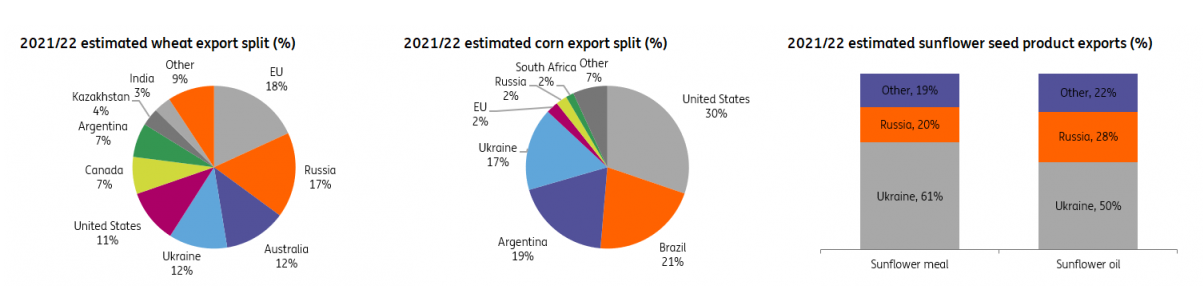
Russia accounts for 25% of the global natural gas market and 11% of the crude oil market. It is also the largest wheat exporter (China and India are still the largest producers, but Russia exports appreciably more). Together with Ukraine, also a powerhouse of agricultural exports, the two now enemies account for 29% of international annual wheat sales.
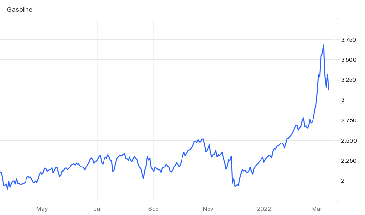
Wheat prices were already nearly double the five-year average shortly before the invasion; after February 24, they rose by another 30%. Today we are at a staggering 53% increase in wheat prices in just the last few months. We are at a 14-year peak. And the countries that import the most from Russia and Ukraine (such as Egypt or Indonesia) will bear the brunt of this crisis.
Together, Russia and Ukraine’s exports account for 12% of the world’s traded calories. The two countries account for almost 30 percent of global wheat exports, almost 20 percent of corn exports, and more than 80 percent of the world supply of sunflower oil. However, the compounded effect of embargo and devastation in the two countries will surely exert tremendous influence on the global economic outlook for years to come.
We need to be realistic about the coming months and years. Corn (where Ukraine accounts for 13% of global exports) and wheat will be severely hit by the war and its aftermath. This will compound all the pre-existing factors (transportation costs, supply chain slowdown, continuing weather disruptions, energy costs), none of which will trend down. Fertilizer prices have also gone up exponentially, and Russia – the largest exporter – has banned fertilizer exports at the beginning of March. The effects will be ultimately reflected in the cost of raw materials.
Ukraine and Russia have all but banned grains exports – either for security reasons or to protect internal needs. On top of this, the last harvests collected in Ukraine are now sitting in bins where ventilation and temperature controls have been affected by power cuts.
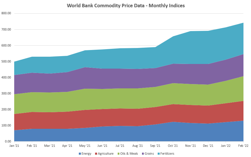
At the end of February, World Bank data already showed upward movement for nearly all categories; whatever was not trending up at that time is catching up fast. The last time things looked like this, experts warn, was in 2008-2009 – and social unrest followed around the world, to serious global consequences.
However, the perspective is not catastrophic and there is room to conserve profitability. The essential is to intervene with fast, targeted action that favors smart optimization, localization, and long-term planning.
What can feed producers do?
Most feed producers will be caught in the middle of all rising costs, from raw materials to transport and energy. Where, then, can they look for shelter when the storm hits?
Optimize feed costs without losing performance
One of the first things feed producers will focus on will be cutting down feed costs. At this point, it is essential that this basic optimization does not impact animal health and performance. Here is what should be kept in mind.
Preserve feed material and feed quality
Whatever raw materials you choose to use, minimizing losses and maintaining quality should be the first step. Losses caused by storage are often the easiest to mitigate.
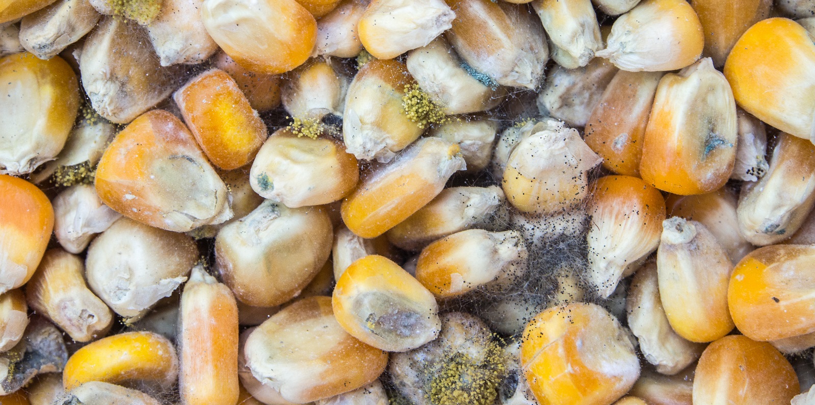
Quick intervention #1: Use mold inhibitors and mitigate the impact of mycotoxins
Compensate for lost nutrients (protein content, digestibility)
Freight costs will continue to cause pressure on transported raw materials, driving producers to local/regional options. When you replace one feed ingredient with a cheaper one, the first effects will be on the active principle and on the digestibility of the feed. Often something you are taking out of the diet cannot be replaced 1:1.
Quick intervention #2: Maximize the use of enzymes to ensure high feed digestibility; for poultry, pigments can replace corn-derived coloration (to control color variability)
Compensate for stress caused by diet changes
Adjusting the feed composition doesn’t only have effects on paper.
Even if you choose the best replacements, adjust the balance, compensate for loss of digestibility and optimize everything in every possible way, one thing remains:
The animal receives a new diet.
New diets are textbook stressors. But sometimes the nutritionist or the producer is so stressed that it is easy to overlook the stress placed inside the animal. Since animal efficiency is key for productivity, it is essential that the effects of diet stress are mitigated for the animal.
Quick intervention #3: Precautionary use of gut-health mitigating additives; also consider palatable feed materials and taste enhancers
Optimize production costs without losing quality
To optimize costs on the production floor, there are three essential areas where feed producers can act:
Saving on energy costs and reducing the carbon footprint
Reducing losses on the production floor
Increasing throughput without increasing manpower
To answer these challenges, there are solutions that can operate individually. More importantly in such times, there are products that can impact all three areas without negatively influencing the quality of output. One such solution, for instance, can decrease energy costs, increase throughput and pellet quality, and reduce fines.
Quick intervention #4: Choose a solution that satisfies 3/3 of your issues
Conclusion
Climate change will continue to wreak havoc on the predictability of harvests. Freight costs are projected to keep rising. And the costs of war and (hopefully) reconstruction will take a toll on the cost of living and cost of doing business around the world, for years to come.
In the storm that has already started, it is unwise to take shelter for a while and hope for good weather soon. Cutting down on ingredients here and additives there won’t keep profitability high in the long run. Feed producers must look at all aspects – from feed storage and composition to process improvement – and consider holistic measures that protect animals and profitability at the same time.
Mycotoxin interactions amplify damages – What are the right solutions?
Contamination with multiple mycotoxins is the rule for animal feeds, rather than the exception. Trial data shows that producers can prevent negative effects on animal health and performance by using high-performing toxin binders.
Multiple mycotoxins contaminate animal feed – problems and solutions
Mycotoxins pose an exceptional challenge for feed and animal producers. Generated by common molds, they occur in a great variety and numbers. Difficult to diagnose, mycotoxicosis in farm animals shows in a range of acute and chronic symptoms: decreased performance, feed refusal, poor feed conversion, reduced body weight gain, immune suppression, reproductive disorders, and residues in animal food products.
Regulatory mycotoxin thresholds don’t account for interactions
Regulatory thresholds for permissible mycotoxin levels in feed are derived from toxicological data on the effects of exposure of a certain species, at a certain production stage, to a single mycotoxin. This makes practical sense: while aflatoxins are carcinogens, fumonisins attack the pulmonary system in swine, for example. Mycotoxins also affect poultry in a different way than cattle, and broilers in a different way than breeders or laying hens, to mention more cases.
The problem is that, in reality, individual mycotoxin challenges are the exception. Animal diets are usually contaminated by multiple mycotoxins at the same time (Monbaliu et al., 2010; Pierron et al., 2016). Since 2014, EW Nutrition has conducted more than 50,000 mycotoxin tests on both raw material and finished feeds samples, across the globe. 85% of these samples were contaminated with more than one mycotoxin and one third positive for four or more mycotoxins.
How does contamination with multiple mycotoxins occur in animal feed?
The concurrent appearance of mycotoxins in feed can be explained as follows: each mold species has the capacity to produce several mycotoxins simultaneously. Each species, in turn, may infest several raw materials, leaving behind one or more toxic residue. In the end, a complete diet is made up of various raw materials with individual mycotoxin loads, resulting in a multitude of toxic challenges for the animals.
If animals were exposed to only one mycotoxin at a time, following the regulatory guidelines on maximum challenge levels would usually be enough to keep them safe. However, several studies have shown that the effects of exposure to multiple mycotoxins can differ greatly from the effects observed in animals exposed to a single mycotoxin (Alassane-Kpembi et al., 2015 & 2017). The simultaneous presence of mycotoxins may be more toxic than one would predict based on the known effects of the individual mycotoxins involved. This is because mycotoxins interact with each other. The interactions can be classified into three main different categories: antagonistic, additive, and synergistic (Grenier and Oswald, 2011).
Types of mycotoxin interactions
Additivity occurs when the effect of the combination equals the expected sum of the individual effects of the two toxins.
Synergistic interactions of two mycotoxins lead to a greater effect of the mycotoxin combination than would be expected from the sum of their individual effects. Synergistic actions may occur when the single mycotoxins of a mixture act at different stages of the same mechanism. A special form of synergy, sometimes called potentiation, occurs when one or both of the mycotoxins do not induce significant effects alone but their combination does. Fumonisin alone, for example, requires high levels to exerts effects on broiler performance. When aflatoxin is also in the feed, the effects are higher than those of aflatoxin alone (Miazzo et al., 2005)
Antagonism can be observed when the effect of the mycotoxin combination is lower than expected from the sum of their individual effects. Antagonism may occur when mycotoxins compete with one another for the same target or receptor site. In an in-vitro study using human colon carcinoma cells (HCT116), Bensassi and collaborators (2014), found that DON and Zearalenone individually caused a marked decrease of cell viability in a dose-dependent manner; when combined, the effect was drastically reduced.
Most of the mycotoxin mixtures lead to additive or synergistic effects. The actual consequences for the animal will depend on its species, age, sex, nutritional status, the dose and duration of exposure as well as environmental factors. What is clear is that mycotoxin interactions pose a significant threat to animal health and critically impede risk assessment.
From awareness to action: risk assessment and toxin binders
Given their complex interactions, the toxicity of combinations of mycotoxins cannot merely be predicted based upon their individual toxicities. Mycotoxin risk assessments have to consider that even low levels of mycotoxin combinations can harm animal productivity, health, and welfare. Feed and animal producers need to be aware of which raw materials are likely to be contaminated with which mycotoxins, be able to accurately link them to the risk they pose for the animal and consequently take actions before the problems appear in the field.
Trials demonstrate effectiveness of toxin mitigation solutions
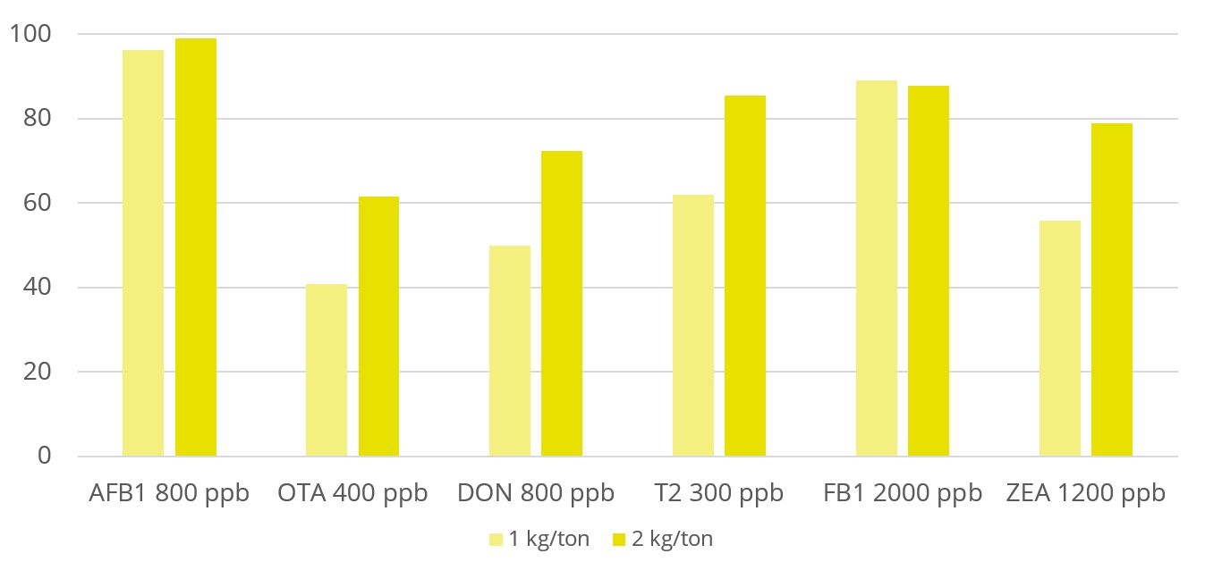
Toxin binders that are effective against a broad spectrum of mycotoxins significantly reduce the risks of mycotoxin exposure. In vitro trial data shows that EW Nutrition’s cost-effective toxin-mitigating product Solis Max shows a high mitigation capacity, even at low inclusion rates (Figure 1). Importantly, Solis Max helps to reduce various mycotoxins’ negative effects on performance without any negative effects on nutrient absorption.
Figure 1: Solis Max shows mitigation capacity in in vitro trial (%)
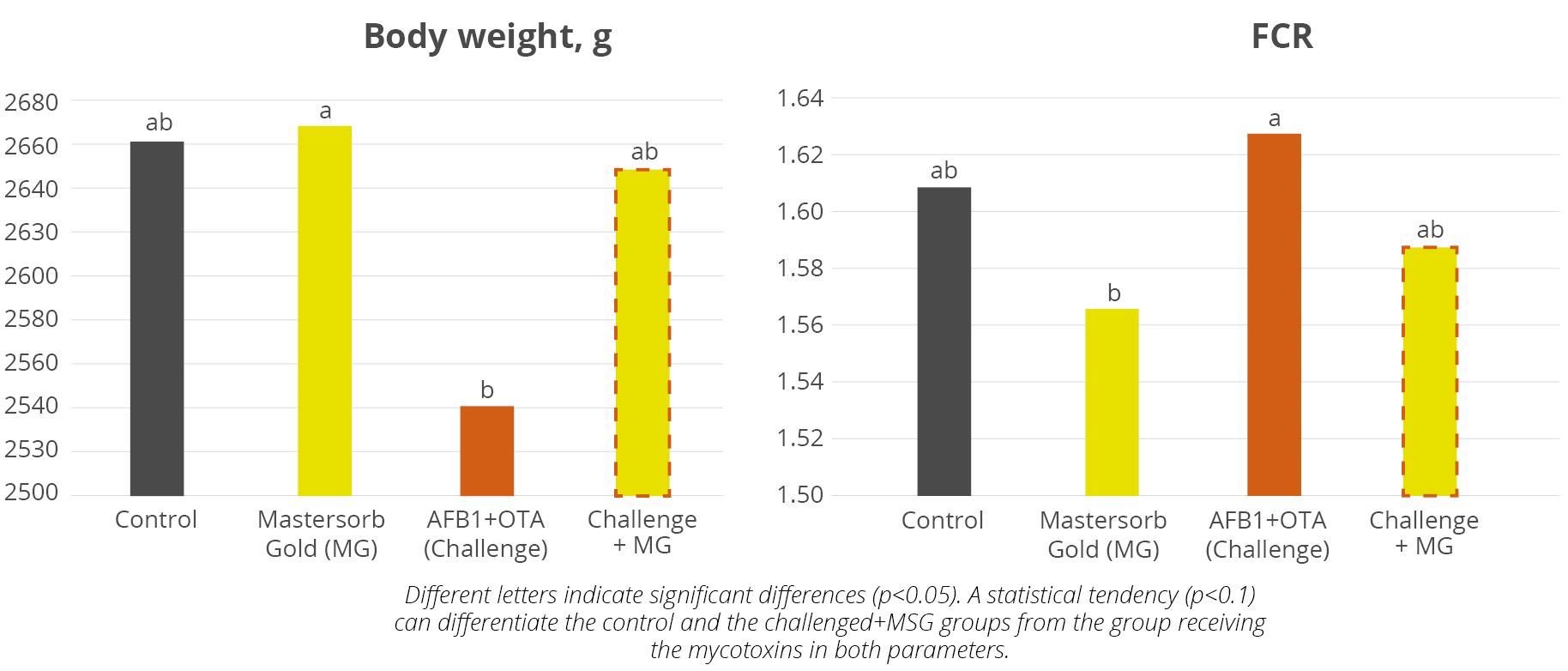
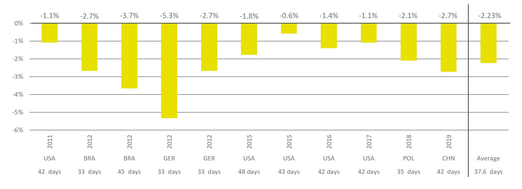
In a recent trial of 416 day-old Vencobb-430 broilers, premium product Mastersorb Gold has demonstrated its ability to support animals coping with multiple mycotoxin challenges. For broilers challenged with 200 ppb AFB1 and 350 ppb OTA, Mastersorb Gold supplementation resulted in 4.3% higher average daily weight gain than the challenged group, a higher body weight on day 42 and a 2% better feed conversion (Figure 2), which means a total recovery of the performance when compared with the non-challenged control.
Figure 2: Mastersorb Gold improves body weight and FCR of broilers challenged with AFB1 and OTA
Liver health also improved: after 21 days, broilers receiving Mastersorb Gold showed lower AST (-20%) and ALT (-50%) levels compared to the challenged group. Mycotoxin-induced stress was also lower, as evidenced by a 25% lower H/L ratio and 20% reduced white blood cell count for the Mastersorb Gold group. All of the mentioned biomarkers were similar to the non-challenged control, showing the preventive effects of Mastersorb Gold on health and performance.
Proactive management: tackle multiple mycotoxin challenges head on
Mycotoxins interactions are the norm, not the exception. Yet, regulatory standards currently only cover the effects of individual mycotoxins, leaving productions exposed to risks of additive and synergistic mycotoxin interactions animals’ health and performance. Luckily, management options are available: Careful risk evaluation explicitly includes the threat of multiple contaminations. And producers can proactively ensure better health, welfare and productivity of their animals by investing in the right toxin mitigation solution for their business.
References
Alassane-Kpembi, Imourana, Olivier Puel, and Isabelle P. Oswald. “Toxicological Interactions between the Mycotoxins Deoxynivalenol, Nivalenol and Their Acetylated Derivatives in Intestinal Epithelial Cells.” Archives of Toxicology 89, no. 8 (August 2015): 1337–46. https://doi.org/10.1007/s00204-014-1309-4.
Alassane-Kpembi, Imourana, Gerd Schatzmayr, Ionelia Taranu, Daniela Marin, Olivier Puel, and Isabelle Paule Oswald. “Mycotoxins Co-Contamination: Methodological Aspects and Biological Relevance of Combined Toxicity Studies.” Critical Reviews in Food Science and Nutrition 57, no. 16 (November 2017): 3489–3507. https://doi.org/10.1080/10408398.2016.1140632.
Bensassi, Fatma; Gallerne, Cindy; Sharaf el dein, Ossama; Rabeh Hajlaoui, Mohammed; Lemaire, Christophe and Bacha, Hassen. “In vitro investigation of toxicological interactions between the fusariotoxins deoxynivalenol and zearalenone” Toxicon 84 (2014): 1-6. https://doi.org/10.1016/j.toxicon.2014.03.005.
Grenier, B., and I. Oswald. “Mycotoxin Co-Contamination of Food and Feed: Meta-Analysis of Publications Describing Toxicological Interactions.” World Mycotoxin Journal 4, no. 3 (May 5, 2011): 285–313. https://doi.org/10.3920/wmj2011.1281.
Miazzo, R., M.F. Peralta, C. Magnoli, M. Salvano, S. Ferrero, S.M. Chiacchiera, E.C.Q. Carvalho, C.A.R. Rosa, and A. Dalcero. “Efficacy of Sodium Bentonite as a Detoxifier of Broiler Feed Contaminated with Aflatoxin and Fumonisin.” Poultry Science 84, no. 1 (January 2005): 1–8. https://doi.org/10.1093/ps/84.1.1.
Monbaliu, Sofie, Christof Van Poucke, Christ’l Detavernier, Frédéric Dumoulin, Mario Van De Velde, Elke Schoeters, Stefaan Van Dyck, Olga Averkieva, Carlos Van Peteghem, and Sarah De Saeger. “Occurrence of Mycotoxins in Feed as Analyzed by a Multi-Mycotoxin LC-MS/MS Method.” Journal of Agricultural and Food Chemistry 58, no. 1 (2010): 66–71. https://doi.org/10.1021/jf903859z.
Pierron, Alix, Imourana Alassane-Kpembi, and Isabelle P. Oswald. “Impact of Mycotoxin on Immune Response and Consequences for Pig Health.” Animal Nutrition 2, no. 2 (2016): 63–68. https://doi.org/10.1016/j.aninu.2016.03.001.
Global mycotoxin challenges: 2021 report
By Technical Team, EW Nutrition
Climate around the globe has changed, increasing atmospheric temperatures and carbon dioxide levels. This change favors the growth of toxigenic fungi in crops and thus increases the risk of mycotoxin contamination. When contaminating feed, mycotoxins exert adverse effects in animals and could be transferred into products such as milk and eggs.
*** Please download the full article for detailed information
Click to see the full-size image
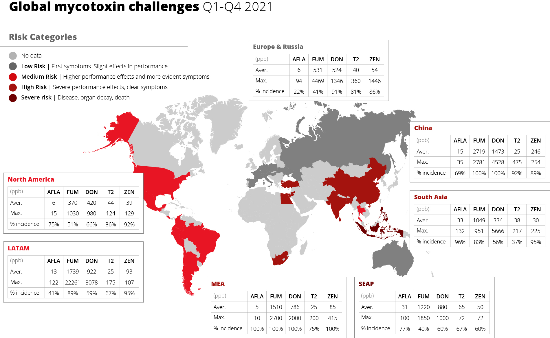
Mycotoxins: a worldwide challenge in 2021
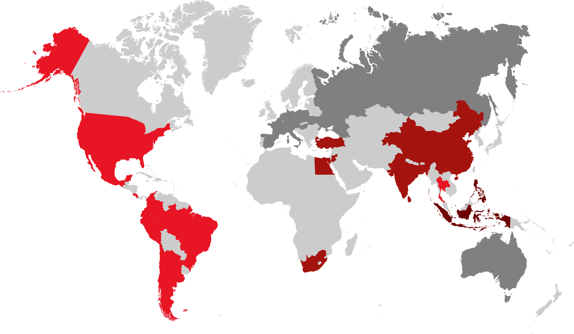
Amongst naturally occurring mycotoxins, the five most important ones are aflatoxin, ochratoxin, deoxynivalenol, zearalenone, and fumonisin. Their incidence varies with the different climates, the prevalence of plant cultures, the occurrence of pests, and the handling of harvest and storage. Worldwide, farmers faced various and sometimes extremely high mycotoxin contamination in their feed materials in 2021. In the following, we show the major challenges in five main regions.
Asia faced high aflatoxin contamination
In Asia, high temperatures and humidity favor Aspergillus growth in grains. As a result, 95 % of the samples in South Asia and three-quarters of the samples in the China and the SEAP region (Indonesia, Philippines, Vietnam) showed aflatoxin contamination. The average contamination being higher than the threshold for all farm animals represents an increased risk for their health and performance.
In China and the SEAP region, also DON and T-2 were highly prevalent. Showing an incidence of more than 60%, they pose a severe risk when combined with aflatoxin.
Fumonisins afflicted the LATAM region
In Mexico, Central and South America, fumonisin contamination prevailed with an incidence of almost 90% at average levels that can be considered risky for swine and dairy. Together with incidence levels of around 60% found for DON and T2, fumonisin may act synergically in the animals, raising the risk for health and performance.
The Fusarium species linked to these mycotoxin contaminations occur in the grains on the field. Amongst others, insect damage, droughts during growing, and rain at silking favor their development.
Trichothecenes prevailed in North America
Contamination with trichothecenes (DON and T2) is the rule in the United States. The interaction of these mycotoxins is at least additive. The damage they cause to the gut opens the door to dysbiosis and disease, decreasing performance and profitability.
Also in this case, the responsible molds for the contamination are Fusarium species that develop when grains are in the field. As with fumonisins, the molds are favored by insect damage, moderate to warm temperatures and rainfall.
Fusarium toxins contaminated grain in the MEA region
Fusarium toxins such as Fumonisin, DON, and T2 prevail in the region of Egypt, Jordan, and South Africa. In combination, these mycotoxins have additive effects at the intestinal level, which increases the risk of dysbiosis in poultry.
A challenging year with long-term repercussions
Since mycotoxin contamination affects animal health, measures must be taken to provide the best protection. Besides improving agricultural practices in the field, smart in-feed solutions and mold inhibitors can be used in stored grain. These measures help producers preserve feed quality after a troubled year for crops around the world.
Appetizing eggs with natural pigmentation: The new-generation solution
By Dr. Inge Heinzl, Editor, EW Nutrition
Eggs are an unparalleled source of nutrition for humans. Apart from being tasty and easy to cook, they are an essential ingredient for pasta, cakes, ice cream, and more. More importantly, they provide high-value proteins with amino acids we cannot produce, various B-vitamins, fat-soluble vitamins, and trace elements.
Assessing the value of the egg
The quality characteristics of eggs are usually divided into external features, such as:
· albumen weight
· Haugh unit (a measure of egg protein quality)
· yolk height,
· yolk diameter,
· albumen pH,
· yolk pH
· yolk color
For consumers, yolk color is probably the most important criterion for egg quality. Higher color intensity often is taken as indicating the good health of the laying hen.
Depending on the region or on the culture, people prefer more yellow or more orange yolks. In countries with traditional corn feeding, e.g., Mexico, they often like a deep yellow. In Northern Europe, consumers prefer a lighter yellow; in Southern Europe, more gold-orange yolks (see table 1).
Country
Yolk color fan value*
Belgium
12-13
Denmark
9-10
Finland
9-10
France
11-12
Germany
11-14
Greece
11
Italy
12-13
Netherlands
7-9
Austria
12-14
Portugal
12-14
Spain
11-14
Sweden
9-10
United Kingdom
10-11
Table 1. Egg pigmentation preferences – variation across European countries
* Values range from 1 (very pale yellow) to 16 (intense orange)
Egg yolk color is achieved via feed
The typical color of the yolk depends on pigments that are ingested with the feed. Corn and alfalfa meal provide the yellow pigments lutein and zeaxanthin, belonging to the xanthophylls, a sub-group of carotenoids. The golden-orange color is provided by red pigments from chili or paprika (Grashorn, 2008). Egg yolks start changing color about 48 h after the application of xanthophylls.
To reach an optimal yolk coloration in egg production, diets should be supplemented with yellow and red xanthophylls. Yellow xanthophylls achieve a correct yellow base coloration. The main yellow pigments used in poultry feeding are apoester, a synthetic carotenoid, and saponified marigold extracts, a natural alternative containing lutein and zeaxanthin. For the redness, paprika or chili offer natural sources; canthaxanthin is a nature-identical red xanthophyll.
For a long time, synthetic colorants were the substances of choice in the poultry industry because they provide consistently predictable results and high product stability. However, consumers’ preferences concerning food have shifted; demand favors natural over synthetic food ingredients. Moreover, current EU regulations restrict these synthetic molecules’ inclusion level due to their potentially harmful effects on human health if applied in excessive doses.
Carotenoid
Maximum inclusion level
Apoester (ethyl ester of β-apo-8’-carotenoic acid)
5 ppm
Canthaxanthin (β,β-Carotene-4,4′-dione)
8 ppm
Table 2. Maximum concentration allowed in feed for poultry production
Fortunately, there is already a natural, highly efficient option to replace apoester.
Lutein: a natural colorant, antioxidant, and provider of health benefits
One of these natural compounds is lutein, a lipophilic pigment. It is extracted from marigold petals, which contain up to 8.5 mg/g wet weight. Lutein is always accompanied by its isomer zeaxanthin.
Lutein – the yolk colorant
The use of xanthophylls such as lutein and zeaxanthin enables producers to safely control the color of the egg yolk and the broiler skin. In poultry, the carotenoids are deposited in high quantities in the epidermis, the fatty tissue, and the egg yolk. According to Grashorn (2016), between 4.4-23 % of dietary lutein and 23 % of dietary zeaxanthin are deposited in the egg yolk.
Lutein – the antioxidant protects the egg lipids
Another critical characteristic of lutein is its antioxidant effect. Egg yolks contain a high fat content. Therefore, they are very susceptible to lipid oxidation. Lutein, acting as an antioxidant, can prevent or at least limit lipid oxidation during egg processing. Kljak et al. (2021) compared different sources of pigments (basil, calendula, dandelion, marigold, and an industrial product containing canthaxanthin) concerning their antioxidant capacity. In this trial, marigold improved the yolks’ oxidative stability by 75 % compared to the control, with canthaxanthin showing no antioxidant effect. Kljak et al. attributed this effect to the carotenoids in the marigold extract.
Lutein – a value-added ingredient
Lutein and its isomer are nutritionally valuable and, therefore, welcome ingredients of the eggs. Once more, due to their antioxidant effects, they play an essential role in preventing and reducing cataracts and age-related eye dysfunctionalities in humans and animals (Landrum & Bone, 2001; Wang et al., 2016).
However, the amounts of antioxidant pigments in a standard egg are not very high (approx. 400 µg/egg). Compared to the total amount of antioxidants ingested, their importance for humans is only limited (Grashorn, 2008). The situation is different for functional eggs, which are widely sold in certain English-speaking countries. These eggs are enriched with n-3 fatty acids and with antioxidants such as ß-carotene (approx. 150 IE/egg).
Can natural pigments be as effective as synthetic apoester?
The precondition for the deposition of lutein in the egg or the skin is its absorption in the intestine. This absorption makes the difference between the synthetic apoester and the traditional yellow natural xanthophylls (lutein/zeaxanthin). In the case of traditional yellow xanthophylls, about three parts of the product are necessary to achieve the same effectiveness as one part of apoester.
With special technology owned by EW Nutrition, it is possible to improve the absorption of natural carotenoids and, therefore, the efficacy of lutein products. Only about 1.25 parts are then needed to replace one part of apoester.
Trial 1: A new generation of pigment products as effective as apoester
A trial was conducted in Spain to compare the effectiveness of apoester and a new generation natural pigment in combination with canthaxanthin.
For the trial, 288 layers (Hy-Line Brown, 39 weeks of age) were divided into 12 groups with 8 replications and 3 hens per replication. The trial consisted of a 7-week xanthophyll depletion and a 4-week experimental phase. The products included in the trial were a natural lutein product produced with a unique absorption-improving technology (Colortek Yellow, CTY), the synthetic xanthophyll apoester, and canthaxanthin. Three yolk color fan (YCF) targets were tested (10, 11, and 12).
For canthaxanthin, 1.5, 2.0, and 3.0 ppm were used. Within these groups of three different canthaxanthin concentrations, different concentrations of Colortek Yellow and apoester were applied to an otherwise xanthophyll-free diet:
The colors of the egg yolks were measured with the help of the DSM egg yolk color fan.
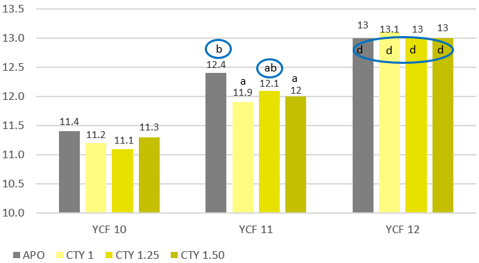
Figure 1 shows that Colortek Yellow at a 1.25 fold concentration as apoester (3.13 ppm) provided the same result as apoester regarding YCF target 11 (= canthaxanthin concentration of 2.00 ppm). In the case of YCF target 12 (= canthaxanthin concentration of 3.00 ppm), the same yolk color as apoester could be achieved using Colortek Yellow at a 1.25 or 1.5-fold concentration as apoester. Furthermore, it could be seen that the recommendations for apoester were overestimated and yielded color results 1 point above the target.
Figure 1. Egg yolk color values achieved by the use of apoester (APO) and different concentrations of Colortek Yellow (CTY) * a, b, c, d: different superscripts mean statistical difference (P<0.05)
Can lutein be as stable as synthetic pigments like apoester?
Another potential disadvantage of natural pigments is lower stability. By accelerating saponification in a continuous process, producing a product with low moisture and a high content of xanthophylls is possible. This process leads to higher stability of the product and prolongs the shelf life.
Trial 2: New generation pigment shows better stability than apoester
In this trial, the stability of products containing either a new generation natural colorant (Colortek Yellow) or apoester was tested. A vitamin-mineral premix containing 12.5 % choline chloride and one of the tested products were stored in closed bags at 30 °C and 75 % relative humidity. The recovery of the substances was tested after one, two, and three months.
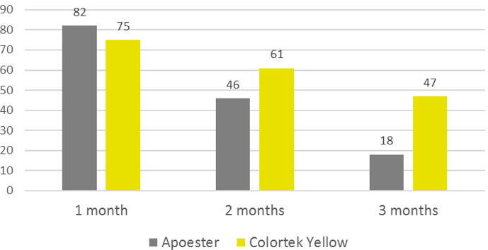
The trial shows higher recovery rates for Colortek Yellow than for apoester at a longer storage time (Figure 2). This new technology, therefore, provides natural pigments with higher stability than products containing synthetic apoester.
Figure 2. Recovery rates of apoester and Colortek Yellow after different times of storage (%)
New-generation natural pigments beat traditional synthetic options
The trend towards natural food ingredients also affects egg yolk color: consumers want natural alternatives to get their preferred yolk color, and regulators are imposing ever stricter limits on synthetic additives. Natural pigments have historically had two limiting characteristics compared to synthetic ones, their lower absorption and their lower stability. Due to new technologies, natural pigmentation products such as Colortek Yellow can now offer absorption rates comparable to apoester and even higher stability – making them the optimal replacement for synthetic colorants.
References
Grashorn, M. “Eiqualität.” In Legehuhnzucht und Eiererzeugung. Empfehlungen für die Praxis (special issue 322) edited by W. Brade, G. Flachowsky, and L. Schrader, 18-33. Landbauforschung – vTI Agriculture and Forestry Research, 2008
Grashorn, M. “Feed additives for influencing chicken meat and egg yolk color.” In Handbook on Natural Pigments in Food and Beverages. Industrial Applications for Improving Food Color, edited by R. Carle and R.M. Schweiggert, 283-302. Woodhead Publishing, 2016.
Kljak, K., K. Carović-Stanko, I. Kos, Z. Janječić , G. Kiš, M. Duvnjak, T. Safner, and D. Bedeković. “Plant carotenoids as pigment sources in laying hen diets: effect on yolk color, carotenoid content, oxidative stability and sensory properties of eggs.” Foods 10, no. 4 (2021):721
Wang, W., J., C. Moore, J. Jackson, and K. Narfström. “Antioxidant supplementation increases retinal responses and decreases refractive error changes in dogs.” J. Nutr. Sci. 5 e18 (2016): 7 pages
Climate across the globe has changed, with rising atmospheric temperatures and carbon dioxide levels. This change favors the growth of toxigenic fungi in crops and thus increases the risk of mycotoxin contamination. When contaminating feed, mycotoxins exert adverse effects in animals and could be transferred into products such as milk and eggs.
95% of the samples were contaminated with at least one mycotoxin
EW Nutrition constantly analyzes feed and raw material samples for their mycotoxin contamination. We report challenges from the most common mycotoxins hindering animal health around the globe.
Worldwide, more than 4,000 analyses on more than 1,000 samples were performed between June – October of the present year. The samples covered grain and by-products commonly used in animal feed worldwide. Figure 1 shows the percentage of the samples tested for which a positive result was found, detailing the number of mycotoxins per sample.
Click to enlarge
The number of mycotoxins analyzed per sample can vary based on regional risk-evaluation, including weather conditions, raw material origin and past frequency of positives. However, a minimum number of samples per region is always analyzed for the full spectrum, in order to monitor and corroborate the risk level.
3 or more mycotoxins per sample
95% of the samples were contaminated with at least one mycotoxin. In Europe and Latin America, most samples were analyzed for up to five mycotoxins, and were found contaminated with at least two. In South Asia, three mycotoxins were regularly analyzed per sample and most samples were positive for two. Worldwide, it is common to find samples with 3 or more mycotoxins, indicating that, even in raw materials, poly-contamination is the rule.
Aflatoxin: Main concern for South Asia
From all samples tested positively for mycotoxin contamination, 55% were contaminated with Aflatoxins. In all regions, the maximum levels lay over the thresholds for dairy and poultry. In Europe, less than 20% of the samples were contaminated with Aflatoxin. In Europe and the USA, the average contamination is low, hence this toxin can hardly be considered an issue for animal production in those areas (Figure 2).
In South Asia, where high temperatures and humidity are prevalent, Aflatoxin was detected in more than 95% of the samples and the average contamination is over all thresholds. Management strategies, such as the use of mold inhibitors for stored grain and toxin binders in feed, are necessary in this area to keep animals healthy and productive.
Click to enlarge
Impact
Aflatoxins have a negative impact on animal performance, as they affect the function of liver and kidney, alter the immune function, and impair protein synthesis. This affects weight gain, feed efficiency and mortality. Carryover into milk, eggs and edible organs is possible with high or chronic intake of the toxin.
Fumonisins: Main concern for LATAM, also global
Fumonisin was found in 70% of the samples globally and roughly in 90% of the samples coming from Latin America (figure 3). Moreover, in LATAM, more than 50% of the results have values over the threshold for dairy and swine, and 14% over the threshold for poultry, making it a great concern in the area. South Asia is the second concern area, with a high proportion of contaminated samples (80%) and 14% of them representing a danger for poultry production.
Click to enlarge
Impact
The main issue with the typical contamination levels of fumonisins – often considered of low risk – is their capacity to disrupt gut health. As their absorption is low, fumonisins interact with other toxins and the gut barrier components, including those affecting immunity and the microbiome. They are known to decrease the available surface for nutrient digestion and absorption, and to increase the risk and incidence of gut-related diseases. As a result, lower productivity is expected in animals exposed to even low levels of this toxin.
Deoxynivalenol (DON): Present worldwide
All across the regions, the maximum tested levels lay over the threshold for dairy, poultry, and swine. This trichothecene was found in more than 70% of the samples analyzed worldwide. In the United States, more than 75% of the positive tested samples showed a contamination with DON and the average of the positives exceeded the thresholds for swine and poultry.
The region with highest maximum values is LATAM, followed by South Asia, and the region with the highest frequency of positives in analyzed samples is Europe. Thus, it can be concluded that the worldwide frequency and levels in which DON is found represent a high risk for production animals.
Click to enlarge
Impact
Deoxynivalenol shows a broad spectrum of toxic effects in animals. In poultry and swine, for instance, this mycotoxin is related to lesions in the gastrointestinal tract and alterations in the immune response. This, in turn, leads to lower productivity and poor feed efficiency. DON also interacts with the microbiome, and it is known that it favors the colonization of coliform bacteria in pigs.
Ruminants can tolerate 10–20 times more DON than, for example, pigs. The majority of ingested DON is converted into the less toxic de-epoxy DON, but the degradation rate is influenced by different factors such as the diet, where high starch decrease the process. Moreover, DON also has a detrimental effect on rumen microorganisms, impacting its fermentative capacity.
T2: A danger for poultry producers word-wide
Average levels of T2 were over the threshold for poultry in all regions, with a high presence (>70% of the analyzed samples) in Europe, the US & LATAM.
Click to enlarge
Impact
T-2 s is a potent inhibitor of protein synthesis, which affects actively dividing cells, such as the lining of the gastrointestinal tract, skin, and immune cells. The consequences include weight loss or poor weight gain, diarrhea, skin and beak lesions, and decreased production.
T-2 is de-epoxidated in the rumen to HT-2 and neosolaniol, which are significantly less toxic than the parent toxin. In acidotic animals, rumen detoxification of T-2 toxin is impaired and animals may show gastroenteritis and intestinal hemorrhages.
Zearalenone: 80% positive tests globally
More than 80% of all samples tested for this mycotoxin were found positive. The maximum contaminations lay over the thresholds for dairy and swine. These high levels found should not be ignored, considering feedstuffs for long living and reproduction animals.
Click to enlarge
Impact
Especially in pig breeding, Zearalenone is an important issue, due to its high absorption and rapid biotransformation into more estrogenic components. Its structural similarity with 17β-estradiol leads this toxin to impair reproductive performance in cows and sows.
Recent studies point to interactions of Zearalenone with immune cells and organs in animals, leading to alterations in cell viability, proliferation, and functionality. Consequences are alterations of the immune response, enhancing the effects of other challenges.
A bad year for crops could be a bad year for production animals
The high mycotoxin contamination found so far in 2021 is partially explained by climate events, such as high temperature and humidity. Temperate zones such as Europe or parts of the USA tend to have higher contaminations compared with previous years.
Multiple mycotoxins co-occur, increasing their impact on animals. Certain combinations of mycotoxins are known to have synergistic or additive effects, aggravating their adverse effects.
To safeguard animal performance, it is important to continually strive for low levels of contamination and to manage the risk of mycotoxins through the use effective tools to measure, interpret, and manage the risk. MasterRisk can aid in the interpretation of mycotoxin risks, weighing in the animal species, age, purpose, as well as the mycotoxin exposure and interactions.
The 3 critical factors for successful pigmentation
By Predrag Persak, Regional Technical Manager, EW Nutrition
We eat with our eyes. Depending on our cultural background and our experience, we prefer foods that have a certain appearance. Moreover, we regulate our taste and health expectations based on this appearance. In that equation, color plays an essential role. Think of healthy-looking salad, fruit, eggs, meat, and more. Certain foods are more appetizing and appear healthier – and, in many cases, are indeed so – when they display a certain color.
For poultry producers, skin color and the yolk color of table eggs are of major concern. This concern is driven by the market (in certain regions, skin and yolk pigmentation heavily affect buying preferences), by regulations, and by an interest in using all options to increase product quality with natural solutions.
Where does poultry pigmentation come from?
Birds cannot synthesize pigments; they must take them up with their feed. Natural pigments have, besides their pigmenting properties, an antioxidant role in the bird’s organism. Unfavorable conditions can heavily influence the outcome of pigmentation. For producers looking to achieve reliable and consistent coloration, results are often unpredictable and disappointing.
Knowing the factors that affect pigmentation will help us to better understand how to achieve the desired level of pigmentation – or to identify, in hindsight what went wrong and when. In general, three different factors are decisive for efficient pigmentation:
The quality of the product (type, content, and stability of the pigment)
The amount of pigment ingested/absorbed/deposited
The persistence of the pigment in the final product
1. Product quality is essential
The first point to be considered is the quality of the product you use, including type, content, and stability of the pigment in the product and the feed.
Content and quality of active substances determine efficacy
Concerning type and content, what matters more than the total amount of carotenoids is the level of active substances. The trans-isomers have higher efficiency than the cis-isomers and are decisive for pigmentation.
Natural pigments originate from natural sources that often vary due to growth conditions, harvest, and handling. Therefore, producers need to control incoming materials and conduct proper formulation during the production process. This is crucial in order to obtain an adequate level of pigments for appropriate pigmentation.
Adequate measures ensure the stability of the pigment in the product
Natural pigments are sensitive to light and air; they are easily oxidized. Also in the feed formulation there are many substances (e.g. oxidized forms of trace elements, choline, chloride) enhancing the oxidation of the pigments. Some precautions can be taken to protect natural pigments from oxidation:
Use of adequate package materials preventing the exposure to light and air
Use of antioxidants in the product as well as in the feed formulation
With these measures in place, the pigments are given adequate protection to ensure their stability.
2. Pigment intake, absorption, and deposition affect pigmentation
Every factor reducing the amount of pigment reaching its target deteriorates the quality of pigmentation. Below are the crucial factors producers need to take into account.
Feed intake is correlated to pigment intake
Assuming that the pigment is homogeneously distributed in the feed, feed intake directly determines the intake of pigment. Consequently, anything that affects feed intake also affects pigment intake and pigmentation. To that end, what is also decisive is particle size and homogeneous distribution of the pigment in the product.
The energy concentration in the feed is also a critical factor. Antinutrients, unpleasant taste, or inconsistent feed structure negatively influence feed intake.
Feed intake is also influenced by other elements:
the animal’s health status
environmental conditions
the availability of water
the housing system (free-range, farm)
feeding management factors (length of the feeding lines, separation of the feed in silo bins or through the feeding lines etc.).
Saponification plays a role in pigment absorption
Through saponification, the natural, esterified form of the pigment gets broken down and the pigment is separated from the fatty acid molecule. This step is necessary to enable the pigment to pass the intestinal wall. The higher the saponification, the better the bioavailability of the pigment.
Besides improving bioavailability, saponification also influences the particle size and the homogeneous distribution of the pigment particles in the product.
Some feed materials and nutrients influence pigment absorption
If pigments are used, it is essential to know that some feed materials or nutrients have a beneficial or adverse effect on the absorption or deposition of the pigments. The inclusion of saturated, low-digestible fats or fat sources decreases pigment absorption and, therefore, the efficacy of pigmentation, whereas unsaturated fats (oils) facilitate it. The addition of oil up to 5% linearly increases pigment deposition in the egg.
Nutrients such as Calcium or Vitamin A also change pigment absorption. In the case of calcium, the level and the source are decisive. High levels of fast soluble limestone or calcium levels higher than 4 % will decrease the absorption. Also, increased levels of Vitamin A are critical for the effectiveness of deposition, as Vitamin A and the pigment use the same transporters. This fact is very important in broilers if vitamin A addition is applied through the water.
Mycotoxins affect feed intake and absorption
The presence of mycotoxins in feed, especially DON, will reduce feed intake due to the bad taste. The gut health-impacting effect of the mycotoxins will increase the passage rate of the feed and will prevent adequate absorption through the intestinal wall. Additionally, the liver function is negatively impacted by the mycotoxins. This results in an affected serum transport and a lower storage capacity for the pigments, leading to lower deposition in the tissue.
Impacted gut health is bad for pigmentation, too
Good gut health is essential for good pigmentation, including the uptake/absorption of pigments, their deposition, but also already existing pigmentation. All health challenges that negatively affect digestion and absorption, such as dysbiosis, negatively influence pigment availability and pigmentation. In such cases, products or strategies improving digestibility and gut integrity can be a solution.
Specific diseases such as NCD, Coryza, helminthiasis, as well as coccidiosis are an important consideration. The first three diseases lower pigment deposition; coccidiosis, however, has multiple impacts. It not only affects digestion and absorption and, therefore, the ongoing pigmentation but also decreases the already existing one.
Coccidia cause damage to the intestinal wall and affect its activity, resulting in a lower absorption. Additionally, the animals lose weight due to an insufficient supply of energy. The consequence is a degradation of fat tissue where the pigments are stored. Furthermore, coccidiosis means oxidative stress for the animal – triggering a reaction of the organism. As pigments also serve as antioxidants, they are removed from the fatty tissues and used as antioxidants.
Within three days post-infection, pigment levels in the subcutaneous tissues, but also in the serum and the liver, drop to 0. Coccidiosis outbreaks occur more frequently in alternative housing systems, affecting broilers, but also laying hens. Paying close attention to coccidiosis and having a proper anticoccidial program in place is obligatory for good pigmentation.
3. Pigmentation ends when the final products are on the shelf
For the end consumer, an attractive color in the final products (such as pasta or the broiler carcass) is essential. Producers of these final products request to put more pigments into the feed, but is this always the solution? As described before, there are a lot of factors possibly impacting the process of pigmentation during animal production on the farm.
However, also in the pasta factory or in the slaughterhouse, pigmentation of the final products can be impacted. In the pasta factory, oxidizing enzymes can destroy the pigments making the pasta pale and unattractive. If they have issues with Salmonella in the slaughterhouse, the birds may be scalded in slightly hotter water. The defeathering afterward can cause the loss of the upper layer of the skin with the pigments.
These examples show why pigmentation is not just the responsibility of the animal producer, but rather continues up to the moment when the pasta or meat is ready for the consumer.
Control these 3 factors for best pigmentation results
Pigmentation is a dynamic process that requires knowledge and attention. The better we control the influences, the more consistent and predictable the outcome. To that end, it is essential to use the product with the best quality, the best amount of pigment that can be not just ingested, but also absorbed and deposited, and with the best persistence in the final product and along its shelf life.
Keeping everything under control is not always possible or is extremely difficult. That is why choosing the right product is a vital link that will allow us to pay more attention to those things that we can find difficult to manage.
To meet all these demands, Colortek Yellow B is the best natural yellow pigment on the market. This highly concentrated natural yellow evidences optimal flowability, homogeneous mixing in feed and high stabilit, for reliable and consistent results. In addition, it boasts high bioavailability and is produced in the EU in a state-of-the art facility, with FAMI-QS certification and strict control of undesirable substances.
Reducing apo-esters: What are the alternatives?
By Technical Team, EW Nutrition
A year ago, the European Commission announced regulation (EU) 2020/1400 – restricting the use of ethyl ester of β-apo-8’-carotenoic acid (generally known as ‘apo-ester’). Starting on 26 October 2021, this legislation restricts the use of apo-ester in poultry feed to 5 mg/kg for laying hens and 15 mg/kg for broilers.
As apo-esters is a synthetic pigment – not naturally occurring in nature – this measure was taken because the authorities could not guarantee safety upon exposure to the user. Limiting the concentration in feed would reduce this risk to acceptable levels, according to the legislators’ decision.
Why use apo-esters in the first place?
Apo-ester is a synthetic yellow colorant, with good stability in premixtures and complete feed. It also has a high deposition rate in the yolk, making it an effective egg yolk colorant.
Its ability to be applied through premix facilitates the proper dispersion in the final feed, which is relevant if micro-dosing systems are lacking in the feed mill.
Why was the legislative change necessary?
The legislative change which limits the use of synthetic apo-ester is based on the precautionary principle and in line with a broader market trend: away from synthetic (non-natural) components, towards the use of naturally occurring alternatives.
The alternative to apo-ester
Natural yellow pigments, typically based on lutein and zeaxanthin produced from marigold oleoresin, are available in the market and can be used to reach the egg yolk pigmentation desired by the consumer. In contrast to apo-ester, these natural solutions are functional antioxidants, further contributing to the egg’s nutritious composition.
Challenges for natural alternatives
However, stability in premixtures and complete feed can be a challenge, with inconsistent yolk coloration as a risk. Safety can also be an issue, so it is important to ask for Quality Control measures routinely applied to avoid contamination with undesired substances (e.g., dioxins). To limit the risk of producing eggs with insufficient yolk coloration, it is important to select natural pigments with excellent stability and deposition efficiency.
What is the best natural alternative to apo-ester?
EW Nutrition’s natural pigment Colortek® Yellow B, produced with a proprietary technology, withstands the harsh conditions in premixtures, while the unique saponification process provides unparalleled deposition rates.
Moreover, Colortek® Yellow B is the most concentrated natural pigment on the market, making it the perfect premix-delivered colorant in the egg industry. If you want to produce all-natural eggs without worrying about the stability of the product or the reliability of your egg coloration, please contact your local EW Nutrition person.
Encapsulation: How a modern phytogenic feed additive makes all the difference
By Technical Team, EW Nutrition
Secondary plant extracts have been shown to improve digestion, have positive effects on intestinal health, and offer protection against oxidative stress in various scientific studies in recent years. Their use as a feed additive has become established and various mixtures, adapted to the various objectives, are widely available.
However, their use in pelleted feed has been criticized for some time. In particular, an unsatisfactory reproducibility of the positive influences on performance parameters is the focus of criticism. The causes invoked for the loss of quantifiable benefits are inadequately standardized raw materials, as well as uncontrollable and uneven losses of the valuable phytomolecules contained during compound feed production.
Delivery mechanisms influence product benefits
The animal production industry has long attempted to reduce its need for antibiotic drugs to an indispensable minimum. As a result, more natural and nature-identical feed additives have been used for preventive health maintenance. These categories include numerous substances that are known in human nutrition in the field of aromatic plants and herbs, or in traditional medicine as medicinal herbs.
The first available products of these phytogenic additives were simply added to compound feed. The desired parts of the plant were, like spices and herbs in human nutrition, crushed or ground into the premix. Alternatively, liquid plant extracts were placed on a suitable carrier (e.g. diatomaceous earth) beforehand in order to then incorporate them into the premix. These procedures are usually less than precise and may be responsible for the difficult reproducibility of positive results mentioned at the beginning.
Another negative factor that should not be underestimated is the varying concentration and composition of the active substances in the plant. This composition is essentially dependent on the site conditions, such as weather, soil, community and harvest time [Ehrlinger, 2007]. In an oil obtained from thyme, the content of the relevant phenol thymol can therefore vary between 30% and 70% [Lindner, 1987]. These extreme fluctuations are avoided with modern phytogenic additives through the use of nature-identical ingredients.
Effective encapsulation is key to stability
The loss of valuable phytomolecules under discussion can also be traced back to the natural origin of the raw materials. Some phytomolecules (e.g. cineole) are volatile even at low temperatures. In regular medicinal use, this effect is mainly employed with cold products. Thus essential oils, such as of mint and eucalyptus, can be added to hot water and inhaled via the resultant steam.
In the process of pelleting in compound feed production, temperatures between 60°C and 90°C are common, depending on the type of production. The process can last for several minutes until the cooling process is over. Sensitive additives can be easily inactivated or volatilized during this step.
A technical solution for the preservation of temperature-sensitive additives is using a protective cover. This is, for instance, an already established practice for enzymes. Such so-called encapsulation is already used successfully in high-quality products with phytogenic additives. The volatile substances should be protected by a coating with fat or starch so that the majority (>70%) of the ingredients can also be found after pelleting.
Unfortunately, complete protection is not possible with this capsule, as this simple protective cover can be broken open by mechanical pressure during grinding and pelletizing. New types of microencapsulation further reduce losses. In a sponge-like type of microencapsulation, if a capsule is destroyed, only a small proportion of the chambers filled with volatile phytomolecules are damaged.
High protection and recovery with Ventar D
A new type of encapsulation, developed by EW Nutrition for use in feed, delivers further optimization. Results show that the technology implemented in Ventar D ensures very high recovery rates of the sensitive phytomolecules even under demanding pelleting conditions.
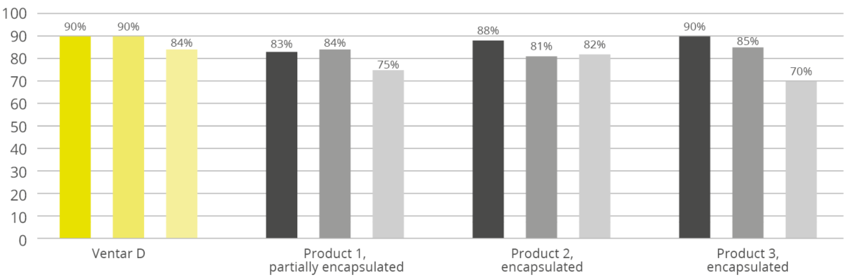
In a comparative study with encapsulated products established on the market, Ventar D was able to achieve the highest recovery rates in all three tested scenarios (70°C, 45 sec; 80°C, 90 sec; 90°C, 180 sec). In the stress test at a temperature of 90°C for 180 seconds, at least 84% of the valuable phytomolecules were recovered, while the comparison products varied between 70% and 82%. A constant recovery rate of 90% was achieved for Ventar D under simpler conditions.
Phytomolecule recovery rates under processing conditions, relative to mash baseline (100%)
Site-specific release of active ingredients
The major gastrointestinal pathogens (like Clostridia spp., Salmonella spp., E. coli, etc.) are present across the gastrointestinal tract after the proventriculus. This leads to infection or lesions at different sites of preference, reaching up to ceca. Any feed-based solution should have a profound antimicrobial effect. It is, however, also crucial that active ingredients are released across the gastrointestinal tract, for a better contribution to intestinal health.
The unique, innovative delivery system used for Ventar D specifically addresses this point, which many traditional coating technologies do not. Other encapsulation technologies tend to release the active ingredient either too early or too late (depending on the coating composition). The active ingredients in Ventar D reach across sites in the gastrointestinal tract and exert antimicrobial effects, supporting optimal gut health and improving performance.
Economically and ecologically sustainable
In the past, the losses mentioned in compound feed production and especially in pelleting were described as largely unavoidable. To obtain the desired effect of the valuable phytomolecules in the finished product, higher use of products was recommended and thus increased costs to the end-users and the associated CO2 footprint, lowering sustainability overall.
The modern encapsulation technology used in Ventar D now offers significantly better protection for the valuable phytomolecules and, in addition to the economic advantage, also offers more efficient use of the resources required for production.
References
Hashemi, S. R .; Davoodi, H .; 2011; Herbal plants and their derivatives as growth and health promoters in animal nutrition; Vet Res Commun (2011) 35: 169-180; DOI 10.1007 / s11259-010-9458-2; Springer Science + Business Media BV, 2011
Ehrlinger, M., 2007: Phytogenic additives in animal nutrition. Inaugural dissertation. Munich: Veterinary Faculty of the Ludwig Maximilians University in Munich.
Lindner, U., 1987: Aromatic plants – cultivation and use. Contribution to the special show – Medicinal and Spice Plants (Federal Garden Show 1987), Teaching and Research Institute for Horticulture Auweiler-Friesdorf, Düsseldorf.
Nutrition and feeding in ABF poultry production
By T.J. Gaydos
Management practices and feed additive selection are often discussed when working in antibiotic-free (ABF) poultry production. Nutrition is another critical component of any agricultural animal system. Working with a qualified nutritionist will help ensure that the diet is correctly formulated with high-quality ingredients.
5 nutrition tips for antibiotic-free poultry production
1. Consider feed form and delivery
Feed form and delivery are nearly as important as the nutrient content of the formulation. If feed form or handling is improper and feed separates, is improperly mixed, or oxidized, the birds will not appreciate the effort that went to develop a balanced diet. A durable pellet or crumble is important to allow all birds to have equal access to a nutritionally complete diet with every bite.
Additionally, if the finished feed or individual ingredients are not stored properly, they may not have the same value that is attributed to them in the formulation process. Other than correct nutrient formulation, three parts of the diet that should be considered are feed additives, mycotoxin contamination, and lipid oxidation.
2. Prevent oxidative stress
The impact of oxidative stress on the intestinal mucosa, immune system, and performance is well-documented across species. Oxidized fat sources reduce the available energy, but equally significant to bird health is the reduction in vitamin availability, resulting in increased oxidative stress for the animal. Protecting the sources of fat and the finished feed is important to spare fat-soluble vitamins, specifically vitamin E.
Oxidized fat can also irritate the intestinal mucosa leading to decreased absorption of nutrients. The process of breaking down macromolecules during digestion and converting them to forms useful for further metabolism is a significant contributor to oxidative stress. The immune system is also a great contributor to oxidative stress. Immune cells use reactive oxygen species to kill pathogens that are phagocytosed.
A large portion of the immune system is located in the GI tract in order to protect the animal from pathogens crossing from the gut into the animal. In addition to being a contributor to oxidative stress, the immune system can be negatively impacted by oxidized feed (Liang et al., 2015). The combination of metabolic and immune activity in the intestines puts it at a high risk of damage from oxidative stress. It is vital to protect fat sources with synthetic or natural antioxidants; reducing the incoming stress from oxidized fat should be a priority to improve poultry health.
3. Mitigate mycotoxin risks
Another risk to bird health and mucosal integrity is mycotoxins. Diets containing mycotoxins may damage the mucosa of the GI tract directly or may damage other organs leading to significant health challenges and decreases in performance. Some mycotoxins or compounds created by fungi can disrupt the intestinal microflora by acting on bacterial cells, as many fungal metabolites are antimicrobial.
The best approach to managing mycotoxins is eliminating them from the system by purchasing high-quality grain and storing it appropriately. It is impossible to completely eliminate all risks of receiving ingredients contaminated with mycotoxins. An internal program should be developed to test the incoming ingredients and finished feed regularly for mycotoxins.
Knowing the challenging ingredient sources may help reduce the risk to highly susceptible birds like Breeders or chicks through dilution in formulation or the addition of toxin binders and/or enzymes. Several toxins may be found in a feed stuff and many of the mycotoxins are synergistic in their deleterious effects (Murugesan et al., 2015). Different binders have varying affinity for different mycotoxins; closely examining the product literature can help to choose the correct product to mitigate risk.
4. Choose optimal additives
Choosing the correct feed additive program for intestinal health, food safety, and growth performance depends on the specific challenges in the complex. When selecting a feed additive that is not FDA approved, it is important to base the decision as much as possible on scientific evidence through peer-reviewed research.
In addition to published data, internal testing within the production system is also helpful to ensure the product matches the local challenge. In a market saturated with “natural” products, it is essential to find a supplier that is trustworthy and is engaged in the success of the complex and health of the birds, not only in selling products. A partnership will be much more successful in the long term than only a buy/sell arrangement.
5. Manage expectations
When considering removing antibiotics from a program, the temptation is to expect natural products to completely replace the efficacy of antibiotics. This is an unreasonable expectation. The success of a transition to ABF production relies on modifying management practices as well. The vast majority of program success is related to attention to the details of husbandry, biosecurity, and sanitation. The remaining opportunity to improve health rests on the additive program.
References
Liang, Fangfang, Shouqun Jiang, Yi Mo, Guilian Zhou, and Lin Yang. “Consumption of Oxidized Soybean Oil Increased Intestinal Oxidative Stress and Affected Intestinal Immune Variables in Yellow-Feathered Broilers.” Asian-Australasian Journal of Animal Sciences 28, no. 8 (2015): 1194–1201. https://doi.org/10.5713/ajas.14.0924.
Murugesan, G.R., D.R. Ledoux, K. Naehrer, F. Berthiller, T.J. Applegate, B. Grenier, T.D. Phillips, and G. Schatzmayr. “Prevalence and Effects of Mycotoxins on Poultry Health and Performance, and Recent Development in Mycotoxin Counteracting Strategies.” Poultry Science 94, no. 6 (2015): 1298–1315. https://doi.org/10.3382/ps/pev075.
How animal nutrition can contribute to sustainability
By Dr. Inge Heinzl, Editor, EW Nutrition
Nowadays, the whole world is talking about sustainability. Many efforts aim to maintain our world for future generations, creating a balance between our current needs and those of our children, grandchildren, and great-grandchildren. The right animal nutrition choices play a crucial role in achieving the challenging aim of sustainable animal production.
Animal nutrition solutions can support producers’ sustainability contributions, from animal welfare to antibiotic reduction
Sustainability – an old concept now set out in writing
The idea of sustainability is not new. Already the first humans lived sustainably, taking only as much as they needed and the environment could cope with, using all parts of the animals they killed. The German Hannss Carl von Carlowitz (1645-1714) coined the term sustainability in his oeuvre “Sylvicultura oeconomica” to counter a threatening raw material crisis. Wood was one of the most important raw materials. Besides heating, it was used for shipbuilding and mining. This was the reason that extensive areas in Europe were deforested and became deserted. Observing the impending disaster, von Carlowitz ” (1713) stated that only as many trees should be felled as can grow back through planned reforestation, sowing, and planting.
The Brundtland Report (1987), a document created by the World Commission on Environment and Development, is reckoned to be the starting signal for worldwide discussions about sustainability. In 2015, the result of a meeting of 193 members of the United Nations was the Agenda 2030 with 17 sustainable development goals for a “world we want” that should be achieved by 2030.
Sustainable Development Goals (SDG) of the Agenda 2030, fixed by the UN in 2015
How can the feed sector contribute to sustainability?
The animal nutrition industry’s sustainability efforts play into different SDGs, notably no. 2, zero hunger, no. 3, good health and well-being, no. 12, responsible consumption and production, no. 13, climate action, no. 14, life below water, and no. 15, life on land. In addition to the overarching goal of fostering higher animal welfare (cf. Keeling et al., 2019), the feed sector’s measures center on three areas:
Optimal use of feed resources, which includes optimizing feed conversion, preserving feed quality, and using alternative ingredients
Preserving the environment by reducing ammonia and methane emissions and energy requirements
Reducing antibiotics usage to maintain their efficacy for future generations
1. Make best use of available resources
One of the 17 points on the list of the United Nations is “responsible consumption and production”. For the feed industry, this means making the most out of available feed sources. Improvements in feed conversion, the maintenance of feed quality, and the use of alternative ingredients are all part of this.
Optimize FCR to utilize the available feed best
The feed conversion rate shows the amount of feed consumed in relation to the outputs produced, such as weight gain, eggs, or milk. The better or lower the feed conversion rate (FCR), the less feed you need to achieve your target, and the higher the yield. Products that improve feed conversion, therefore, can help to save resources.
Good feed conversion or an optimal utilization of nutrients depends on gut health. Only a healthy gut can digest the feed and absorb the nutrients adequately. Hence, products to improve feed conversion often do so by improving gut health.
Phytomolecules: proven to improve feed conversion
Herbs and their active components have been used in human and veterinary medicine for thousands of years to treat digestive tract diseases. Nowadays, products based on phytomolecules help improve feed conversion through their digestive, anti-inflammatory, and antimicrobial effects on the intestinal tract.
How do these three characteristics contribute to a better FCR?
Phytomolecules stimulate the secretion of digestive juices and the motility of the gut
Their antimicrobial effect supports a “healthy” balance in the microbiome, preventing damages of the gut wall by harmful microbes and, therefore, maintaining an optimal nutrient absorption
Their anti-inflammatory properties also contribute to good nutrient absorption and reduce endogenous nutrient loss
FCR improvements in broilers thanks to ACTIVO found in several studies
As phytomolecules are often volatile, EW Nutrition offers encapsulated phytomolecule-based products for the feed (ACTIVO product line). During episodes of elevated enteric challenge, e.g., weaning or following feed change, a liquid solution (ACTIVO LIQUID) can be applied via the waterline.
Enzymes help to make nutrients available
Some feed materials are hard to digest for certain animals. For example, pigs’ digestive systems do not have the enzymes required to break down non-starch polysaccharides (NSPs), such as cellulose, hemicellulose (ß-glucans and xylans), pectins or oligosaccharides. However, pig feed ingredients usually contain these substances.
Besides the non-usability of NSPs, the cage effect is a further problem. Cellulose and hemicellulose, water-insoluble NSPs, encage nutrients such as proteins or digestible carbohydrates. Encaged nutrients cannot be reached by the digestive enzymes and don’t become available to the animal.
Xylanases are available on the market to degrade structural substances in the feed and make them, as well as the nutrients they encaged, available for the organism.
Maintain the quality of your feed materials
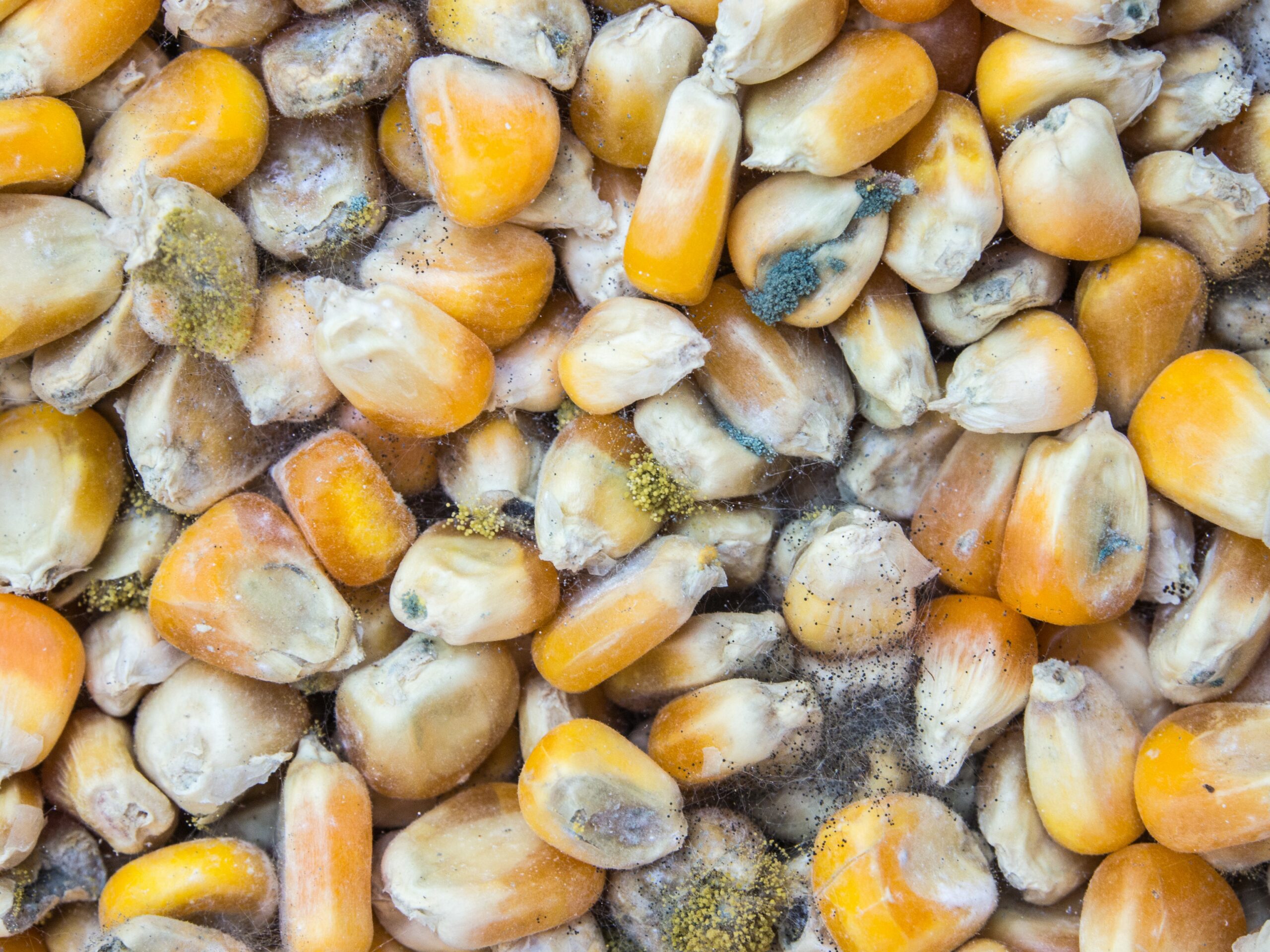
Another possibility to save resources is the maintenance of feed quality. Bad weather conditions at harvest or incorrect storage can downgrade feed quality due to the development of molds and their mycotoxins or the oxidation of nutrients. Products mitigating the adverse effects of toxins, acidifiers that reduce microbial load, and antioxidants can help to keep your feed quality on a high level – or to re-establish it.
Mitigate the adverse effects of mycotoxins
Feed materials contaminated with mycotoxins harm animals in different manners and should not be used without further treatment. Mycotoxins are not visible – even if no molds are visible, mycotoxins might be present. Additionally, they are pH- and thermo-stable, meaning that mycotoxins produced in the raw materials on the field remain in the finished feed. As mycotoxins often do not cause apparent, specific symptoms but manifest in decreased performance, feed refusal or lower feed intake, and higher disease susceptibility, it is difficult to notice contamination.
Products such as SOLIS or MASTERSORB contain clay minerals (bentonite and montmorillonite) that adsorb the toxins. MASTERSORB GOLD and MASTERSORB FM also include toxin-adsorbing yeast cell walls and herbal substances to help protect the liver.
Feed spoilage through molds, yeasts, and mycotoxins wastes precious resources
Reduce microbes in the feed with acidifiers
Acidifiers based on organic acids counter harmful microbes in the feed in two ways. Most pathogenic bacteria are susceptible to low pH. The proliferation of, e.g., E. coli, Salmonella, and Clostridium perfringens is minimized at pH < 5 (cf. Fuller 1977). Acidic-tolerant beneficial bacteria such as Lactobacilli or Bifidobacterium, however, survive.
Other than antimicrobial activity, organic acids also cause a significant reduction in ammonia (Eriksen et al., 2014). This finding could be due to a reduction in the microbial deamination of amino acids, which would then be available for absorption, resulting in increased nitrogen digestibility and reduced ammonia excretion, as observed in monogastrics fed organic acids (Pearlin et al., 2020).
The acidifier product lines ACIDOMIX, FORMYCINE, and PRO-STABIL all help protect feed from contamination with pathogenic microorganisms.
Protect the feed’s nutrients from oxidation
The oxidation of nutrients in the feed decreases its nutritional value and, thereby, the value of the whole diet. Fat, proteins, fat-soluble vitamins, pigments, and other biologically active molecules, including sugars and phospholipids, can get oxidized. Metal ions and other pro-oxidative factors can affect the ingredients of the feed during mixing, storage, and feeding. The oxidation of fats and fat-soluble vitamins results in color changes or odors and – this is even more serious – in the production of harmful substances such as aldehydes and ketones. An oxidized feed can lead to oxidative stress in the animals, reduce their immunity, productivity, and livability.
To protect valuable ingredients, the timely addition of effective antioxidants such as STABILON is recommended.
Use alternatives to natural protein sources
Soybeans are an excellent source of protein in animal nutrition. During the last 50 years, soy production has increased from 27 million tons to 269 million tons, causing environmental degradation of forests and savannas (WWF, 2021). The use of alternative protein sources helps protect our environment.
Ruminants partly cover their protein requirements with the help of rumen bacteria. These bacteria can turn nitrogen from urea into bacterial protein, provided they receive enough energy available from carbohydrates. Thanks to its encapsulation, PROTE-N, a feed-grade urea-based nitrogen source, slowly releases nitrogen into the rumen, synchronized with the energy supply. PROTE-N affords producers a degree of independence from soybean protein without compromising nutritional quality.
Reducing soybeans in ruminant feeds helps to lower their environmental impact
2. Preserve the environment
Animal production generates gases such as ammonia and methane that negatively impact the environment. Measures to reduce these gases help to protect plants, animals, us, and our globe.
Reduce ammonia by improving protein digestion
Besides nitrogen oxides, ammonia is one of the primary sources of nitrogen pollution. Ammonia damages ecological systems through acidification and nutritional oversupply. Fast-growing plants that need high amounts of nitrogen or plants that tolerate low soil pH proliferate, whereas more susceptible plants disappear, decreasing biodiversity. According to Max-Planck-Gesellschaft (2017), reducing ammonia emissions by 50 % could prevent 250.000 deaths caused by fine dust worldwide per year.
Improved protein digestion in animals reduces their ammonia production. Decreasing the intestinal pH through using organic acid-based products such as ACIDOMIX or FORMYCINE is essential for the activation and correct functioning of the enzymes responsible for protein digestion.
Reduce methane, the second most abundant greenhouse gas
Together with CO2, N2O, and three fluorinated gases, methane belongs to the greenhouse gases listed in the Kyoto protocol. Being over 25 times more potent than carbon dioxide at trapping heat in the atmosphere, it dramatically affects the earth’s temperature and the climate system (United States Environmental Protection Agency). Methane is a final product of feed fermentation in the rumen and is produced by methanogenic bacteria. Ruminants can produce 250-500 L methane per day (Johnson & Johnson, 1995).
Reducing methane production in ruminants is a critical step towards climate protection. Herbal substances can change the microbiome, leading to improved protein and fiber degradation and reduced methane production (Ku-Vera et al., 2020). ACTIVO PREMIUM is a phytomolecules-based product for ruminants that helps reduce their methane emissions.
Energy savings
To preserve the environment, reducing energy needs is also an important topic. Using the surfactant SURF-ACE in the pelletizing process, feed mills can cut 10-15 % of their energy consumption or produce up to 10-15 % higher pellet output without increasing their energy consumption. When moisture is added together with the surfactant, the emulsion of the dietary fat and the added water leads to better general lubrication of the machinery and improved press throughput.
Feed mill efficiency is key to animal nutrition’s carbon footprint
3. Reduce antibiotic use in animal production to keep this tool effective
Point 3 on the UN’s Agenda 2030 is good health and well-being. For many years, antibiotics, a very effective weapon, have been used to fight bacterial diseases. However, the occurrence of resistance is increasing. One of the reasons is the inappropriate use of antibiotics. These substances are often used preventively or for viral diseases against which they are ineffective. Also, the use of antibiotics as growth promoters at low dosages in animal production strongly contributed to the development of antimicrobial resistance.
Limiting antibiotic use to therapeutic treatment is possible through good farm management and feed supplements that support animals’ gut health, immune systems, and respiratory health. For this purpose, solutions ranging from phytomolecules (ACTIVO products, GRIPPOZON) to egg immunoglobulins (GLOBIGEN products, PROTEGG), products mitigating the impact of toxins (MASTERSORB products, SOLIS), beta-glucans/MOS (BGMOS), and acidifiers (ACIDOMIX, FORMYCINE) are available.
The feed sector has the tools to achieve more sustainability!
The animal nutrition industry provides many products to support animal producers in coping with their main challenges, including the shift to more sustainable production practices. Solutions exist to save feed resources, better protect the environment, and keep antibiotic tools effective. As an additional reward, implementing sustainability solutions leads to healthy animals with high performance. Let’s all help to preserve this planet for our next generations!
References
Eriksen, J., Nørgaard, J. V., Poulsen, H. D., Poulsen, H. V., Jensen, B. B., & Petersen, S. O. (2014). Effects of Acidifying Pig diets on emissions of AMMONIA, methane, and sulfur FROM Slurry during storage. Journal of Environmental Quality, 43(6), 2086–2095. https://doi.org/10.2134/jeq2014.03.0108
Fuller, R. (1977). The importance of lactobacilli in maintaining normal microbial balance in the crop. British Poultry Science, 18(1), 85–94. https://doi.org/10.1080/00071667708416332
Johnson, K. A., & Johnson, D. E. (1995). Methane emissions from cattle. Journal of Animal Science, 73(8), 2483–2492. https://doi.org/10.2527/1995.7382483x
Keeling, Linda, Håkan Tunón, Gabriela Olmos Antillón, Charlotte Berg, Mike Jones, Leopoldo Stuardo, Janice Swanson, Anna Wallenbeck, Christoph Winckler, and Harry Blokhuis. “Animal Welfare and the United Nations Sustainable Development Goals.” Frontiers in Veterinary Science 6 (October 10, 2019). https://doi.org/10.3389/fvets.2019.00336.
Ku-Vera, J. C., Jiménez-Ocampo, R., Valencia-Salazar, S. S., Montoya-Flores, M. D., Molina-Botero, I. C., Arango, J., Gómez-Bravo, C. A., Aguilar-Pérez, C. F., & Solorio-Sánchez, F. J. (2020). Role of secondary plant metabolites on enteric methane mitigation in ruminants. Frontiers in Veterinary Science, 7. https://doi.org/10.3389/fvets.2020.00584
Pearlin, B. V., Muthuvel, S., Govidasamy, P., Villavan, M., Alagawany, M., Ragab Farag, M., Dhama, K., & Gopi, M. (2020). Role of acidifiers in livestock nutrition and health: A review. Journal of Animal Physiology and Animal Nutrition, 104(2), 558–569. https://doi.org/10.1111/jpn.13282
United Nations. (n.d.). How your company can advance each of THE SDGS: UN Global Compact. How Your Company Can Advance Each of the SDGs | UN Global Compact. https://www.unglobalcompact.org/sdgs/17-global-goals.
von Carlowitz, H. C. (1713). Sylvicvltvra oeconomica, oder, Hausswirthliche Nachricht und Naturmässige Anweisung zur Wilden BAŬM-ZŬCHT: Nebst gründlicher darstellung, wie Zu FÖRDERST durch Göttliches Benedeyen Dem allenthalben und insgemein einreissenden Grossen Holtz-mangel: Vermittelst Säe-pflantz- und Versetzung Vielerhand Bäume zu prospiciren …: Worbey zugleich eine Gründliche nachricht von den in Churfl. Sächss. Landen gefundenen Turff Dessen Naturliche beschaffenheit, Grossen NÜTZEN, Gebrauch und nutzlichen verkohlung, Aus Liebe Zu BEFÖRDERUNG des Algemeinen Bestens beschrieben. Verlegts Johann Friedrich Braun.