Endotoxins in 250 words

E coli

Dr Inge Heinzl, EW Nutrition

Endotoxins are… toxic, of course. The part “endo” in their name means that they are part of the bacterial cell, or, to specify it, they are part of the outer membrane of Gram-negative bacteria such as E. coli, Salmonella, Shigella, Klebsiella, and Pseudomonas.

When do they occur?

Always. Endotoxins are released with the lysis of bacteria, e.g., at the end of their life cycle, due to the effective immune defense of the host or treatments. The other possibility is bacterial growth as the membrane gets restructured and the endotoxins (or lipopolysaccharides -LPS-) are liberated.

What is the problem?

The “normal” occurrence (animals and humans always have Gram-negative bacteria in their gut) does not matter, because gut cells do not have receptors to recognize them as a danger in their apical side. However, when the barrier function is compromised, they pass into the bloodstream.

The liver still detoxifies small amounts. The problem comes with higher amounts of endotoxins in the bloodstream. Then, they provoke a strong immune reaction, feed intake drops, and nutritional resources are shifted from growth and production to immunity. These performance drops affect the profitability of the farmer.

What can be done?

Use broad-spectrum toxin binders that

    • contain clay minerals showing high affinity and selectivity against endotoxins
    • contain yeast cell walls, which, in addition to their binding capacity, support the immune response through macrophage activation and are involved in modulating microflora and bacterial load from the intestine
    • provide adequate liver protection.


Decoding the connection between stress, endotoxins, and poultry health

Poultry

By Marisabel Caballero, Global Technical Manager Poultry, EW Nutrition

Stress can be defined as any factor causing disruptions to homeostasis, which triggers a biological response to regain equilibrium. We can distinguish four major types of stressors in the poultry industry:

  • Technological: related with management events and conditions
  • Nutritional: involving nutritional disbalances, feed quality and feed management
  • Pathogenic: comprising health challenges.
  • Environmental: changes in environment conditions

In practical poultry production, multiple stress factors occur simultaneously. Their effects are also additive, leading to chronic stress. The animals are not regaining homeostasis and continuously deviate the use of resources through inflammation and the gut barrier-function, thus leading to microbiome alteration. As a consequence, welfare, health, and productivity are compromised.

What are endotoxins?

Bacterial lipopolysaccharides (LPS), also known as endotoxins, are the main components of the outer membrane of all Gram-negative bacteria and are essential for their survival. LPS have direct contact with the bacteria’s surroundings and function as a protection mechanism against the host’s immunological response and chemical attacks from bile salts, lysozymes, or other antimicrobial agents.

Gram-negative bacteria are part of animals’ microbiota; thus, there are always LPS in the intestine. Under optimal conditions, this does not affect the animals, because intestinal epithelial cells are not responsive to LPS when stimulated from the apical side. In stress situations, the intestinal barrier function is impaired, allowing the passage of endotoxins into the blood stream. When LPS are detected by the immune system either in the blood or in the basolateral side of the intestine, inflammation and changes in the gut epithelial structure and functionality occur.

The gut is critically affected by stress

Even when there is no direct injury to the gut, signals from the brain can modify different functions of the intestinal tract, including immunity. Stress can lead to functional disorders, as well as to inflammation and infections of the intestinal tract. Downstream signals act via the brain–gut axis, trigger the formation of reactive oxygen and nitrogen species as well as local inflammatory factors, and circulating cytokines, affecting intestinal homeostasis, microbiome, and barrier integrity.

Stress then results in cell injury, apoptosis, and compromised tight junctions. For this reason, luminal substances, including toxins and pathogens, leak into the bloodstream. Additionally, under stress, the gut microbiome shows and increment on Gram-negative bacteria (GNB). For instance, a study by Minghui Wang and collaborators (2020) found an increase of 24% in GNB and lower richness, in the cecum of pullets subjected to mild heat stress (increase in ambient temperature from 24 to 30°C).

Both these factors, barrier damage and alterations in the microbiome, facilitate the passage of endotoxins into the blood stream, which promotes systemic chronic inflammation.

What categories of stress factors trigger luminal endotoxins’ passage into the bloodstream?

Technological stress

Various management practices and events can be taken as stressors by the animals’ organism. One of the most common examples is stocking density, defined as the number of birds or the total live weight of birds in a fixed space. High levels are associated with stress and loss of performance.

A study from the Chung-Ang University in 2019 found that broilers with a stocking density of 30 birds/m2 presented two times more blood LPS than birds kept at half of this stocking density. Moreover, the body weight of the birds in the high-density group was 200g lower than the birds of the low-density group. The study concluded that high stocking density is a factor that can disrupt the intestinal barrier.

Nutritional stress

The feed supplied to production animals is designed to contribute to express their genetic potential, though some feed components are also continuous inflammatory triggers. Anti-nutritional factors, oxidized lipids, and mycotoxins induce a low-grade inflammatory response.

For instance, when mycotoxins are ingested and absorbed, they trigger stress and impair immunity in animals. Their effects start in gastrointestinal tract and extend from disrupting immunity to impairing the intestinal barrier function, prompting secondary infections. Mycotoxins can increase the risk of endotoxins in several ways:

A study conducted by EW Nutrition (Figure 1) shows an increase in intestinal lesions and blood endotoxins after a mycotoxin challenge of 200pbb of Aflatoxin B1 + 360ppb Ochratoxin in broilers at 21 days of age. The challenged birds show two times more lesions and blood endotoxins than the ones in the unchallenged control. The use of the right mitigation strategy, a product based on bentonite, yeast cell walls, and phytogenics (EW Nutrition GmbH) successfully prevented these effects as it not only mitigates mycotoxins, but also targets endotoxins in the gut.

Figure 1
Figure 1 Blood LPS and intestinal lesion score of broilers challenged with 200ppb AFB1 + 350 ppb OTA from 1 to 21 days of age without and with an anti-toxin product from EW Nutrition GmbH (adapted from Caballero et al., 2021)

Pathogenic stress

Intestinal disease induces changes in the microbiome, reducing diversity and allowing pathogens to thrive. In clinical and subclinical necrotic enteritis (NE), the intestinal populations of GNB, including Salmonella and E.coli also increases. The lesions associated with the pathogen compromise the epithelial permeability and the intestinal barrier function, resulting in translocation of bacteria and LPS (Figure 5) into the bloodstream and internal organs.

Figure 2
Figure 2 Increase in systemic LPS (vs a healthy control) after a NE challenge (adapted from Chen et al., 2015, Li et al., 2018 & Liu at al., 2018)

Environmental stress

Acute and chronic heat and cold stress increases gut permeability, by increasing intestinal oxidative stress and disrupting the expression of tight junction proteins. This results in the damage and destruction of intestinal cells, inflammation, and imbalance of the microbiota. An increased release and passage of endotoxins has been demonstrated in heat stress (Figure 3), as well as a higher expression of TLR-4 and inflammation.

Figure 3
Figure 3 Systemic LPS increase (in comparison with a non-stressed control) after different heat stress challenges in broilers: 16°C increased for 2, 5 and 10 hours (Huang et al., 2018); 9°C increased for 24 and 72 hours (Nanto-Hara et al., 2020); 10°C continuously for 3 and 10 days, and 15°C 4 hours daily for 3 and 10 days (Alhenaky et al., 2017)


Zhou and collaborators (2021) showed that 72 hours of low temperature treatment in young broilers increased intestinal inflammation and expression of tight junction proteins, while higher blood endotoxins indicate a disruption of the intestinal barrier. As a consequence, the stress decreased body gain and increased the feed conversion rate.

An experiment conducted by EW Nutrition GmbH with the objective of evaluating the ability of a toxin mitigation product to ameliorate heat-stress induced LPS. For the experiment, 1760 Cobb 500 pullets were divided into two groups, and each was placed in 11 pens of 80 hens, in a single house. One of the groups received feed containing 2kg/ton of the product from the first day. From week 8 to week 12, the temperature of the house was raised 10°C for 8 hours every day.

Throughout the heat stress period, blood LPS (Fig 4) was lower in the pullets receiving the product, which allowed lower inflammation, as evidenced by the lower expression of TLR4 (Fig. 5). Oxidative stress was also mitigated with the help of the combination of phytomolecules in the product, obtaining 8.5% improvement on serum total antioxidant capacity (TAC), supported by an increase in in superoxide dismutase (SOD glutathione peroxidase (GSH) and a decrease in malondialdehyde (MDH).

Fig

Figures 4-5
Figures 4 and 5 – Blood LPS and expression of toll-like receptor 4 (TLR4) in lymphocites of pullets before (wk 6) and during heat stress (wk 9 and 10). (*) indicates significant differences (P<0,05), and (‡) a tendency to be different against the control group (P<0,1).

In practice: there is no silver bullet

In commercial poultry production, a myriad stressors may occur at the same time and some factors trigger a chain of events that work to the detriment of animal health and productivity. Reducing the solution to the mitigation of LPS is a deceitfully simplistic approach. However, this should be part of a strategy to achieve better animal health and performance. In fact, EW Nutrition’s toxin mitigation product alone helped the pullets to achieve 3% improvement in body weight and 9 points lower cumulative feed conversion (Figure 6).

Figure 6
Figure 6

Keeping the animals as free of stress as possible is a true priority for poultry producers, as it promotes animal health as well as the integrity and function of the intestinal barrier. Biosecurity, good environment, nutrition and good management practices are crucial; the use of feed additives to reduce the consequences of unavoidable stress also critically supports the profitability of poultry operations.


Masked mycotoxins – particularly dangerous for dairy cows

By Si-Trung Tran, SEAP Regional Technical Manager, EW Nutrition

Marisabel Caballero, Global Technical Manager Poultry, EW Nutrition, and
Inge Heinzl, Editor, EW Nutrition

Mycotoxins are secondary metabolites of fungi, commonly found as contaminants in agricultural products. In some cases, these compounds are used in medicine or industry, such as penicillin and patulin. In most cases, however, they are considered xenobiotics that are toxic to animals and humans, causing the disease collectively known as mycotoxicosis. The adverse effects of mycotoxins on human and animal health have been documented in many publications. Aflatoxins (AFs) and deoxynivalenol (DON, vomitoxin) are amongst the most critical mycotoxins affecting milk production and -quality.

Aflatoxins do not only affect cows

Aflatoxins (AFs) are highly oxygenated, heterocyclic difuranocoumarin compounds produced by Aspergillus flavus and Aspergillus parasiticus. They colonize crops, including many staple foods and feed ingredients. Within a group of over 20 AFs and derivatives, aflatoxin B1 (AFB1), B2, G1, and G2 are the most important naturally occurring compounds.

Among the aflatoxins, AFB1 is the most widespread and most toxic to humans and animals. Concern about mycotoxin contamination in dairy products began in the 1960s with the first reported cases of contamination by aflatoxin M1 (AFM1), a metabolite of AFB1 formed in the liver of animals and excreted in the milk.

There is ample evidence that lactating cows exhibit a significant reduction in feed efficiency and milk yield within a few days of consuming aflatoxin-contaminated feed. At the cellular level, aflatoxins cause degranulation of endoplasmic membranes, loss of ribosomes from the endoplasmic reticulum, loss of nuclear chromatin material, and altered nuclear shapes. The liver, as the organ mainly dealing with the decontamination of the organism, gets damaged, and performance drops. Immune cells are also affected, reducing immune competence and vaccination success (Arnold and Gaskill, 2023).

DON reduces cows’ performance

Another mycotoxin that can also reduce milk quality and affect metabolic parameters, as well as the immune function of dairy cows, is DON. DON is produced by different fungi of the Fusarium genus that infect plants. DON synthesis is associated with rainy weather from crop flowering to harvest. Whitlow and co-workers (1994) reported the association between DON and poor performance in dairy herds and showed decreased milk production in dairy cows fed 2.5 mg DON/kg. However, in cows fed 6 to 12 mg DON/kg dry matter for 10 weeks, no DON or its metabolite DOM-1 residues were detected in milk.

Masked mycotoxins hide themselves during analysis

Plants suffering from fungal infestations and thus confronted with mycotoxins convert the harmful forms of mycotoxins into less harmful or harmless ones for themselves by conjugation to sulfates, organic acids, or sugars. Conjugated mycotoxins cannot always be detected by standard analytical methods. However, in animals, these forms can be released and transformed into parent compounds by enzymes and microorganisms in the gastrointestinal tract. Thus, the feed may show a concentration of mycotoxins that is still below the limit value, but in the animal, this concentration is suddenly much higher. In dairy cows, the release of free mycotoxins from conjugates during digestion may play an important role in understanding the silent effects of mycotoxins.

Fusarium toxins, in particular, frequently occur in this “masked form”. They represent a serious health risk for animals and humans.

Aflatoxins first show up in the milk

Masked aflatoxins may also play a role in total aflatoxin contamination of feed materials. Research has harvested little information on masked aflatoxins that may be present in TMR ingredients. So far, metabolites such as Aflatoxin M2 have been identified (Righetti, 2021), which may reappear later in milk as AFM1.

DON-related symptoms without DON?

Sometimes, animals show DON-related symptoms, with low levels detected in the feed or raw materials. Besides sampling errors, this enigma could be due to conjugated or masked DON, which is structurally altered DON bound to various compounds such as glucose, fatty acids, and amino acids. These compounds escape conventional feed analysis techniques because of their modified chemical properties but can be released as their toxic precursors after acid hydrolysis.

Masked DON was first described in 1984 by Young and co-workers, who found that the DON content of yeast-fermented foods was higher than that of the contaminated wheat flour used in their production. The most plausible reason for this apparent increase was that the toxin from the wheat had been converted to a compound other than DON, which could be converted back to DON under certain conditions. Since this report, there has been much interest in conjugated or masked DON.

Silage: masked DON is a challenge for dairy producers

Silage is an essential feed for dairy cows, supporting milk production. Most silage is made from corn and other grains. The whole green plant is used, which can be infected by fungi. Since infection of corn with Fusarium spp. and subsequent DON contamination is usually a major problem in the field worldwide, a relatively high occurrence of this toxin in silage must be expected. The ensiling process may reduce the amount of Fusarium fungi, but the DON formed before ensiling is very stable.

Corn Silage

Silage samples show DON levels of concern

It is reasonable to assume that the DON biosynthesized by the fungi was metabolized by the plants to a new compound and thus masked DON. Under ensiling conditions, masked DON can be hydrolyzed, producing free DON again. Therefore, the level of free DON in the silage may not reflect the concentration measured in the plants before ensiling.

A study analyzed 50 silage samples from different farms in Ontario, Canada. Free DON was found in all samples, with levels ranging from 0.38 to 1.72 µg/g silage (unpublished data). Eighty-six percent of the samples contained DON at concentrations higher than 0.5 µg/g. Together with masked DON, it poses a potential threat to dairy cattle.

Specific hydrolysis conditions allow detection

However, in the natural ensiling process, the conditions for hydrolysis of masked DON are not optimal. The conditions that allow improved analysis of masked DON were recently described. This method detected masked DON in 32 of 50 silage samples (64%) along with free DON, increasing DON concentration by 23% in some cases (unpublished data).

Mycotoxins impact humans and animals

Aflatoxins, as well as DON, have adverse effects. In the case of DON, the impact on the animal is significant; in the case of aflatoxin, the possible long-term effects on humans are of higher relevance.

DON has more adverse effects on the animal and its performance

Unlike AFs, DON may be found in milk at low or trace concentrations. It is more associated with negative effects in the animal, altered rumen fermentation, and reduced flow of usable protein into the duodenum. For example, milk fat content was significantly reduced when cows were fed 6 µg DON/kg. However, the presence of DON also indicates that the feed probably contains other mycotoxins, such as zearalenone (ZEA) (estrogenic mycotoxin) and fusaric acid (pharmacologically active compound). All these mycotoxins may interact to cause symptoms that are different or more severe than expected, considering their individual effects. DON and related compounds also have immunosuppressive effects, resulting in increased somatic cell counts in milk. The U.S. FDA has established an action level for DON in wheat and wheat-derived products intended for cows, which is 5µg DON/g feed and the contaminated ingredient must not exceed 40% of the ration.

Aflatoxins decrease milk quality and pose a risk to humans

Aflatoxins are poorly degraded in the rumen, with aflatoxicol being the main metabolite that can be reconverted to AFB1. Most AFs are absorbed and extensively metabolized/hydrolyzed by enzymes found mainly in the liver. This results in the formation of AFM1, a part of which is conjugated to glucuronic acid and subsequently excreted in the bile. The other part enters the systemic circulation. It is either excreted in urine or milk. AFM1 appears within 12-48 hours after ingestion in cow’s milk. The excreted amount of AFM1 in milk from dairy cows usually ranges from 0.17% to 3% of the ingested AFB1. However, this carryover rate may vary from day to day and from one milking to the next in individual animals, as it is influenced by various factors, such as feeding regime, health status, individual biotransformation capacity, and, of course, by actual milk production. Carryover rates of up to 6.2% have been reported in high-yielding dairy cows producing up to 40 liters of milk per day.

In various experiments, AFM1 showed both carcinogenic and immunosuppressive effects. Accordingly, the International Agency for Research on Cancer (IARC) classified AFM1 as being in Group 2B and, thus, possibly carcinogenic in humans. The action level of 0.50 ppb and 0.05 ppb for AFM1 in milk is strictly adhered to by the U.S. Food and Drug Administration (FDA) and the European Food Safety Authority (EFSA), respectively.

Trials show the high adsorption capacity of Solis Max

A trial was conducted at an independent laboratory located in Spain. The evaluation of the performance of Solis Max was executed with the following inclusion levels:

  • 0.10% equivalent to 1.0 kg of Solis Max per ton of feed
  • 0.20% equivalent to 2.0 kg of Solis Max per ton of feed

A phosphate buffer solution at pH 7 was prepared for the trial to simulate rumen conditions. Each mycotoxin was tested separately, preparing solutions with known contamination (final concentration described in the table below). The contaminated solutions were divided into 3 parts: A positive control, 0.10% Solis Max and 0.20% Solis Max. All samples were incubated at 41°C for 1 hour, centrifuged, and the supernatant was analyzed for the mycotoxin added to determine the binding efficacy. All analyses were carried out by high-performance liquid chromatography (HPLC) with standard detectors.

Mycotoxin Contamination Level (ppb)
Aflatoxin B1 800
DON 800
Fumonisin B1 2000
ZEA 1200

Results:
The higher concentration of Solis max showed a higher adsorption rate for most mycotoxins. The high dose of Solis Max adsorbed 99% of the AFB1 contamination. In the case of DON, more than 70% was bound. For fumonisin B1 and zearalenone, Solis max showed excellent binding rates of 87.7% and 78.9%, respectively (Figure 1).

FigureFigure 1: Solis Max showed a high binding capacity for the most relevant mycotoxins

Another trial was conducted at an independent laboratory serving the food and feed industry and located in Valladolid, Spain.

All tests were carried out as duplicates and using a standard liquid chromatography/mass spectrometry (LC/MS/MS) quantification. Interpretation and data analysis were carried out with the corresponding software. The used pH was 3.0, toxin concentrations and anti-mycotoxin agent application rates were set as follows (Table 1):

TableTable 1: Trial set-up testing the binding capacity of Solis Plus 2.0 for several mycotoxins in different contamination levels

Results:

Under acidic conditions (pH3), Solis Plus 2.0 effectively adsorbs the three tested mycotoxins at low and high levels. 100% binding of aflatoxin was achieved at a level of 150ppb and 98% at 1500ppb.In the case of fumonisin, 87% adsorption could be reached at 500ppb and 86 for a challenge with 5000ppb. 43% ochratoxin was adsorbed at the contamination level of 150ppb and 52% at 1500ppb.

FigureFigure 2: The adsorption capacity of Solis Plus 2.0 for three different mycotoxins at two challenge levels

Mycotoxins – Effective risk management is of paramount importance

Although the rumen microflora may be responsible for conferring some mycotoxin resistance to ruminants compared to monogastric animals, there are still effects of mycotoxins on rumen fermentation and milk quality. In addition, masked mycotoxins in feed present an additional challenge for dairy farms because they are not readily detectable by standard analyses.

Feeding dairy cows with feed contaminated with mycotoxins can lead to a reduction in milk production. Milk quality may also deteriorate due to an adverse change in milk composition and mycotoxin residues, threatening the innocuousness of dairy products. Dairy farmers should therefore have feed tested regularly, consider masked mycotoxins, and take action. EW Nutrition’s MasterRisk tool provides a risk evaluation and corresponding recommendations for the use of products that mitigate the effects of mycotoxin contamination and, in the end, guarantee the safety of all of us.


Toxin Mitigation 101: Essentials for Animal Production

By Monish Raj, Assistant Manager-Technical Services, EW Nutrition
Inge Heinzl, Editor, EW Nutrition  

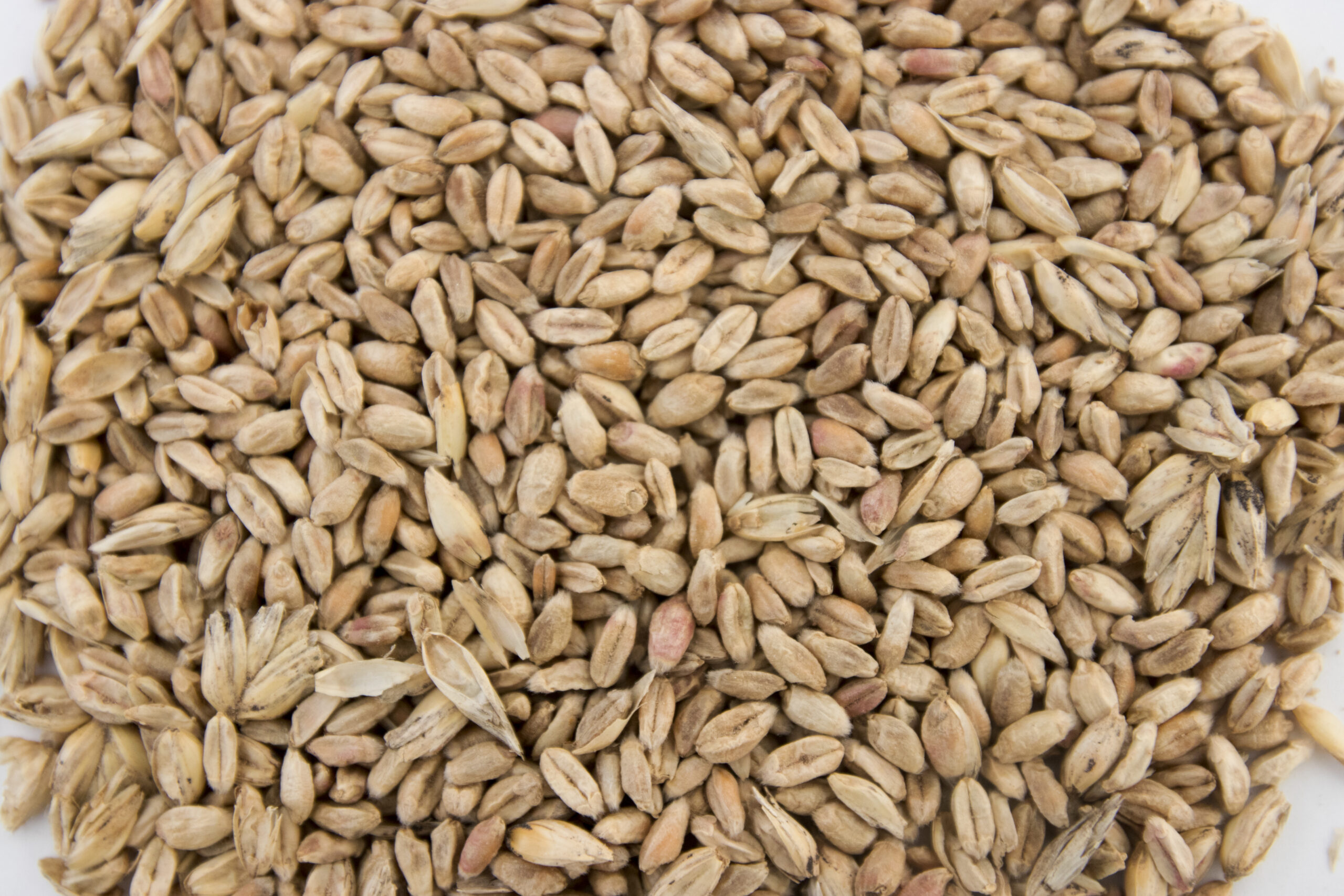
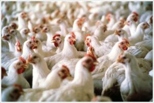
Mycotoxins, toxic secondary metabolites produced by fungi, are a constant and severe threat to animal production. They can contaminate grains used for animal feed and are highly stable, invisible, and resistant to high temperatures and normal feed manufacturing processes. Mycotoxin-producing fungi can be found during plant growth and in stored grains; the prevalence of fungi species depends on environmental conditions, though in grains, we find mainly three genera: Aspergillus, Penicillium, and Fusarium. The most critical mycotoxins for poultry production and the fungi that produce them are detailed in Fig 1.

FigureFigure 1: Fungi species and their mycotoxins of worldwide importance for poultry production (adapted from Bryden, 2012).

The effects of mycotoxins on the animal are manifold

When, usually, more than one mycotoxin enters the animal, they “cooperate” with each other, which means that they combine their effects in different ways. Also, not all mycotoxins have the same targets.

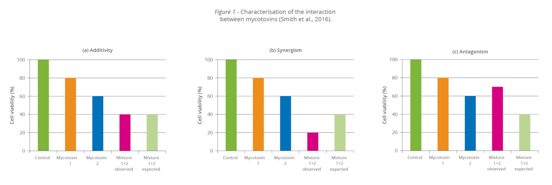
The synergistic effect: When 1+1 ≥3

Even at low concentrations, mycotoxins can display synergistic effects, which means that the toxicological consequences of two or more mycotoxins present in the same sample will be higher than the sum of the toxicological effects of the individual mycotoxins. So, disregarded mycotoxins can suddenly get important due to their additive or synergistic effect.

Table 1: Synergistic effects of mycotoxins in poultry

Synergistic interactions
DON ZEN T-2 DAS
FUM * * *
NIV * * *
AFL * *

Table 2: Additive effects of mycotoxins in poultry

Additive interactions
AFL T2 DAS MON
FUM + + + +
DON + +
OTA + +

Recognize the effects of mycotoxins in animals is not easy

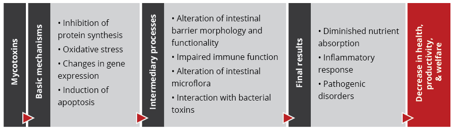
The mode of action of mycotoxins in animals is complex and has many implications. Research so far could identify the main target organs and effects of high levels of individual mycotoxins. However, the impact of low contamination levels and interactions are not entirely understood, as they are subtle, and their identification requires diverse analytical methods and closer observation.

With regard to the gastrointestinal tract, mycotoxins can inhibit the absorption of nutrients vital for maintaining health, growth, productivity, and reproduction. The nutrients affected include amino acids, lipid-soluble vitamins (vitamins A, D, E, and K), and minerals, especially Ca and P (Devegowda and Murthy, 2005). As a result of improper absorption of nutrients, egg production, eggshell formation, fertility, and hatchability are also negatively influenced.

Most mycotoxins also have a negative impact on the immune system, causing a higher susceptibility to disease and compromising the success of vaccinations. Besides that, organs like kidneys, the liver, and lungs, but also reproduction, endocrine, and nervous systems get battered.

Mycotoxins have specific targets

Aflatoxins, fumonisins, and ochratoxin impair the liver and thus the physiological processes modulated and performed by it:

  • lipid and carbohydrate metabolism and storage
  • synthesis of functional proteins such as hormones, enzymes, and nutrient transporters
  • metabolism of proteins, vitamins, and minerals.

For trichothecenes, the gastrointestinal tract is the main target. There, they hamper digestion, absorption, and intestinal integrity. T-2 can even produce necrosis in the oral cavity and esophagus.

Figure Main Targets Of Important MycotoxinsFigure 2: Main target organs of important mycotoxins

How to reduce mycotoxicosis?

There are two main paths of action, depending on whether you are placed along the crop production, feed production, or animal production cycle. Essentially, you can either prevent the formation of mycotoxins on the plant on the field during harvest and storage or, if placed at a further point along the chain, mitigate their impact.

Preventing mycotoxin production means preventing mold growth

To minimize the production of mycotoxins, the development of molds must be inhibited already during the cultivation of the plants and later on throughout storage. For this purpose, different measures can be taken:

Selection of the suitable crop variety, good practices, and optimal harvesting conditions are half of the battle

Already before and during the production of the grains, actions can be taken to minimize mold growth as far as possible:

  • Choose varieties of grain that are area-specific and resistant to insects and fungal attacks.
  • Practice crop rotation
  • Harvest proper and timely
  • Avoid damage to kernels by maintaining the proper condition of harvesting equipment.

Optimal moisture of the grains and the best hygienic conditions are essential

The next step is storage. Here too, try to provide the best conditions.

  • Dry properly: grains should be stored at <13% of moisture
  • Control moisture: minimize chances of moisture to increase due to condensation, and rain-water leakage
  • Biosecurity: clean the bins and silos routinely.
  • Prevent mold growth: organic acids can help prevent mold growth and increase storage life.

Mold production does not mean that the war is lost

Even if molds and, therefore, mycotoxins occur, there is still the possibility to change tack with several actions. There are measures to improve feed and support the animal when it has already ingested the contaminated feed.

1.    Feed can sometimes be decontaminated

If a high level of mycotoxin contamination is detected, removing, replacing, or diluting contaminated raw materials is possible. However, this is not very practical, economically costly, and not always very effective, as many molds cannot be seen. Also, heat treatment does not have the desired effect, as mycotoxins are highly heat stable.

2.    Effects of mycotoxins can be mitigated

Even when mycotoxins are already present in raw materials or finished feed, you still can act. Adding products adsorbing the mycotoxins or mitigating the effects of mycotoxins in the organism has been considered a highly-effective measure to protect the animals (Galvano et al., 2001).

This type of mycotoxin mitigation happens at the animal production stage and consists of suppressing or reducing the absorption of mycotoxins in the animal. Suppose the mycotoxins get absorbed in the animal to a certain degree. In that case, mycotoxin mitigation agents help by promoting the excretion of mycotoxins, modifying their mode of action, or reducing their effects. As toxin-mitigating agents, the following are very common:

Aluminosilicates: inorganic compounds widely found in nature that are the most common agents used to mitigate the impact of mycotoxins in animals. Their layered (phyllosilicates) or porous (tectosilicates) structure helps “trap” mycotoxins and adsorbs them.

  • Bentonite / Montmorillonite: classified as phyllosilicate, originated from volcanic ash. This absorbent clay is known to bind multiple toxins in vivo. Incidentally, its name derives from the Benton Shale in the USA, where large formations were discovered 150 years ago.
    Bentonite mainly consists of smectite minerals, especially montmorillonite (a layered silicate with a larger surface area and laminar structure).
  • Zeolites: porous crystalline tectosilicates, consisting of aluminum, oxygen, and silicon. They have a framework structure with channels that fit cations and small molecules. The name “zeolite” means “boiling stone” in Greek, alluding to the steam this type of mineral can give off in the heat). The large pores of this material help to trap toxins.

Activated charcoal: the charcoal is “activated” when heated at very high temperatures together with gas. Afterward, it is submitted to chemical processes to remove impurities and expand the surface area. This porous, powdered, non-soluble organic compound is sometimes used as a binder, including in cases of treating acute poisoning with certain substances.

Yeast cell wall: derived from Saccharomyces cerevisiae. Yeast cell walls are widely used as adsorbing agents. Esterified glucomannan polymer extracted from the yeast cell wall was shown to bind to aflatoxin, ochratoxin, and T-2 toxin, individually and combined (Raju and Devegowda 2000).

Bacteria: In some studies, Lactic Acid Bacteria (LAB), particularly Lactobacillus rhamnosus, were found to have the ability to reduce mycotoxin contamination.

Which characteristics are crucial for an effective toxin-mitigating solution

If you are looking for an effective solution to mitigate the adverse effects of mycotoxins, you should keep some essential requirements:

  1. The product must be safe to use:
    1. safe for the feed-mill workers.
    2. does not have any adverse effect on the animal
    3. does not leave residues in the animal
    4. does not bind with nutrients in the feed.
  2. It must show the following effects:
    1. effectively adsorbs the toxins relevant to your operation.
    2. helps the animals to cope with the consequences of non-bound toxins.
  3. It must be practical to use:
    1. cost-effective
    2. easy to store and add to the feed.

Depending on

  • the challenge (one mycotoxin or several, aflatoxin or another mycotoxin),
  • the animals (short-cycle or long-living animals), and
  • the economical resources that can be invested,

different solutions are available on the market. The more cost-effective solutions mainly contain clay to adsorb the toxins. Higher-in-price products often additionally contain substances such as phytogenics supporting the animal to cope with the consequences of non-bound mycotoxins.

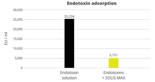
Solis – the cost-effective solution

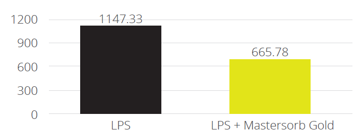
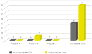
In the case of contamination with only aflatoxin, the cost-effective solution Solis is recommended. Solis consists of well-selected superior silicates with high surface area due to its layered structure. Solis shows high adsorption of aflatoxin B1, which was proven in a trial:

FigureFigure 3: Binding capacity of Solis for Aflatoxin

Even at a low inclusion rate, Solis effectively binds the tested mycotoxin at a very high rate of nearly 100%. It is a high-efficient, cost-effective solution for aflatoxin contamination.

Solis Max 2.0: The effective mycotoxin solution for sustainable profitability

Solis Max 2.0 has a synergistic combination of ingredients that acts by chemi- and physisorption to prevent toxic fungal metabolites from damaging the animal’s gastrointestinal tract and entering the bloodstream.

Figure

Figure 4: Composition and effects of Solis Max 2.0

Solis Max 2.0 is suitable for more complex challenges and longer-living animals: in addition to the pure mycotoxin adsorption, Solis Max 2.0 also effectively supports the liver and, thus, the animal in its fight against mycotoxins.

In an in vitro trial, the adsorption capacity of Solis Max 2.0 for the most relevant mycotoxins was tested. For the test, the concentrations of Solis Max 2.0 in the test solutions equated to 1kg/t and 2kg/t of feed.

FigureFigure 5: Efficacy of Solis Max 2.0 against different mycotoxins relevant in poultry production

The test showed a high adsorption capacity: between 80% and 90% for Aflatoxin B1, T-2 Toxin (2kg/t), and Fumonisin B1. For OTA, DON, and Zearalenone, adsorption rates between 40% and 80% could be achieved at both concentrations (Figure 5). This test demonstrated that Solis Max 2.0 could be considered a valuable tool to mitigate the effects of mycotoxins in poultry.

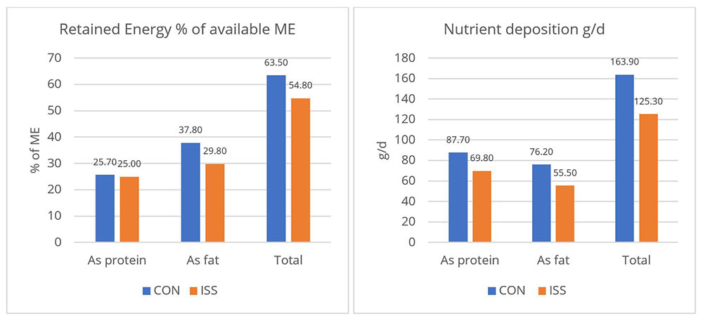
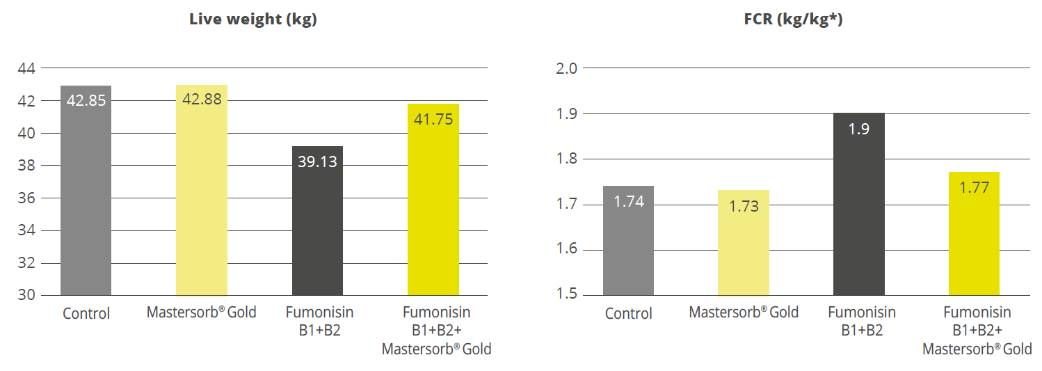
Broiler trial shows improved performance in broilers

Protected and, therefore, healthier animals can use their resources for growing/laying eggs. A trial showed improved liver health and performance in broilers challenged with two different mycotoxins but supported with Solis Max 2.0.

For the trial, 480 Ross-308 broilers were divided into three groups of 160 birds each. Each group was placed in 8 pens of 20 birds in a single house. Nutrition and management were the same for all groups. If the birds were challenged, they received feed contaminated with 30 ppb of Aflatoxin B1 (AFB1) and 500 ppb of Ochratoxin Alpha (OTA).

Negative control: no challenge no mycotoxin-mitigating product
Challenged group: challenge no mycotoxin-mitigating product
Challenge + Solis Max 2.0 challenge Solis Max 2.0, 1kg/t

The body weight and FCR performance parameters were measured, as well as the blood parameters of alanine aminotransferase and aspartate aminotransferase, both related to liver damage when increased.

Concerning performance as well as liver health, the trial showed partly even better results for the challenged group fed with Solis Max 2.0 than for the negative, unchallenged control (Figures 6 and 7):

  • 6% higher body weight than the negative control and 18.5% higher body weight than the challenged group
  • 12 points and 49 points better FCR than the negative control and the challenged group, respectively
  • Lower levels of AST and ALT compared to the challenged group, showing a better liver health

The values for body weight, FCR, and AST, even better than the negative control, may be owed to the content of different gut and liver health-supporting phytomolecules.

FigureFigure 6: Better performance data due to the addition of Solis Max 2.0

FigureFigure 7: Healthier liver shown by lower values of AST and ALT

Effective toxin risk management: staying power is required

Mycotoxin mitigation requires many different approaches. Mycotoxin mitigation starts with sewing the appropriate plants and continues up to the post-ingestion moment. From various studies and field experience, we find that besides the right decisions about grain crops, storage management, and hygiene, the use of effective products which mitigate the adverse effects of mycotoxins is the most practical and effective way to maintain animals healthy and well-performing. According to Eskola and co-workers (2020), the worldwide contamination of crops with mycotoxins can be up to 80% due to the impact of climate change and the availability of sensitive technologies for analysis and detection. Using a proper mycotoxin mitigation program as a precautionary measure is, therefore, always recommended in animal production.

Toxin Risk ManagementFigure

EW Nutrition’s Toxin Risk Management Program supports farmers by offering a tool (MasterRisk) that helps identify and evaluate the risk and gives recommendations concerning using toxin solutions.


Stop endotoxins from decreasing animal performance

e coli shutterstock 347266496

By Marisabel CaballeroGlobal Technical Manager Poultry, EW Nutrition

Find out why endotoxemia threatens animal production and how intelligent toxin mitigation solution SOLIS MAX can support endotoxin management.

Figure 1: Structure of Lipopolysaccharide

The quick guide to endotoxins (LPS) and what to do about them

Lipopolysaccharides (LPS) are a constant challenge for animal production. LPS, which are also known as endotoxins, are the major building blocks of the outer walls of Gram-negative bacteria (see figure 1). Throughout its life cycle, a bacterium releases these molecules upon cell death and lysis. When endotoxins are released into the intestinal lumen of chickens or swine, or in the rumen of polygastric animals, they can cause serious damage to the animal’s health and performance by over-stimulating their immune system.

LPS may induces inflammation and fever, lowering feed intake, and redirecting nutritional resources to the immune response, which results in hindered animal performance.

Endotoxins depress animal performance

One of the biggest issues caused by endotoxemia is that animals reduce their feed intake and show a poor feed conversion rate (FCR). Why does this happen? The productive performance of farm animals (producing milk, eggs, or meat) requires nutrients. An animal also requires a certain baseline amount of nutrients for maintenance, that is, for all activities related to its survival.

As a result of inflammation, endotoxemia leads to a feverish state. Maintenance needs to continue; hence, the energy required for producing heat will be diverted from the nutrients usually spent on production of milk, eggs, meat, etc., and performance suffers. This is amplified because the immune reaction also requires resources (e.g., energy, amino acids, etc. to produce more immune cells).

The inflammation response can result in mitochondrial injury to the intestinal cells, which alter the cellular energy metabolism. This is reflected in changes to the levels in adenosine triphosphate (ATP), the energy “currency” of living cells. A study by Li et al. (2015) observed a respective reduction of 15% and 55% in the ATP levels of the jejunum and ileum of LPS-challenged broilers, compared to the unchallenged control group.

piglet study by Huntley, Nyachoti, and Patience (2017) found that LPS-challenged pigs retained 15% less of the available metabolizable energy and showed 25% less nutrient deposition (figure 2). These results illustrate how animal performance declines during endotoxemia.

  • Control treatment (CON) = Pigs fed by a basal diet
  • Immune system stimulation treatment (ISS) = Pigs given LPS (E. coli serotype 055:B5) injection

Figure 2: Retained Energy as % of ME intake and nutrient deposition of pigs in metabolic cages (adapted from Huntley, Nyachoti, and Patience, 2017)

A loss of energy retained due to a reduction in available metabolizable energy leads to losses in performance as the amount of energy available for muscle production and fat storage will be lower. Furthermore, the decrease in feed intake creates a further energy deficit concerning production needs.

Endotoxin tolerance

The repeated exposure to LPS leads to the production of anti-inflammatory cytokines, as a reaction of the body to prevent tissue damage due to the excessive inflammation. This immunosuppression during stress may lead to an increased risk of secondary infection and poor vaccination titers.

LPS tolerance, also known as CARS (compensatory anti-inflammatory response syndrome) essentially depresses the immune system to control its activity. This “regulation” can be extremely dangerous as an excessive depression of the immune system leaves the organism exposed to the actual pathogens.

The way forward: Natural endotoxin mitigation with SOLIS MAX

The quantity of Gram-negative bacteria in an animal intestine is considerable; therefore, the danger of immune system over-stimulation through endotoxins cannot be taken lightly. Stress factors – that are not uncommon in animal production – affect the microbiome (favoring gram-negative bacteria) and also decrease the intestinal barrier function, which leads to the passage of LPS into the bloodstream

Animals suffering from endotoxemia are subject to severe metabolic dysfunctions. If they do not perish from septic shock (and most of them do not), they are still likely to show performance losses. Moreover, they at great risk of immunosuppression caused by CARS, the immune system “overdrive” discussed above.

Fortunately, research shows that EW Nutrition’s SOLIS MAX effectively binds bacterial toxins, helping to prevent these scenarios.

In vitro trial shows SOLIS MAX’ effectiveness against bacterial endotoxins

Binding endotoxins in the gastrointestinal tract, especially during stress situations in animal production, can help to mitigate the negative impact of LPS on the animals. It reduces the endotoxins passing into the bloodstream and entering the organism.

SOLIS MAX is a synergistic combination of natural plant extracts, yeast cell walls, and natural clay minerals. An in vitro study conducted at a research facility in Germany evaluated its binding performance for LPS derived from E. coli.

To test the efficacy of SOLIS MAX in binding endotoxins, 0.1% (w/v) of SOLIS MAX was resuspended in endotoxin-free water, with and without a challenge of 25,2568 EU/ml. After one hour, the solutions were centrifuged and the supernatants tested for LPS using Endo-LISA test kits.

The results show that 1 mg of SOLIS MAX adsorbs 20 endotoxin units (EU) of E. coli endotoxin, which corresponds – for this challenge – to an 80% adsorption rate (figure 3).

Figure 3: SOLIS MAX effectively adsorbs E. coli endotoxins

Endotoxin solution SOLIS MAX: Stabilize gut health, support performance

The detrimental impact of LPS can be mitigated by using a high-performance solution such as SOLIS MAX. To prevent negative health and performance outcomes for the animal it is important to stabilize the challenged intestinal barrier and to support the balance of the gut microbiome. Binding endotoxins before they can exert their damaging impact is the primary objective, which SOLIS MAX achieves through the intelligent interaction of natural plant extracts. This can be expected to yield positive results in terms of production levels and the prevention of secondary infections, preserving animal health and farms’ economic viability.


References

Adib-Conquy, Minou, and Jean-Marc Cavaillon. “Compensatory Anti-Inflammatory Response Syndrome.” Thrombosis and Haemostasis 101, no. 01 (2009): 36–47. https://doi.org/10.1160/th08-07-0421.

Huntley, Nichole F., C. Martin Nyachoti, and John F. Patience. “Immune System Stimulation Increases Nursery Pig Maintenance Energy Requirements.” Iowa State University Animal Industry Report 14, no. 1 (2017). https://doi.org/10.31274/ans_air-180814-344.

Li, Jiaolong, Yongqing Hou, Dan Yi, Jun Zhang, Lei Wang, Hongyi Qiu, Binying Ding, and Joshua Gong. “Effects of Tributyrin on Intestinal Energy Status, Antioxidative Capacity and Immune Response to Lipopolysaccharide Challenge in Broilers.” Asian-Australasian Journal of Animal Sciences 28, no. 12 (2015): 1784–93. https://doi.org/10.5713/ajas.15.0286.


Mycotoxin Monitoring Update: Fall 2021 Essentials

img 0355

By Vinil Samraj Padmini, Global Category Manager Feed Quality, and Marisabel Caballero, Global Technical Manager Poultry, EW Nutrition

Climate across the globe has changed, with rising atmospheric temperatures and carbon dioxide levels. This change favors the growth of toxigenic fungi in crops and thus increases the risk of mycotoxin contamination. When contaminating feed, mycotoxins exert adverse effects in animals and could be transferred into products such as milk and eggs.

Mycotoxin Monitoring

95% of the samples were contaminated with at least one mycotoxin

EW Nutrition constantly analyzes feed and raw material samples for their mycotoxin contamination. We report challenges from the most common mycotoxins hindering animal health around the globe.

Worldwide, more than 4,000 analyses on more than 1,000 samples were performed between June – October of the present year. The samples covered grain and by-products commonly used in animal feed worldwide. Figure 1 shows the percentage of the samples tested for which a positive result was found, detailing the number of mycotoxins per sample.

Mycotoxins per sample world-wide
Click to enlarge

The number of mycotoxins analyzed per sample can vary based on regional risk-evaluation, including weather conditions, raw material origin and past frequency of positives. However, a minimum number of samples per region is always analyzed for the full spectrum, in order to monitor and corroborate the risk level.

3 or more mycotoxins per sample

95% of the samples were contaminated with at least one mycotoxin. In Europe and Latin America, most samples were analyzed for up to five mycotoxins, and were found contaminated with at least two. In South Asia, three mycotoxins were regularly analyzed per sample and most samples were positive for two. Worldwide, it is common to find samples with 3 or more mycotoxins, indicating that, even in raw materials, poly-contamination is the rule.

Aflatoxin: Main concern for South Asia

From all samples tested positively for mycotoxin contamination, 55% were contaminated with Aflatoxins. In all regions, the maximum levels lay over the thresholds for dairy and poultry. In Europe, less than 20% of the samples were contaminated with Aflatoxin. In Europe and the USA, the average contamination is low, hence this toxin can hardly be considered an issue for animal production in those areas (Figure 2).

In South Asia, where high temperatures and humidity are prevalent, Aflatoxin was detected in more than 95% of the samples and the average contamination is over all thresholds. Management strategies, such as the use of mold inhibitors for stored grain and toxin binders in feed, are necessary in this area to keep animals healthy and productive.

Aflatoxin: Main concern for South Asia
Click to enlarge

Impact

Aflatoxins have a negative impact on animal performance, as they affect the function of liver and kidney, alter the immune function, and impair protein synthesis. This affects weight gain, feed efficiency and mortality. Carryover into milk, eggs and edible organs is possible with high or chronic intake of the toxin.

Fumonisins: Main concern for LATAM, also global

Fumonisin was found in 70% of the samples globally and roughly in 90% of the samples coming from Latin America (figure 3). Moreover, in LATAM, more than 50% of the results have values over the threshold for dairy and swine, and 14% over the threshold for poultry, making it a great concern in the area. South Asia is the second concern area, with a high proportion of contaminated samples (80%) and 14% of them representing a danger for poultry production.

Main concern for LATAM, also global
Click to enlarge

Impact

The main issue with the typical contamination levels of fumonisins – often considered of low risk – is their capacity to disrupt gut health. As their absorption is low, fumonisins interact with other toxins and the gut barrier components, including those affecting immunity and the microbiome. They are known to decrease the available surface for nutrient digestion and absorption, and to increase the risk and incidence of gut-related diseases. As a result, lower productivity is expected in animals exposed to even low levels of this toxin.

Deoxynivalenol (DON): Present worldwide

All across the regions, the maximum tested levels lay over the threshold for dairy, poultry, and swine. This trichothecene was found in more than 70% of the samples analyzed worldwide. In the United States, more than 75% of the positive tested samples showed a contamination with DON and the average of the positives exceeded the thresholds for swine and poultry.

The region with highest maximum values is LATAM, followed by South Asia, and the region with the highest frequency of positives in analyzed samples is Europe. Thus, it can be concluded that the worldwide frequency and levels in which DON is found represent a high risk for production animals.

Deoxynivalenol (DON): Present worldwide
Click to enlarge

Impact

Deoxynivalenol shows a broad spectrum of toxic effects in animals. In poultry and swine, for instance, this mycotoxin is related to lesions in the gastrointestinal tract and alterations in the immune response. This, in turn, leads to lower productivity and poor feed efficiency. DON also interacts with the microbiome, and it is known that it favors the colonization of coliform bacteria in pigs.

Ruminants can tolerate 10–20 times more DON than, for example, pigs. The majority of ingested DON is converted into the less toxic de-epoxy DON, but the degradation rate is influenced by different factors such as the diet, where high starch decrease the process. Moreover, DON also has a detrimental effect on rumen microorganisms, impacting its fermentative capacity.

T2: A danger for poultry producers word-wide

Average levels of T2 were over the threshold for poultry in all regions, with a high presence (>70% of the analyzed samples) in Europe, the US & LATAM.

T2: A danger for poultry producers word-wide
Click to enlarge

Impact

T-2 s is a potent inhibitor of protein synthesis, which affects actively dividing cells, such as the lining of the gastrointestinal tract, skin, and immune cells. The consequences include weight loss or poor weight gain, diarrhea, skin and beak lesions, and decreased production.

T-2 is de-epoxidated in the rumen to HT-2 and neosolaniol, which are significantly less toxic than the parent toxin. In acidotic animals, rumen detoxification of T-2 toxin is impaired and animals may show gastroenteritis and intestinal hemorrhages.

Zearalenone: 80% positive tests globally

More than 80% of all samples tested for this mycotoxin were found positive. The maximum contaminations lay over the thresholds for dairy and swine. These high levels found should not be ignored, considering feedstuffs for long living and reproduction animals.

Zearalenone: 80% positive tests globally
Click to enlarge

Impact

Especially in pig breeding, Zearalenone is an important issue, due to its high absorption and rapid biotransformation into more estrogenic components. Its structural similarity with 17β-estradiol leads this toxin to impair reproductive performance in cows and sows.

Recent studies point to interactions of Zearalenone with immune cells and organs in animals, leading to alterations in cell viability, proliferation, and functionality. Consequences are alterations of the immune response, enhancing the effects of other challenges.

A bad year for crops could be a bad year for production animals

The high mycotoxin contamination found so far in 2021 is partially explained by climate events, such as high temperature and humidity. Temperate zones such as Europe or parts of the USA tend to have higher contaminations compared with previous years.

Multiple mycotoxins co-occur, increasing their impact on animals. Certain combinations of mycotoxins are known to have synergistic or additive effects, aggravating their adverse effects.

To safeguard animal performance, it is important to continually strive for low levels of contamination and to manage the risk of mycotoxins through the use effective tools to measure, interpret, and manage the risk. MasterRisk can aid in the interpretation of mycotoxin risks, weighing in the animal species, age, purpose, as well as the mycotoxin exposure and interactions.


From sub-acute ruminal acidosis to endotoxins: Prevention for lactating cows

udder 562980 1920 pixabay

by Technical Team, EW Nutrition

Sub-acute acidosis (SARA) is linked to high levels of ruminal LPS. The LPS cause inflammation and contribute to different metabolic conditions and diseases. Various strategies and solutions can be applied to modulate the rumen microbiota and prevent this risk.

lactating cows

In sub-acute rumen acidosis (SARA), the quantity of free lipopolysaccharides (LPS) coming from Gram- bacteria increases considerably. These LPS cross the ruminal wall and intestine, passing into the bloodstream. The negative consequences on the health of the animal are then reflected in decreased productive and reproductive performance.

The LPS are released during the lysis of GRAM- bacteria which die due to the low pH, and these bacteria are mainly responsible for the production of propionic acid for the energy yield that is obtained. It is essential to preserve ruminal balance between Gram+ and Gram- such that there is no excess of LPS.

Nutritional needs of lactating cows with SARA

In the first phase of lactation (from 1 week after calving to 80 – 100 days of lactation), the cow needs a high energy level to meet the large demand for milk production. This energy demand is often not fully satisfied and feed intake falls short. This deficit leads to the need to provide as much energy as possible per feed ration.

Imagine a 650 kg live weight cow, producing about 35 kg of milk per day with a fat percentage of 3.7 and a protein percentage of 3.2. To achieve this production level and fulfill its maintenance requirements, this animal needs a feed intake of 22 kg of dry matter (DM) per day, with an energy level of 21 UFL equal to 36,000 Kcal/day of NE l (Net Energy Lactation)).

To obtain an energy supply of this type, it is necessary to provide rations with a high content of cereals rich in nonstructured carbohydrates (NSC). This will allow the animals to obtain the maximum efficacy in getting the NE I from the metabolizable energy  (ME) expressed as kl*.

*kl expresses the effectiveness in passing from EM to EN l net of the heat dissipated by the animal, therefore kl = ENl/EM (Van Es 1978).

Compared to a diet rich in NDF (Neutral Detergent Fiber), this type of diet promotes and stimulates certain strains of bacteria to the detriment of others, shifting the balance towards a greater population of bacteria that produce propionic acid instead those which produce acetic acid. This change also determines a greater share of Gram- compared to Gram+.

What is rumen acidosis?

Rumen acidosis is that “pathology” whereby the volume of SCFA (Short Chain Fatty Acids) produced by the rumen bacteria is greater than the ability of the rumen itself to absorb and neutralize them. Rumen acidosis is mainly caused by the amylolytic and saccharolytic bacteria (Streptococcus bovis; Selenomonas ruminantium, Bacteroides amylophilus, Bacteroides ruminicola and others) responsible for the production of lactic acid. Unlike the other most representative volatile fatty acids (acetic, butyric and propionic), lactic acid has a lower pKa: 7 (3.9 versus 4.7). This means that for the same amount of molecules produced, lactic acid releases a number of ions H+ in the fluid ten times greater than other VFAs, with evident effects on the pH.

Ruminal acidosis can be characterized as acute or subacute. During acute ruminal acidosis, the pH in the rumen drops below 4.8 and remains low for an extended period of time. Acute acidosis leads to complete anorexia, abdominal pain, diarrhea, lethargy, and eventually death. However, the prevalence of acute acidosis in dairy is very low.

Consequences of rumen acidosis

In such situations, a series of negative consequences can be triggered in the lactating cow. Investigations (for instance, using fistulated cows) can reveal, among others, the following alteration in the rumen:

  • Shift in total microbiome rumen profile (density; diversity; community structure)
  • Shift in protozoa population (increase in ciliates protozoa after 3 weeks of SARA; increase in the GNB population)
  • Shift in fungi population (decreasing the fungi population with high fibrolytic enzymes, which are sensitive to low pH)
  • Rise in LPS rumen concentration (increasing the GNB strain and their lysis)
  • Influence on the third layer of Stratified Squamous Epithelium (SSE) (desmosomes and tight junctions)
  • Lower ruminal fiber degradation (reduction in the number of cellulolytic bacteria which are less resistant to acid pH)
  • Reduction of the total production of fatty acids (propionic, acetic, butyric), therefore less available energy
  • Lower rumen motility (also as a consequence of the smaller number of protozoa)
  • The increased acid load damages the ruminal epithelium
  • Acid accumulation increases the osmotic pressure of the rumen inducing an higher flux of water from the blood circulation into the rumen, causing swelling and rupture of rumen papilla as well as a greater hemoconcentration

The last points are extremely important, as it enables an easier passage of fluids from the blood to the pre-stomachs, greatly influencing the fermentation processes.

Furthermore, with diets low in NDF, the level of chewing and salivation is certainly lower, with a consequent lower level of salivary buffers that enter the rumen and which would maintain an appropriate pH under normal conditions.

Rumen sub-acute and acute acidosis: a fertile ground for LPS

Studies inducing SARA in dairy cows have shown that feeding high levels of grain causes the death and cell lysis of Gram- bacteria, resulting in higher concentration of free LPS in the rumen. In a trial conducted by Ametaj et al., in 2010 (Figure 1), a lower ruminal pH and an increase in the concentration of LPS in the rumen fluid -measured as ng / ml (nanograms / milliliter)-, was the result of increasing of NSC present in the diet (% of grains).

Rumen endotoxins
Figure 1. The increase in the level of endotoxins in the rumen is directly correlated with an increase in ration concentrates


In the rumen, the presence of Gram- is very significant, however the dietary changes towards high energy concentrates, reduce the substates necessary for them to thrive, leading to their lysis and favoring gram-positive bacteria (Gram+). Gram+ also produce bacteriocins against a wide variety of bacteria, including many Gram-. Figure 2 shows the influence of ruminal pH in the population of different bacteria, many of which are are crucial for the production of SCFA and therefore of energy. 

Gram bacteria influenced by pH
Figure 2. Activity of main bacteria in the rumen in function of pH (Daniele Cevolani Edizioni Agricole di New Business Media srl 2020)


It is therefore necessary to pay close attention to the energy level of the ration as an energy input (generally around 1500 – 1700 Kcal/kg of DM intake). At the same time, we need to ensure that the animal does receive and ingest that daily amount of DM. If ingestion is negatively influenced by acidosis (clinical or sub-clinical), this can lead to endotoxemia, with harmful consequences for the animal’s health and production performance.

We can therefore note that the level of LPS (endotoxins) present in the rumen is directly correlated with the pH of the rumen itself and with a symptomatologic picture dating back to SARA. This occurs when the mortality and lysis of Gram- bacteria (GNB) is high and through the consequent imbalance created with diets containing excess fermentable starches, compared to diets with higher fiber content.

In fact, it was shown that the transition from a concentrated fodder ratio of 60:40 to a more stringent ratio of 40:60 caused the level of free LPS in the rumen to go from 410 to 4.310 EU / ml.

Endotoxemia: Pathological consequences in dairy cows

Once the LPS enter the bloodstream, they are transported to the liver (or other organs) for the detoxification. However, sometimes this is not enough to neutralize all the endotoxins present in blood. The remaining excess can cause issues such as the modification of the body’s homeostasis or cause that cascade of inflammatory cytokines responsible for the most common pathologies typical in cows in the first phase of lactation. The most common symptoms are the increase of somatic cells in milk or claws inflammation.

Pro-inflammatory cytokines as TNF, IL6 and IL8 induced by LPS-related inflammation are able to stimulate the production of ACTH (adrenocorticotropic hormone).

ACTH, together with cortisol and the interleukins, inhibit the production of GnRH and LH, with serious effects on milk production. The productivity and the fertility of the animal are thus compromised.

Moreover, prostaglandins are as well stimulated by LPS, and are linked with fever, anorexia and ruminal stasis. This not only limits the amount of energy available for production and maintenance functions, but also induces a higher susceptibility to disease and adds-up to the emergence of other metabolic conditions, such as laminitis and mastitis.

Preventing rumen acidosis

The solution to these massive risks is a prudent and proactive approach by the nutritionist towards all situations that can cause a rapid increase of Gram- in the rumen. It is therefore necessary to avoid cases of clinical and sub-clinical acidosis (SARA) in order to avoid the issues listed above. This would also help avoid stressful conditions for the animal that would lead to decreased performance and health.

To maintain balance and a healthy status of the animal, the use of additives such as phytomolecules and binders is suggested in the first phase of lactation, starting from 15 days before giving birth.

Activo Premium (a mix of phytogenic substances) has given excellent results in decreasing the acetic/propionic acid ratio, while safeguarding the population of Gram+ bacteria. This is in contrast to treatments with ionophores, which, as is well known, interfere with the Gram+ population.

Case study. Acetic acid:propionic acid ratio with Activo Premium

In a study conducted at the the University of Lavras and the Agr. Res. Comp. of Minas Gerais (both Brazil), 30 Holstein cows were allocated to two groups considering parity and milk production. One group was fed the standard feed (control), the other group received standard feed containing 150mg of Activo Premium/kg of dietary dry mass (DM). The following parameters were measured or calculated: intake of DM and milk production, milk ingredients such as fat, protein, lactose every week, body weight and body condition score every two weeks, and ruminal constituents (ph and SCFAs) through oesophaeal samples at day 56.

Activo Premium was able to decrease the ratio between acetic acid and propionic acid, and at the same time maintain the level of Gram+ bacteria in the rumen, thus reducing the risk of endotoxins. The same trial carried out at the University of Lavras demonstrated how the performance of the animals was superior in the group fed with Activo Premium compared to the control group (see below).

Figure 3. Effect of Activo Premium on ruminal constituents


Figure 4. Effect of Activo Premium on animal performance


Solution: Preserve Gram+ bacteria levels while decreasing free LPS

We have therefore seen how important it is to decrease the acetic:propionic ratio in the rumen to obtain a greater share of available energy. However, the level of endotoxins in the rumen must remain low in order to avoid those problems of endotoxemia linked to very specific pathologies typical of “super productive cows”. These pathologies (always linked to inflammatory manifestations) can be prevented by decreasing the level of free LPS in the rumen with a product that can irreversibly bind the LPS and thus make them inactive.

In a trial with porcine intestinal cells (IPEC-J2) challenged by E. coli LPS, a decrease in the intensity of inflammation was observed when Mastersorb Gold was added. This decrease could be shown through a lower amount of phosphorylated NF-kB in an immunofluorescence trial, as well as through the reduced production of Interleukin (IL)-8 in the cells measured by ELISA.  

The fact that pig intestine tissue was used does not affect the adsorption concept. In this case, these intestinal cells are only a vehicle to demonstrate that in an aqueous solution containing 50 ŋg of LPS / ml and in the same solution with the addition of Mastersorb Gold, the level of LPS actually active is decreased, as a part of the LPS was tied up by Mastersorb. The solution with a lower level of LPS gave minor “inflammatory” reactions to intestinal cells, and this can be statistically reported in dairy cows.

Immunofluorescence in PEG-J2
Figure 5. Immunofluorescence in PEG-J2: Challenge with LPS without (in the middle) and with Mastersorb Gold (right)


IL-8 AP secretion
Figure 6. IL-8 AP secretion after incubation with LPS 0111:B4 for 24h without and with Mastersorb Gold


Conclusions

To demonstrate how the decrease in the level of LPS in the rumen is directly correlated with inflammatory states in general, a trial with a total of 60 dairy cows shows that the inclusion of 25g of Mastersorb Premium/animal/day increases milk yield and improves milk quality by decreasing somatic cell count. Adsorbing substances contained in Mastersorb Premium tie up the LPS produced in the rumen in different cow lactation phases.

Normally, the rise in the level of somatic cells in milk depends on etiological agents such as Streptococcus spp, Staphylococcus spp, mycoplasma and more. LPS stress is not the sole agent responsible for raising somatic cell counts, but also other factors among which:

  • Lactation stage and age of the animal
  • Season of the year (in summer the problem is increased)
  • Milking plant (proper maintenance)
  • General management and nutrition

 However, by reducing the level of LPS, Mastersorb provides an important aid to decrease somatic cell count.

somatic cell count
Figure 7. Effect of Mastersorb Premium on somatic cell count


Prevent escalation with rumen balance

In the end, ruminant producers are, like all livestock operations, interested in producing healthy animals that can easily cope with various stressors. Ensuring a proper diet, adjusted to the energy requirements of various production stages, is a first step. Providing the animal with the ingredients that modulate the microbiota and reduce the negative impact of stress in the rumen is the next essential step in efficient production.


A complex battlefield: mycotoxins in the gastrointestinal tract

shutterstock 494829349 fusarium mycotoxins 1 scaled

Most grains used as feed raw materials are susceptible to mycotoxin contamination. These toxic secondary metabolites are produced by fungi before or after harvest and cause severe economic losses all along agricultural value chains. For livestock, negative consequences include acute effects such as impaired liver and kidney function, vomiting, or anorexia, as well as chronic effects such as immunosuppression, growth retardation, and reproductive problems. Mycotoxin management is, therefore, of the utmost priority for animal producers worldwide.

But how is it that mycotoxins cause such damage in the first place? This article delves into the complex processes that take place when mycotoxins come into contact with the gastrointestinal tract (GIT). The intestinal epithelium is the first tissue to be exposed to mycotoxins, and often at higher concentrations than other tissues. A deeper understanding of how mycotoxins affect the GIT allows us to appreciate the cascading effects on animal health and performance, why such damage already occurs at contamination levels well below official safety thresholds – and what we can do about it.

The intestinal epithelium: the busy triage site for nutrients and harmful substances

When mycotoxins are ingested, they encounter the GIT’s intestinal epithelium (Figure 1). This single layer of cells lining the intestinal lumen serves two conflicting functions: firstly, it must be permeable enough to allow the absorption of nutrients. On the other hand, it constitutes the primary physiological barrier against harmful agents such as viruses, microorganisms, and toxins.

Within the intestinal epithelium, several types of highly specialized cells are involved in epithelial regeneration, nutrient absorption, innate defense, transport of immunoglobulins, and immune surveillance. The selective barrier function is maintained due to the formation of complex networks of proteins that link adjacent cells and seal the intercellular space. Besides, the intestinal epithelium is covered with mucus produced by goblet cells, which isolates its surface, preventing the adhesion of pathogens to the enterocytes (intestinal absorptive cells).

Due to its dual involvement in digestive and immune processes, the intestinal epithelium plays a pivotal role in the animal’s overall health. Importantly, the epithelium is directly exposed to the entire load of ingested mycotoxins. Hence their effects can be problematic even at low concentrations.

Figure 1: The intestinal epithelium

The intestinal epithelium


Problematic effects of mycotoxins on the intestinal epithelium

Most mycotoxins are absorbed in the proximal part of the gastrointestinal tract (Table 1). This absorption can be high, as in the case of aflatoxins (~90%), but also very limited, as in the case of fumonisins (<1%); moreover, it depends on the species. Importantly, a significant portion of unabsorbed toxins remains within the lumen of the gastrointestinal tract.

Some of the mycotoxins that enter the intestinal lumen can be bio-transformed into less toxic compounds by the action of certain bacteria. This action, however, predominantly happens in the large intestine – therefore, no detoxification takes place before absorption in the upper parts of the GIT. Part of the absorbed mycotoxins can also re-enter the intestine, reaching the cells from the basolateral side via the bloodstream. Furthermore, they re-enter through enterohepatic circulation (the circulation of substances between the liver and small intestine). Both actions increase the gastrointestinal tract’s overall exposure to the toxins.

Table 1: Rate and absorption sites of different mycotoxins

Rate and absorption sites of different mycotoxins

Adapted from: Biehl et al., 1993; Bouhet & Oswald, 2007; Devreese et al., 2015; Ringot et al., 2006

The damaging impact of mycotoxins on the intestinal epithelium initially occurs through:

  • A decrease in protein synthesis, which reduces barrier and immune function (Van de Walle et al., 2010)
  • Increased oxidative stress at the cellular level, which leads to lipid peroxidation, affecting cell membranes (Da Silva et al., 2018)
  • Changes in gene expression and the production of chemical messengers (cytokines), with effects on the immune system and cellular growth and differentiation (Ghareeb et al., 2015)
  • The induction of programmed cell death (apoptosis), affecting the reposition of immune and absorptive cells (Obremski & Poniatowska-Broniek, 2015)

Importantly, studies based on realistic mycotoxin challenges (e.g., Burel et al., 2013) show that the mycotoxin levels necessary to trigger these processes are lower than the levels reported as safe by EFSA, the Food Safety Agency of the European Union. The ultimate consequences range from diminished nutrient absorption to inflammatory responses and pathogenic disorders in the animal (Figure 2).

Figure 2: Mycotoxins’ impact on the GIT and consequences for monogastric animals

Mycotoxins’ impact on the GIT and consequences for monogastric animals

1. Alteration of the intestinal barrier‘s morphology and functionality

The mycotoxins DON, fumonisin, and T2 induce a reduction in the rate of epithelial cell proliferation and differentiation. This causes a decrease in the height and the surface of the intestinal villi, which in turn leads to a reduction in nutrient absorption. Additionally, some nutrient transporters are inhibited by the action of mycotoxins such as DON and T2, for example, negatively affecting the transport of glucose.

Several studies indicate that mycotoxins such as aflatoxin B1, DON, fumonisin B1, ochratoxin A, and T2, can increase the permeability of the intestinal epithelium of poultry and swine (e.g. Pinton & Oswald, 2014). This is mostly a consequence of the inhibition of protein synthesis. As a result, there is an increase in the passage of antigens into the bloodstream (e.g., bacteria, viruses, and toxins). This increases the animal’s susceptibility to infectious enteric diseases. Moreover, the damage that mycotoxins cause to the intestinal barrier entails that they are also being absorbed at a higher rate.

2. Impaired immune function in the intestine

The intestine is a very active immune site, where several immuno-regulatory mechanisms simultaneously defend the body from harmful agents. Immune cells are affected by mycotoxins through the initiation of apoptosis, the inhibition or stimulation of cytokines, and the induction of oxidative stress. Studies demonstrate that aflatoxin, DON, fumonisin, T2, and zearalenone interact with the intestinal immune system in such a manner that the animal’s susceptibility to viral and bacterial infections increases (e.g., Burel et al., 2013). Moreover, by increasing their fecal elimination, the horizontal transmission of pathogens is extended.

For poultry production, one of the most severe enteric problems of bacterial origin is necrotic enteritis, which is caused by Clostridium perfringens toxins. Any agent capable of disrupting the gastrointestinal epithelium – e.g. mycotoxins such as DON, T2, and ochratoxin – promotes the development of necrotic enteritis. The inhibition of the intestinal immune system caused by mycotoxins such as aflatoxin, DON, and T2 also collaborates with the development of this disease.

3. Alteration of the intestinal microflora

The gastrointestinal tract is home to a diverse community of bacteria, fungi, protozoa, and viruses, which lines the walls of the distal part of the intestine. This microbiota prevents the growth of pathogenic bacteria through competitive exclusion and the secretion of natural antimicrobial compounds, volatile fatty acids, and organic acids.

Recent studies on the effect of various mycotoxins on the intestinal microbiota show that DON and other trichothecenes favor the colonization of coliform bacteria in pigs. DON and ochratoxin A also induce a greater invasion of Salmonella and their translocation to the bloodstream and vital organs in birds and pigs – even at non-cytotoxic concentrations. It is known that fumonisin B1 may induce changes in the balance of sphingolipids at the cellular level, including for gastrointestinal cells. This facilitates the adhesion of pathogenic bacteria, increases in their populations, and prolongs infections, as has been shown for the case of E. coli.

From the perspective of human health, the colonization of the intestine of food-producing animals by pathogenic strains of E. coli and Salmonella is of particular concern. Mycotoxin exposure may well increase the transmission of these pathogens, posing a risk for human health.

4. Interaction with bacterial toxins

When mycotoxins induce changes in the intestinal microbiota, this can lead to an increase in the endotoxin concentration in the intestinal lumen. Endotoxins or lipopolysaccharides (LPS) are fragments of Gram-negative bacteria’s cell walls. They are released during bacterial cell death, growth, and division. Hence endotoxins are always present in the intestine, even in healthy animals. Endotoxins promote the release of several cytokines that induce an enhanced immune response, causing inflammation, thus reducing feed consumption and animal performance, damage to vital organs, sepsis, and death of the animals in some cases.

The synergy between mycotoxins and endotoxins can result in an overstimulation of the immune system. The interaction between endotoxins and estrogenic agents such as zearalenone, for example, generates chronic inflammation and autoimmune disorders because immune cells have estrogen receptors, which are stimulated by the mycotoxin. The combination of DON at low concentrations and endotoxins in the intestine, on the other hand, has been shown to engender a decrease in transepithelial resistance and to alter the balance of the microbiota.

What to do? Proactive toxin risk management

To prevent the detrimental consequences of mycotoxins on animal health and performance, proactive solutions are needed that support the intestinal epithelium’s digestive and immune functionality and help maintain a balanced microbiome in the GIT. Moreover, it is crucial for any anti-mycotoxin product to feature both anti-mycotoxin and anti-bacterial toxin properties and that it supports the organs most targeted by mycotoxins, e.g., the liver. EW Nutrition’s Mastersorb® Gold premix is based on the synergistic combination of natural clay minerals, yeast cell walls, and phytomolecules. Its efficacy has been extensively tested, including as a means for dealing with E. coli endotoxins.

Mastersorb® Gold: anti-mycotoxin activity stabilizes performance and strengthens liver health

A field trial conducted in Germany on male Ross 308 broilers showed that for broilers receiving a diet contaminated with DON and zearalenone, adding 1kg of Mastersorb® Gold per ton of feed to their diet led to significant performance enhancements. Not only did they recuperate the mycotoxin-induced weight loss (6% increase relative to the group receiving only the challenge), but they gained weight relative to the control group (which received neither the challenge nor Mastersorb® Gold). Feed conversion also improved by 3% relative to the group challenged with mycotoxins.

A scientific study of crossbred female pigs showed that Mastersorb® Gold significantly reduced the deleterious effects of fumonisin contamination in the feed. The decrease in weight gain and the decline of feed conversion could be mitigated by 6.7% and 13 FCR points, respectively (Figure 3). Also, the sphinganine/sphingosine (Sa/So) ratio, a biomarker for fumonisin presence in the blood serum, could be decreased by 22.5%.

Figure 3: Mastersorb® Gold boosts performance for pigs fed a fumonisin-contaminated diet

Mastersorb® Gold boosts performance for pigs fed a fumonisin-contaminated diet

Another study of crossbred female piglets, carried out at a German university, investigated whether Mastersorb® Gold could support performance as well as liver health under a naturally occurring challenge of ZEA (~ 370ppb) and DON (~ 5000ppb).  Mastersorb® Gold significantly improved weight gain and feed conversion in piglets receiving the mycotoxin-contaminated diet: daily body weight gain was 75g higher than that of a group receiving only the challenge, and the FCR improved by 24% (1.7 vs. 2.25 for the group without Mastersorb® Gold). Moreover, Mastersorb® Gold significantly improved the liver weight (total and relative) and the piglets’ AST levels (aspartate aminotransferase, an enzyme indicating liver damage). A tendency to improve spleen weight and GGT levels (gamma-glutamyl transferase, another enzyme indicative of liver issues) was also evident, all of which indicate that Mastersorb® Gold effectively counteracts the harmful impact of mycotoxin contamination on liver functionality.

In-vitro studies demonstrate Mastersorb® Gold’s effectiveness against mycotoxins as well as bacterial toxins

Animal feed is often contaminated with two or more mycotoxins, making it important for an anti-mycotoxin agent to be effective against a wide range of different mycotoxins. Besides, to prevent mycotoxins damaging the GIT, an effective product should ideally adsorb most mycotoxins in the first part of the animal’s intestine (under acidic conditions). In-vitro experiments at an independent research facility in Brazil showed that an application of 0.2% Mastersorb® Gold binds all tested mycotoxins at rates from 95 to 97% at a pH level of 3, using realistic challenges of 1000ppb (Aflatoxin B1 and ZEA) and 2500ppb (Fumonisin B1 and DON). The binding results achieved for Fumonisin and DON, which are often considered outright “nonbinding,” under challenging close to neutral conditions (pH 6), are particularly encouraging.

Figure 4: Mastersorb® Gold binding capacity against different mycotoxins (%)

Mastersorb Gold binding capacity against different mycotoxins

Concerning its efficacy against endotoxins, an in vitro study conducted at Utrecht University, among other studies, has shown Mastersorb® Gold to be a strong tool against the LPS released by E. coli. For the test, four premium mycotoxin binders were suspended in a phosphate buffer solution to concentrations of 0.25% and 1%. E. coli LPS were suspended to a final concentration in each sample of 50ng/ml. Against this particularly high challenge, Mastersorb® Gold achieved a binding rate of 75% at an inclusion rate of 1%: clearly outperforming competing products, which at best showed a binding rate of 10%.

Conclusion

A healthy gastrointestinal tract is crucial to animals’ overall health: it ensures that nutrients are optimally absorbed, it provides effective protection against pathogens through its immune function, and it is key to maintaining a well-balanced microflora. Even at levels considered safe by the European Union, mycotoxins can compromise different intestinal functions such as absorption, permeability, immunity, and microbiota balance, resulting in lower productivity and susceptibility to disease.

To safeguard animal performance, it is important to continually strive for low levels of contamination in feed raw materials –  and to stop the unavoidable mycotoxin loads from damaging the intestinal epithelium through the use of an effective anti-mycotoxin agent, which also supports animals against endotoxins and boosts liver function. Research shows that Mastersorb Gold is a powerful tool for proactive producers seeking stronger animal health, welfare, and productivity.

By Marisabel Caballero and Sabria Regragui Mazili


The hidden danger of endotoxins in animal production

e coli photo 1

Find out more about endotoxins here

Find out why LPS can cause endotoxemia and how intelligent toxin mitigation solutions can support endotoxin management.

Each E. coli bacterium contains about 100 lipopolysaccharides molecules in its outer membrane

Lipopolysaccharides (LPS) are the major building blocks of the outer walls of Gram-negative bacteria. Throughout its life cycle, a bacterium releases these molecules, which are also known as endotoxins, upon cell death and lysis. The quantity of LPS present in Gram-negative bacteria varies between species and serotypes; Escherichia coli, for example, contain about 100 LPS/bacterial cell. When these are released into the intestinal lumen of chickens or swine, or in the rumen of polygastric animals, they can cause serious damage to the animal’s health and performance by over-stimulating their immune system.

How lipopolysaccharides cause disease

LPS are rather large and structured chemical molecules with a weight of over 100,000 D. They are highly thermostable; boiling in water at 100°C for 30 minutes does not destabilize their structure. LPS consist of three chemically distinct sections: a) the innermost part, lipid A, consisting mostly of fatty acids; b) the core, which contains an oligosaccharide; and c) the outer section, a chain of polysaccharides called O-antigen (Figure 1).

Figure 1: Structure of an LPS

The toxicity of LPS is mainly caused by lipid A; however, both lipid A and O-antigen stimulate the immune system. This happens when the LPS pass the mucosa and enter the bloodstream or when they attack the leukocytes.

The intestinal mucosa is the physical immune barrier that protects the microvilli from external agents (bacteria, free LPS viruses, etc.). Despite its strength (the thickness, for example, amounts to ≈830 µm in the colon and ≈123 µm in the jejunum), vulnerable points exist (cf. Zachary 2017).

LPS can easily come into contact with the cells of the lamina propria (a layer of connective tissue underneath the epithelium) through the microfold (M) cells of the Peyer’s patches (which consist of gut-associated lymphoid tissue). The M cells are not covered by mucus and thus exposed.

Secondly, LPS can also pass through the mucosa, where they become entangled in this gelatinous structure. There, they come into contact with the lymphocytes or can reach the regional lymph nodes through the afferent lymphatic vessels.

Thirdly, LPS might affect the tight junctions, the multiprotein complexes that keep the enterocytes (cells that form the intestinal villi) cohesive. By destabilizing the protein structures and triggering enzymatic reactions that chemically degrade them, LPS can break the tight junctions, reaching the first capillaries and, consequently, the bloodstream.

The presence of endotoxins in the blood, endotoxemia, can trigger problematic immune responses in animals. An innate immune stimulation leads to an increase in the concentration of pro-inflammatory cytokines in the blood and, consequently, to an induced febrile response in the animal: heat production increases, while the available metabolic energy decreases.  As a result, performance suffers, and in the worst-case scenario, septic shock sets in. Furthermore, when LPS compromise intestinal integrity, the risk of secondary infections increases, and production performance may decline.

LPS’ modes of action

How does all of this happen? The physiological consequences of endotoxemia are quite complex. Simplified, the immune system response to LPS in the blood takes three forms:

  • The stimulation of TLR4 (toll-like receptor 4) induces monocytes and macrophages to secrete critical pro-inflammatory cytokines, primarily interleukin (IL) IL-1β, IL-6, IL-8, and tumor necrotic factor (TNF) α and β. TLR4 is a structure on the cell membrane of mainly macrophages and leukocytes, which is activated by the LPS-binding protein (LBP).
  • The complement cascade constitutes about 10% of plasma proteins and determines the chemotaxis and activation of leukocytes. It can form a membrane attack complex (MAC), which perforates the membranes of pathogenic cells, enabling lysis.
  • The Hagemann factor, also known as coagulation factor XII: once stimulated by LPS, it initiates the formation of fibrin (through the intrinsic coagulation pathway), which might lead to thrombosis. The Hagemann factor directly stimulates the transformation of prekallikrein to kallikrein (enzymes involved in regulating blood pressure).

Figure 2: How LPS leads to endotoxemia – 3 modes of action

These three modes of action of inflammatory stimulation lead to important physiological reactions:

  • Pro-inflammatory cytokines (see above) modulate the functional expression of other immune cell types during the inflammatory response;
  • Metabolites of arachidonic acid (prostaglandins, leukotrienes, and lipoxins), intra- and extracellular messengers that influence the coagulation cascade;
  • Synthesis in the blood of bradykinin, a peptide responsible for the typical symptoms of inflammation, such as swelling, redness, heat and pain;
  • PAF (platelet-activating factor), which creates inflammatory effects through narrowing of the blood vessels and constriction of the airways, but also through the degranulation of leukocytes.

The symptoms of endotoxemia are:  hypotension, metabolic acidosis, hemoconcentration, intestinal hemorrhage, fever, activations of neutrophils and endothelial cells, and predisposition to thrombosis.

In case of a progression to septic shock, the following sequence takes place:

1) Reduction in blood pressure and increased heart rate (hemodynamic alterations)

2) Abnormalities in body temperature

3) Progressive hypoperfusion at the level of the microvascular system

4) Hypoxic damage to susceptible cells

Up to here, symptoms follow a (severe) endotoxemia pathogenesis. A septic shock furthermore entails:

5) Quantitative changes in blood levels of leukocytes and platelets

6) Disseminated intravascular coagulation (see Hageman factor)

7) Multi-organ failure

8) Death of animal

If an animal is continously challenged with endotoxins, experiences septic shock, or comes close to it, it risks developing LPS tolerance, also known as CARS (compensatory anti-inflammatory response syndrome). This syndrome essentially depresses the immune system to control its activity. The anti-inflammatory prerogative of CARS is not to interfere directly with the elimination of pathogens but to regulate the “excessive” inflammatory reaction in a hemostatic way. However, this regulation can be extremely dangerous as the syndrome involves a lack of homeostasis control, and an excessive depression of the immune system leaves the organism exposed to the actual pathogens.

Farm animal research on endotoxemia pathogenesis

Lipopolysaccharides are difficult to quantify in the intestine of a live animal. One way to evaluate a possible endotoxemia is to analyze biomarkers present in the bloodstream. The most important one is the LPS themselves, which can be detected in a blood sample taken from the animal via ELISA. Other biomarkers include pro-inflammatory interleukins, such as TNF α and β, IL-6 or IL-8, and fibrin and fibrinogen (though they are not specific to endotoxemia). It is vital to carry out a blood sample analysis to deduce a possible endotoxemia from symptoms and performance losses in the animal.

How the metabolic effects of endotoxemia depress performance

One of the biggest issues caused by endotoxemia is that animals reduce their feed intake and show a poor feed conversion rate (FCR). Why does this happen? The productive performance of farm animals (producing milk, eggs, or meat) requires energy. An animal also requires a certain baseline amount of energy for maintenance, that is, for all activities related to its survival. As a result of inflammation and all those physiological reactions mentioned above, endotoxemia leads to a feverish state. Maintenance needs to continue; hence, the energy required for producing heat will be diverted from the energy usually spent on producing milk, egg, meat, etc., and performance suffers.

The inflammation response can result in mitochondrial injury to the intestinal cells, which alter the cellular energy metabolism. This is reflected in changes to the levels in adenosine triphosphate (ATP), the energy “currency” of living cells. A study by Li et al. (2015) observed a respective reduction of 15% and 55% in the ATP levels of the jejunum and ileum of LPS-challenged broilers, compared to the unchallenged control group. This illustrates the extent to which animals lose energy while they experience (more or less severe) endotoxemia.

Figure 3: Reduction in ATP level in Jejunum and Ileum in broilers (adapted from Li et al., 2015)

A piglet study by Huntley, Nyachoti, and Patience (2017) took this idea further (Figure 4):  3 groups of 10 Yorkshire x Landrace pigs, weighing between 11 and 25 kg, were studied in metabolic cages and in respiratory chambers. This methodology allows for simultaneous measurement of oxygen consumption, CO2 production, energy expenditure, physical activity, and feed/water intake. The study found that LPS-challenged pigs retained 15% less of the available metabolizable energy and showed 25% less nutrient deposition. These results show concrete metabolic consequences caused by the febrile response to endotoxemia we discussed above.

Figure 4: Retained Energy as % of ME intake and nutrient deposition of pigs in metabolic cages (adapted from Huntley, Nyachoti, and Patience, 2017)

Control treatment (CON) = Pigs fed by a basal diet
Immune system stimulation treatment (ISS) = Pigs given LPS (E. coli serotype 055:B5) injection

A loss of energy retained due to a reduction in available metabolizable energy leads to losses in performance as the amount of energy available for muscle production and fat storage will be lower. Furthermore, the decrease in feed intake creates a further energy deficit concerning production needs.

A trial carried out at the University of Illinois examined the effects of repeated injections of 400 μg E. coli LPS on chick performance from 11 to 22 days after hatching. The chicks were fed casein-based diets with graded levels of arginine. LPS administration reduced weight gain (P<0.05) and feed intake, and these effects tended to be worse at higher levels of arginine supplementation (Figure 5). The researchers hypothesize that, in response to endotoxin and elevated cytokine levels, macrophages use more arginine to produce nitric oxide, diverting it from protein production for muscle development.

Figure 5: Effects of LPS on feed intake and body weight gain in chicks fed graded level of arginine (based on Webel, Johnson, and Baker, 1998)

NC = negative control

This data on poultry complements the results for swine, again showing that endotoxin-induced energy losses quantifiably depress animal performance even in milder disease cases.

The way forward: Endotoxin mitigation

Animals suffering from endotoxemia are subject to severe metabolic dysfunctions. If they do not perish from septic shock, they are still likely to show performance losses. Moreover, they at great risk of immunosuppression caused by the immune system “overdrive.” Effective endotoxin mitigating agents can help to prevent these scenarios.

EW Nutrition’s Mastersorb Gold is not only a leading anti-mycotoxin agent; thanks to its specific components, it effectively binds bacterial toxins. An in vitro study conducted at the Hogeschool Utrecht laboratory (part of Utrecht University) evaluated the binding capacity of Mastersorb Gold on LPS compared to three different competitor products. All products were tested at two different inclusion rates. At an inclusion rate of 0.25%, only Mastersorb Gold reduced the toxin load on the solution by 37%. At 1% inclusion, Mastersorb Gold bound 75% of the toxin, while only one competitor product demonstrated any binding (10%).

Lipopolysaccharides are a constant challenge for animal production. The quantity of Gram-negative bacteria in an animal intestine is considerable; therefore, the danger of immune system over-stimulation through endotoxins cannot be taken lightly. Producers need to prioritize the maintenance of intestinal eubiosis in production animals proactively; for instance, through targeted gut health-enhancing additives based on phytomolecules and, possibly, organic acids.

Most importantly, the detrimental impact of LPS can be mitigated by using a high-performance agent such as Mastersorb Gold. To limit losses from an energy point of view yields positive results in terms of production levels and the prevention of secondary infections, preserving animal health and farms’ economic viability.

By Claudio Campanelli, EW Nutrition

References

Adib-Conquy, Minou, and Jean-Marc Cavaillon. “Compensatory Anti-Inflammatory Response Syndrome.” Thrombosis and Haemostasis 101, no. 01 (2009): 36–47. https://doi.org/10.1160/th08-07-0421.

Huntley, Nichole F., C. Martin Nyachoti, and John F. Patience. “Immune System Stimulation Increases Nursery Pig Maintenance Energy Requirements.” Iowa State University Animal Industry Report 14, no. 1 (2017). https://doi.org/10.31274/ans_air-180814-344.

Li, Jiaolong, Yongqing Hou, Dan Yi, Jun Zhang, Lei Wang, Hongyi Qiu, Binying Ding, and Joshua Gong. “Effects of Tributyrin on Intestinal Energy Status, Antioxidative Capacity and Immune Response to Lipopolysaccharide Challenge in Broilers.” Asian-Australasian Journal of Animal Sciences 28, no. 12 (2015): 1784–93. https://doi.org/10.5713/ajas.15.0286.

Mani, Venkatesh, James H Hollis, and Nicholas K Gabler. “Dietary Oil Composition Differentially Modulates Intestinal Endotoxin Transport and Postprandial Endotoxemia.” Nutrition & Metabolism 10, no. 1 (2013): 6. https://doi.org/10.1186/1743-7075-10-6.

Webel, D.M., R.W. Johnson, and D.H. Baker. “Lipopolysaccharide-Induced Reductions in Body Weight Gain and Feed Intake Do Not Reduce the Efficiency of Arginine Utilization for Whole-Body Protein Accretion in the Chick.” Poultry Science 77, no. 12 (1998): 1893–98. https://doi.org/10.1093/ps/77.12.1893.

Zachary, James F. “Chapter 4 – Mechanisms of Microbial Infections.” Essay. In Pathologic Basis of Veterinary Disease, 132–241. St Louis, MO: Mosby, 2017. https://doi.org/10.1016/B978-0-323-35775-3.00004-7.


Mycotoxin interactions: An obstacle to risk assessment

healthy chicks

In animal feed, multi-mycotoxin contamination is found quite frequently and seems to be the rule rather than the exception in practical diets. Here is a quick overview of the known interactions.

What are the most common mycotoxins in feed?

Mycotoxins represent an exceptional challenge for feed and animal producers: they are produced by common molds, occur in a great variety and number, are sporadic or heterogeneous in their distribution, and their effects on farm animals are seldom recognized as mycotoxicosis. Among hundreds of known mycotoxins, aflatoxins, mainly produced by Aspergillus species, ochratoxin A, produced by Aspergillus and Penicillium species, as well as fumonisins, trichothecenes (especially DON and T-2 toxin) and zearalenone, primarily produced by many Fusarium species stand out as the most common contaminants.

Consequences of mycotoxin contamination

Ingestion of these mycotoxins may cause an acute toxicity or chronic disorders, depending on the concentration and duration of exposure. In farm animals, this might manifest as decreased performance, feed refusal, poor feed conversion, reduced body weight gain, immune suppression, reproductive disorders, and residues in animal food products.

Due to their frequent occurrence and their severe toxic properties, many countries appointed legal regulations or guidance for the major mycotoxins to protect animals and human consumers. The current regulations are typically very specific in terms of animal species and even for the production stage considering that mycotoxins affect for example poultry in a different way than cattle and broilers in a different way than breeders or laying hens. The threshold and/or guidance values for each species, however, were determined based on toxicological data from studies investigating a monoexposure leaving out the possibility of any combined effects of mycotoxins.

Multi-contamination: the rule, not the exception

If we were able to ensure that the animals were exposed to only one mycotoxin at a time, following the regulatory guidelines would allow us to protect our animals in most of the cases. Several worldwide surveys show, however, that mycotoxin multicontamination of animal feed is found very frequently* and seems to be the rule rather than the exception in practical diets. The concurrent appearance of mycotoxins in feed can be explained as follows: each mold species has the capacity to produce a number of mycotoxins simultaneously. Each species, in turn, may infest several raw materials leaving behind one or more toxic residue. In the end, a complete diet is made up of various raw materials with individual mycotoxin loads resulting in a multitude of toxic challenges for the animals.

Several researchers showed that the effects observed during multiple mycotoxin exposure can differ greatly from the effects observed in animals exposed to a single mycotoxin, indicating that the simultaneous presence of mycotoxins may be more toxic than predicted from the mycotoxins alone. This is because mycotoxins interact with each other. The interactions can be classified into three main different categories: antagonistic, additive, and synergistic.

Types of mycotoxin interactions

Additivity occurs when the effect of the combination equals the expected sum of the individual effects of the two toxins (Figure 1a).
Synergistic interactions of two mycotoxins lead to a greater effect of the mycotoxin combination than would be expected from the sum of their individual effects (Figure 1b). A special form of synergy, sometimes called potentiation, occurs when one or both of the mycotoxins do not induce effects whereas the combination induces a significant effect.
When the effect of the mycotoxin combination is lower than expected from the sum of their individual effects, antagonism can be observed (Figure 1c). In general, most of the mycotoxin mixtures lead to additive or synergistic effects, highlighting a significant threat to animal health and being the major reason that impedes risk assessment. Synergistic actions may occur when the single mycotoxins of a mixture act at different stages of the same mechanism, e.g. T-2 increases ROS production while AFB1 decreases its clearance when the presence of one mycotoxin increases the absorption of another or decreases its metabolic degradation.

Be aware of contaminations

Given their complex interactions, the toxicity of combinations of mycotoxins cannot merely be predicted based upon their individual toxicities. Knowing that even low levels of mycotoxin combinations can harm animal productivity, health, and welfare, it is useful for feed and animal producers to be aware of present contaminations, to be able to link them to the risk they pose for the animal and consequently take actions before the problems appear in the field.

*References are available on request.

By Marisabel Caballero, Global Technical Manager, Poultry
Published on ALL ABOUT FEED | Reprint 2018.