Mycotoxins in poultry – External signs can give a hint

BROILER ROMANIA

Part 2: Beak/mouth lesions

by Technical Team and Inge Heinzl, Editor EW Nutrition

The second part of this series will focus on oral lesions as signs of mycotoxin exposure. In this segment, we will delve into the appearance and development of oral lesions, their specific locations based on the type of mycotoxin, and how toxin levels and duration of exposure impact these lesions.

A bit of history: oral lesions in poultry and their association with mycotoxin exposure

Exposure to trichothecenes, a specific group of mycotoxins that includes T-2 toxin and scirpenols- such as monoacetoxyscirpenol (MAS), diacetoxyscirpenol (DAS), and triacetoxyscirpenol, has been associated with oral lesions since the early studies related with mycotoxins:

  • After reports of toxicosis in farm animals, Bamburg’s group (1968) aimed to isolate the toxins produced by Fusarium tricintum, then considered the most toxic fungus found in moldy corn in Wisconsin (USA). Their experiments led to the discovery of the T-2 toxin, named after the strain of F. tricintum from which it was isolated. Today, we know that this fungus was wrongly identified; it was F. sporotrichioides (Marasas et al., 1984). However, the toxin remained known as T-2.
  • Wyatt’s group (1972) already described yellowish-white lesions in the oral cavity of commercial broilers in a case report from 1972. The birds also presented lesions on the feet, shanks, and heads, which raised the possibility of contact with the toxin from the litter.
  • In some of the earliest experimental works regarding T-2 toxin in poultry, Christensen (1972) noted the development of oral necrosis in turkey poults consuming increasing levels of feed invaded by tricintum; also Wyatt (1972) found a linear increase in lesion size and severity with increasing toxin concentrations of T-2 in broilers, starting with 1 ppm. He noted that oral lesions occurred without exception in all birds receiving T-2 toxin.
  • Later, Chi and co-workers (1977) tested what later were considered sub-acute levels of T-2 in broiler chickens, finding oral lesions from 0.4 ppm after 5 to 6 weeks of exposure. At higher levels, the lesions appeared after two weeks. In the same year, Speers’ group (1977) concluded that adult laying hens are more tolerant to T-2 than young chicks and also found that another mycotoxin can produce oral lesions in poultry: monoacetoxyscirpenol (MAS).
  • Fast forward, scientific research continued and the effects of T-2 and scirpenols, either alone or in combinations, on performance and oral lesions in poultry are today well known, as studied by Kubena et al. (1989), Ademoyero & Hamilton (1991), Kubena et al. (1994), Diaz et al. (1994), Brake et al. (2000), Schuhmacher-Wolz et al. (2010), Verma & Swamy (2015), Vaccari (2017), and reviewed by Sokolovic et al. (2008), Minafra et al. (2018), Puvača & Ljubojević Pelić (2023), and Vörösházi et al. (2024).

What are oral lesions and how do they develop?

Bild

Oral lesions caused by feed contaminated by T-2 toxin or scirpenols first occur as yellow plaques that develop into raised yellowish-gray crusts with covered ulcers (Hoerr et al., 1982). They also have been described as white in color and sometimes caseous in nature, as well as round and small, pin-point-sized, or large sheets covering a wider part of the mouth (Wyatt et al., 1972; Ademoyero and Hamilton, 1991).

Under the microscope, the lesions show a fibrinous surface layer and intermediate layers with invaginations full of rods and cocci, suggesting that the surrounding microbiota quickly colonizes the lesion. Inflammation immediately ensues as Wyatt’s team (1972) found the underlying tissues filled with granular leukocytes.

Why do T-2 toxins and other trichothecenes cause such lesions?

T-2 toxin and other trichothecenes are known for their caustic nature (evidenced by studies of Chi and Mirocha, 1978; Marasas et al., 1969), and for incidents involving accidental exposure by laboratory personnel (Bamburg et al., 1968, cited in Wyatt et al., 1972).

Induction of necrosis has been proposed as the main toxicity effect based on in vitro experiments on human skin fibroblast models. The findings were a reduction of ATP production in the cell line together with disruption of mitochondrial DNA (mtDNA) but without an increase in reactive oxygen species (ROS) or activity of caspase-3 and caspase-7, which would be the case for apoptosis (Janik-Karpinsa et al., 2022). A further study (Janik-Karpinsa et al., 2023) found that T-2, on the same cell line, reduced the number of mtDNA copies, damaging several genes and hindering its function; consequently, ATP production is inhibited, and cell necrosis ensues.

Meanwhile, an inflammatory response is triggered, and the lesions are colonized by the surrounding microbial flora (Wyatt et al., 1972). Supporting this notion, Hoerr et al. (1981) observed no mouth lesions after directly administering toxins via crop gavage. Enterohepatic recirculation, facilitating the return of toxins to the oral cavity through saliva, can amplify their toxic effects (Leeson et al., 1995).

Oral lesions depend on…

…the toxin

Oral lesions vary depending on the type of toxin involved. The location of lesions is influenced by the specific mycotoxin in the feed. For instance, research by Wyatt et al. (1972) revealed that with T-2 toxin, lesions initially manifest on the hard palate and along the tongue’s margins. Over two weeks, these lesions progress to affect the lingual papillae at the tongue’s root, the underside of the tongue, and the inner side of the lower beak near the midline.

In contrast, Ademoyero and Hamilton (1991) found that scirpenols present a different pattern. A study including 4 mycotoxins at 5 different levels found, after three weeks of exposure, that the lesions caused by triacetoxyscirpenol (TAS) predominantly occurred in the angles of the mouth (53% of the birds in the study), sparing the tongue. On the other hand, diacetoxyscirpenol (DAS) primarily induces lesions inside the upper beak (shown 47% of the broilers), followed by the inside of the lower beak (in 32% of the birds). The lesion distribution for scirpentriol mirrors that of TAS, while monoacetoxyscirpenol (MAS) resembles DAS in its impact.

Chi and Mirocha (1978) conducted a comparative analysis of lesions caused by T-2 toxin and DAS (both 5 ppm). They observed that the severity of DAS-induced lesions was higher, leading to difficulties in mouth closure for some chicks due to encrustations in the mouth angles.

…the contamination level

Different findings regarding the dose dependency of the lesions are available. Wyatt et al. (1972) (Figure 1) showed a relationship between the lesion size and the toxin level. A clear relationship between the severity and incidence of lesions and the amount of T-2 toxin was also demonstrated by Chi et al. (1977) and Speers et al. (1976). This linear relationship in the case of T-2 toxin could be confirmed for the scirpenols TAS, STO, MAS, and DAS by Ademoyero and Hamilton (1991). They demonstrated a distinct dose-response relationship in a trial with the scirpenols STO, TAS (at 5 levels between 0-8 µg/g), MAS, and DAS (at 5 levels between 0-4 µg/g).

Figure 1: Effect of the inclusion rate of T-2 on the lesion size (Wyatt et al., 1972)


Sklan et al. (2001) tested T-2 toxin at more likely levels (0, 110, 530, and 1,050 ppb) in male chickens and found lesions in 90% of the chickens fed 500 ppb T-2 and in 100% of the ones fed 1,000 ppb of T-2 after 10 to 15 days; the higher dosage provoked the lesions of higher severity. When feeding 100 ppb of T-2, mild lesions appeared in 40% of the chickens after 25 and 35 days. Another group led by Sklan (2003) studied four groups of 12 one-day-old male turkey poults fed mash diets with 0 (control), 241, 485, or 982 ppb T-2 toxin for 32/33 days. Feed intake and feed efficiency were not affected, but oral lesions were apparent on day 7. The severity of the lesions plateaued after 7–15 days, and the lesion score was dose-related (see Figure 2). In the same trial, they also tested DAS (0, 223, 429, or 860 ppb) and found a similar dose relationship.

FigureFigure 2: Lesion scores in poults fed T-2 toxin at different inclusion rates and lengths of exposure (Sklan et al., 2003)

A different result is found in the trial conducted by Hoerr et al. (1982), who observed lesions 2-4 days after initiating toxin exposure (T-2 toxin and DAS; 4 and 16 ppm for 21 days) and comparable lesions when feeding 50, 100, or 300 ppm of the same toxins for 7 days. They asserted that the toxin concentration did not influence the time to onset of lesions nor their severity. Most research, however, shows a clear dose-response relation.

…the duration of exposure

On one hand, chronic exposure to low levels of toxins often requires a specific duration before noticeable effects emerge. And on the other hand, symptoms may also diminish due to hormesis, an adaptive response of the organism to moderate, intermittent stress.

With high toxin levels, lesions appear very soon after exposure. For example, Diaz et al. (1994) exposed hens to a diet containing 2 mg DAS/kg feed, finding lesions in 40% of the birds after only 48 h of exposure. Chi and Mirocha (1978) noted lesions after five days with a T-2 level of 5 ppm. At a comparable level (4 ppm), Chi et al. (1977) reported lesions emerging in the second week of exposure, with nearly 75% of chicks experiencing oral lesions by the third week. Sklan et al. (2003) saw lesions already on day 7 when feeding T-2 toxin or DAS at 1 ppm.

When testing lower levels (200 ppb), Sklan et al. (2001) found lesions after 10 days. They became more severe after 15 to 20 days and then, their severity decreased. Hoerr et al. (1982) also confirmed this by reporting that the number and size of the lesions increased until day 14 but decreased thereafter. Both studies confirm the phenomenon of hormesis.

… animal factors

In general, lesions appear with lower levels of toxins in broilers compared with layers and in layers compared with breeders. Turkeys are also less sensitive than broilers (Puvača & Ljubojević Pelić (2023).

Age also has an influence: young birds usually still have a maturing immune system, and the detoxification processes might not be entirely in place. However, their feed intake is lower and for this reason, in studies like Wang and Hogan (2019), higher impact of mycotoxins is found in older chicks.

Furthermore, additional stress factors influence the impact of mycotoxins in animals. Stress factors are cumulative and, when different factors concur, the severity of mycotoxin effects can increase.

Are oral lesions key indicators for implementing effective toxin risk management?

Oral lesions are painful for the animals, distract them from eating, and deteriorate growth performance. Often they are related with mycotoxins; however, when they appear, an investigation of different factors should take place, including mycotoxin analysis, as oral lesions may have other causes. Some of the known causes of oral lesions in poultry are also very fine feed particle size, deficiency of Vitamins A, E, B6 and Biotin, excessive levels of copper sulphate, and some parasite infections.

This article aimed to help with the differential diagnosis by providing a summary of the knowledge we have about the type and shape of the lesions related to mycotoxin contamination, which can help on a differential diagnosis. Checking the feed for mycotoxins and implementing effective toxin management helps prevent their negative effects, keeps the animals healthy, and contributes to animal welfare and, consequently, performance.


References

Ademoyero, Adedamola A., and Pat B. Hamilton. “Mouth Lesions in Broiler Chickens Caused by Scirpenol Mycotoxins.” Poultry Science 70, no. 10 (October 1991): 2082–89. https://doi.org/10.3382/ps.0702082.

Bamburg, J.R., N.V. Riggs, and F.M. Strong. “The Structures of Toxins from Two Strains of Fusarium Tricinctum.” Tetrahedron 24, no. 8 (January 1968): 3329–36. https://doi.org/10.1016/s0040-4020(01)92631-6.

Bamburg, J.R., N.V. Riggs, and F.M. Strong. “The Structures of Toxins from Two Strains of Fusarium Tricinctum.” Tetrahedron 24, no. 8 (January 1968): 3329–36. https://doi.org/10.1016/s0040-4020(01)92631-6.

Brake, J., P.B. Hamilton, and R.S. Kittrell. “Effects of the Trichothecene Mycotoxin Diacetoxyscirpenol on Feed Consumption, Body Weight, and Oral Lesions of Broiler Breeders.” Poultry Science 79, no. 6 (June 2000): 856–63. https://doi.org/10.1093/ps/79.6.856.

Chi, M.S., and C.J. Mirocha. “Necrotic Oral Lesions in Chickens Fed Diacetoxyscirpenol, T—2 Toxin, and Crotocin.” Poultry Science 57, no. 3 (May 1978): 807–8. https://doi.org/10.3382/ps.0570807.

Chi, M.S., C.J. Mirocha, H.J. Kurtz, G. Weaver, F. Bates, and W. Shimoda. “Subacute Toxicity of T-2 Toxin in Broiler Chicks ,.” Poultry Science 56, no. 1 (January 1977): 306–13. https://doi.org/10.3382/ps.0560306.

Christensen, C. M., R. A. Meronuck, G. H. Nelson, and J. C. Behrens. “Effects on Turkey Poults of Rations Containing Corn Invaded by            Fusarium Tricinctum            (CDA.) Sny. & Hans.” Applied Microbiology 23, no. 1 (January 1972): 177–79. https://doi.org/10.1128/am.23.1.177-179.1972.

Diaz, G. J., E. J. Squires, R. J. Julian, and H. J. Boermans. “Individual and Combined Effects of T‐2 Toxin and Das in Laying Hens.” British Poultry Science 35, no. 3 (July 1994): 393–405. https://doi.org/10.1080/00071669408417704.

European Food Safety Authority. “Scientific Opinion on the Risks for Animal and Public Health Related to the Presence of T-2 and HT-2 Toxin in Food and feed1EFSA Panel on Contaminants in the Food Chain (CONTAM).” European Food Safety Authority, 2011. https://www.efsa.europa.eu/en/efsajournal/pub/2481.

Hoerr, F, W Carlton, B Yagen, and A Joffe. “Mycotoxicosis Caused by Either T-2 Toxin or Diacetoxyscirpenol in the Diet of Broiler Chickens.” Fundamental and Applied Toxicology 2, no. 3 (May 1982): 121–24. https://doi.org/10.1016/s0272-0590(82)80092-4.

Hoerr, F. J., W. W. Carlton, and B. Yagen. “Mycotoxicosis Caused by a Single Dose of T-2 Toxin or Diacetoxyscirpenol in Broiler Chickens.” Veterinary Pathology 18, no. 5 (September 1981): 652–64. https://doi.org/10.1177/030098588101800510.

Janik-Karpinska, Edyta, Michal Ceremuga, Magdalena Wieckowska, Monika Szyposzynska, Marcin Niemcewicz, Ewelina Synowiec, Tomasz Sliwinski, and Michal Bijak. “Direct T-2 Toxicity on Human Skin—Fibroblast HS68 Cell Line—in Vitro Study.” International Journal of Molecular Sciences 23, no. 9 (April 29, 2022): 4929. https://doi.org/10.3390/ijms23094929.

Janik-Karpinska, Edyta, Michal Ceremuga, Marcin Niemcewicz, Ewelina Synowiec, Tomasz Sliwiński, and Michal Bijak. “Mitochondrial Damage Induced by T-2 Mycotoxin on Human Skin—Fibroblast HS68 Cell Line.” Molecules 28, no. 5 (March 6, 2023): 2408. https://doi.org/10.3390/molecules28052408.

Kubena, L.F., R.B. Harvey, T.S. Edrington, and G.E. Rottinghaus. “Influence of Ochratoxin A and Diacetoxyscirpenol Singly and in Combination on Broiler Chickens.” Poultry Science 73, no. 3 (March 1994): 408–15. https://doi.org/10.3382/ps.0730408.

Kubena, L.F., R.B. Harvey, W.E. Huff, D.E. Corrier, T.D. Phillips, and G.E. Rottinghaus. “Influence of Ochratoxin A and T-2 Toxin Singly and in Combination on Broiler Chickens.” Poultry Science 68, no. 7 (July 1989): 867–72. https://doi.org/10.3382/ps.0680867.

Leeson, Steven, Gonzalo J. Diaz, and John D. Summers. Poultry metabolic disorders and Mycotoxins. University Books, 1995.

Marasas, W.F.O., J.R. Bamburg, E.B. Smalley, F.M. Strong, W.L. Ragland, and P.E. Degurse. “Toxic Effects on Trout, Rats, and Mice of T-2 Toxin Produced by the Fungus Fusarium Tricinctum (Cd.) Snyd. Et Hans.” Toxicology and Applied Pharmacology 15, no. 2 (September 1969): 471–82. https://doi.org/10.1016/0041-008x(69)90045-3.

Minafra, Cibele, Denise Russi Rodrigues, Isabel Cristina Mores Vaccari, Vinícius Duarte, Fabiana Ramos dos Santos, Weslane Justina da Silva, Alison Batista Vieira Silva Gouveia, Lorrayne Moraes de Paulo, Janaina Borges dos Santos, and Júlia Marixara Souza Silva. “Oral Lesions in Broilers Caused by Corn Mycotoxins: Review – Original: Lesões Orais Em Frangos de Corte Provocadas Por Micotoxinas Do Milho: Revisão.” Pubvet 12, no. 07 (July 17, 2018). https://doi.org/10.31533/pubvet.v12n7a134.1-11.

O., Marasas W F, Paul E. Nelson, and T. A. Toussoun. Toxigenic fusarium species, identity and Mycotoxicology. University Park: Pennsylvania State University Press, 1984.

Puvača, Nikola, and Dragana Ljubojević Pelić. “Problems and Mitigation Strategies of Trichothecenes Mycotoxins in Laying Hens Production.” Journal of Agronomy, Technology and Engineering Management (JATEM) 7, no. 2 (April 1, 2024): 1074–87. https://doi.org/10.55817/isad5453.

Riahi, Insaf, Virginie Marquis, Anna Maria Pérez-Vendrell, Joaquim Brufau, Enric Esteve-Garcia, and Antonio J. Ramos. “Effects of Deoxynivalenol-Contaminated Diets on Metabolic and Immunological Parameters in Broiler Chickens.” Animals 11, no. 1 (January 11, 2021): 147. https://doi.org/10.3390/ani11010147.

Schuhmacher-Wolz, Ulrike, Karin Heine, and Klaus Schneider. “Toxicity of HT-2 and T-2 Toxins.” European Food Safety Authority, 2010. https://www.efsa.europa.eu/en/supporting/pub/en-65.

Sklan, D., E. Klipper, A. Friedman, M. Shelly, and B. Makovsky. “The Effect of Chronic Feeding of Diacetoxyscirpenol, T-2 Toxin, and Aflatoxin on Performance, Health, and Antibody Production in Chicks.” Journal of Applied Poultry Research 10, no. 1 (March 2001): 79–85. https://doi.org/10.1093/japr/10.1.79.

Sklan, D., M. Shelly, B. Makovsky, A. Geyra, E. Klipper, and A. Friedman. “The Effect of Chronic Feeding of Diacetoxyscirpenol and T-2 Toxin on Performance, Health, Small Intestinal Physiology and Antibody Production in Turkey Poults.” British Poultry Science 44, no. 1 (March 2003): 46–52. https://doi.org/10.1080/0007166031000085373.

Sokolović, Marijana, Verica Garaj-Vrhovac, and Borka ŠImpraga. “T-2 Toxin: Incidence and Toxicity in Poultry.” Archives of Industrial Hygiene and Toxicology 59, no. 1 (March 1, 2008): 43–52. https://doi.org/10.2478/10004-1254-59-2008-1843.

Speers, G.M., C.J. Mirocha, C.M. Christensen, and J.C. Behrens. “Effects on Laying Hens of Feeding Corn Invaded by Two Species of Fusarium and Pure T-2 Mycotoxin ,.” Poultry Science 56, no. 1 (January 1977): 98–102. https://doi.org/10.3382/ps.0560098.

Verma, Yamini, and Madhu Swamy. “Clinico-Pathological Effect of FeedingFusarium Sporotrichioidesand t-2 Toxin Contaminated Diet in Broiler Chicken.” Indian Journal of Veterinary Pathology 39, no. 1 (2015): 58. https://doi.org/10.5958/0973-970x.2015.00013.9.

Vörösházi, Júlia, Zsuzsanna Neogrády, Gábor Mátis, and Máté Mackei. “Pathological Consequences, Metabolism and Toxic Effects of Trichothecene T-2 Toxin in Poultry.” Poultry Science 103, no. 3 (March 2024): 103471. https://doi.org/10.1016/j.psj.2024.103471.

Wyatt, R. D., B. A. Weeks, P. B. Hamilton, and H. R. Burmeister. “Severe Oral Lesions in Chickens Caused by Ingestion of Dietary Fusariotoxin T-21.” Applied Microbiology 24, no. 2 (1972): 251–57. https://doi.org/10.1128/aem.24.2.251-257.1972.

Wyatt, R. D., J. R. Harris, P. B. Hamilton, and H. R. Burmeister. “Possible Outbreaks of Fusariotoxicosis in Avians.” Avian Diseases 16, no. 5 (October 1972): 1123. https://doi.org/10.2307/1588839.


Mycotoxins in poultry – External signs can give a hint

Header DSC

Part 1: Impact on Feathering

By Dr. Inge Heinzl, Editor, EW Nutrition


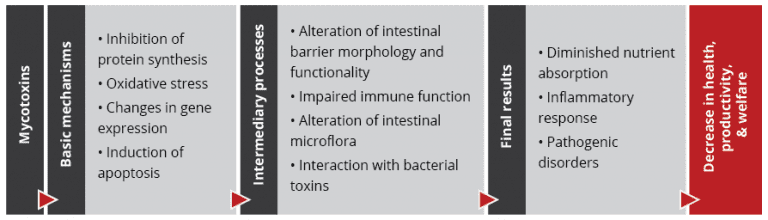
Mycotoxins are known to decrease health and performance in poultry production. Their modes of action, such as reducing protein synthesis and promoting oxidative stress and apoptosis, lead to cell destruction and lower cell replacement, affecting several organs and tissues.

When different stress factors collude, such as high temperatures and humidity, poor ventilation, high stocking density, and management events, the effects of in-feed mycotoxins can reach a higher level, which may include external signs.

The most common and recognized external sign of mycotoxicosis is mouth lesions caused by trichothecenes, which are highly associated with the presence of T-2 in the feed. However, other signs may appear, such as paleness of combs, shanks, and feet, as well as leg problems, ruffled feathers and poor feather coverage, feed passage, and abnormal feces.

In a series of articles, we want to report on external signs facilitating a differential diagnosis of mycotoxin contamination. This is necessarily followed by feed or raw material mycotoxin analysis and strategies to avoid or mitigate the effects of mycotoxin contamination in poultry production. In the first article, we will cover feathers.

A healthy plumage is crucial for growth and reproduction

Feathering is a crucial aspect of poultry health and productivity. Feathers are essential for thermoregulation, locomotion, adequate skin protection, and reproductive success, protecting hens from injury during mating. Inadequate feathering can lead to lower feed efficiency (Leeson and Walsh, 2004) as well as loss in fertility and chick production (Fisher, 2016). Mycotoxins in poultry feed can compromise feather quality in poultry production animals. This first article delves into the relationship between mycotoxins and poor feathering, exploring different mycotoxins and their mechanisms of action.

In which way do mycotoxins compromise feathering?

On the one hand, chronic mycotoxin exposure impairs the digestive process, hindering the absorption and utilization of vital nutrients essential for feather growth. This disruption can lead to malnutrition, directly impacting the quality and health of feathers. On the other hand, mycotoxins also interfere with metabolic processes critical for feather development, such as keratin synthesis (Wyatt et al., 1975;  Nguansangiam, 2004). Enzymatic pathways involved in synthesizing keratin, the protein building block of feathers, are particularly vulnerable to mycotoxin-induced disruptions. The presence of mycotoxins in feed has been associated with the manifestation of sparse feathering and the sticking out of feathers at an unnatural angle (Emous and Krimpen, 2019). In the case of multiple mycotoxins occurring in the feed, even at singularly unimportant concentrations, a negative impact on feathering is possible. Different mycotoxins have different target organs and consequences for the animal, so their ways of compromising feathering also vary. As feathering needs protein availability, all mycotoxins affecting the protein metabolism or the absorption of nutrients also impact the feathering process. Let us look at the most prominent mycotoxins.

1.   T-2 toxin

Due to climate change, T-2 toxins are on the rise. In the US, more than 50% of the tested samples contained T-2 toxin; in Europe, we found it in 31%, and in China, in 82% of the samples (EW Nutrition, 2024). The highest level was found in Europe, with 850 ppb.

Adverse effects of T-2 toxin in goslings were shown by Gu et al. (2023), who exposed the animals to 6 different levels of T-2 toxin, from 0.2 to 2.0 mg T-2 toxin/kg of feed. The goslings showed a sparse covering with short, dry, rough, curly, and gloss-free feathers on their back with dosages ≥0.8 mg/kg. When zooming on, T-2 can cause necroses of the layer of regenerative cells in the feather base, implying malformation or absence of new feathers, as well as structural damage to existing feathers on the base of the ramus and barb ridges (Hoerr et al. (1982), Leeson et al. (1995)).

The effects in feather regenerative cells are dose-dependent, as confirmed by Hoerr et al. (1982), who applied different doses of T-2 toxin (1.5, 2, 2.5, and 3 mg/kg body weight/day) to 7-day-old broilers for 14 days. Delayed feather development, especially at high dosages, was noticed, as well as malformations and opaque bands in the feathers, the latter probably caused by a segmental reduction in diameter.

Manafi et al. (2015) noticed feather malformations when broiler chickens were challenged with 0.5 ppm T-2 toxin in the feed in combination with an inoculation of 2.4×10⁸ cfu Mycoplasma gallisepticum. When the chickens were challenged only with T-2 toxin, the feathers were ruffled, showing that a coincidence of stress factors even aggravates the symptoms.

2.   Aflatoxins

Aflatoxins, produced by certain Aspergillus species, are among the most notorious mycotoxins. Looking at test results of the last year, Aflatoxin shows incidences between 25 (USA) over 40-65% (Europe, LATAM, MEA, and SEAP) up to 84-88% (China and South Asia) with average levels up to 42 ppb in South Asia (EW Nutrition, 2023). However, more information about the concrete impact of aflatoxins on feathering is needed. They may indirectly affect feathering because they impact digestion and the utilization of nutrients or trace minerals such as zinc, which is essential for the feather construction process. Damage to the liver impacts protein metabolism, and keratin is also necessary for feather production.

In other studies, Muhammad et al. (2017) fed 5 mg AFB1/kg to Arbor Acres broilers, and the birds showed ruffled feathers. A significantly lower feather shine was noticed by Saleemi et al. (2020) when they gave the animals 300 μg AFB1/kg of feed, and the birds of Zafar et al. (2017) showed ruffled, broken, dull, and dirty feathers after six weeks of feeding an aflatoxin-contaminated diet.

3.   Ochratoxin

Ochratoxins, commonly produced by Aspergillus and Penicillium fungi, also pose a significant threat to poultry. When looking at the mycotoxin report, this mycotoxin was found in 16% (Europe) to 70% (SEAP) of the samples (EW Nutrition, 2023). Ochratoxins primarily affect feathering by compromising the structural integrity of feathers and causing delayed feathering in broilers (Leeson, 2021).

Several trials have shown the negative impact of ochratoxin on feather quality. Hassan et al. (2010) fed OTA to laying hens and saw a dose-dependent (dosages from 0 to 10 mg/kg feed) occurrence of ruffled and broken feathers in the OTA group, whereas the plumage of the control group was shiny and well-formed. Hameed et al. (2012) also realized dull feathers when feeding 0.4 and 0.8 mg OTA per kg of feed. A further dose-dependent decrease in feather quality was described by Khan et al. (2023) in broiler chicks. He injected them with dosages from 0.1 to 1.7 mg/kg body weight on day 5 of age and saw a deterioration of feather appearance (rippled feathers) in the groups with the higher dosages of 1.3 and 1.7 mg/kg. Abidin et al. (2016) observed a similar dose-dependent deterioration of the feather quality in white Leghorn cockerels when feeding 1 or 2mg OTA/kg feed.

Combinations of aflatoxins and ochratoxins were also tested. Khan et al. (2017) fed moldy feed naturally containing 56 µg OTA and 136 µg AFB1 per kg to layer hens and saw a deterioration of feather quality with increasing feeding time. Qubih (2017) noticed ruffled feathers when feeding a diet naturally contaminated with 800 ppb of OTA and 100 ppb of AFB1.

4.   Scirpenol mycotoxins

Parkhurst et al. (1992) examined the effects of different scirpenol mycotoxins. After feeding graded levels of fusarium mycotoxins to broiler chicks until three weeks of age, they discovered that the impact of scirpenols stretched across the entire feathered body parts and that the degree of feather alteration is dose-dependent. The main alteration was a frayed or even missing web on the medial side of the outer end of the feather due to poor development of the barbs, barbules, and barbicels, and the tip of the feathers became square instead of rounded—the thinner and weaker shafts of the feathers inclined to show an accentuated medial curve.

Figure Feathering Affected By Scirpenol MycotoxinsParkhurst et al. (1992)

Figure 1: Feathering affected by scirpenol mycotoxins

In their trial, Parkhurst and Hamilton realized that 15-monoacetoxyscirpenol (15-MAS) caused the most severe alterations of feathers, and they determined a minimum effective dose (MED) of 0.5 µg/g diet. The MEDs for 4,15-diacetoxyscirpenol (4,15-DAS) and 3,4,15-triacetoxyscirpenol (TAS) were higher, 2 µg/g and > 8 µg/g, respectively.

How can we enable adequate feathering in poultry?

Adequate feathering of poultry is necessary for the animal’s health and welfare and to ensure fertility and productivity. The occurrence of mycotoxins in the feed – and the probability is high! – can cause poor feathering or the development of malformed feathers.

To best equip broilers, layers, and breeders, their feed must contain all nutrients essential for healthy growth and appropriate feathering. As the risk of contamination of the feed materials is very high (see EW Nutrition’s mycotoxin report 2023), it is of crucial importance to have an efficient mycotoxin risk management in place, which includes sampling, analysis of samples, and the use of mycotoxin binders. EW Nutrition offers MasterRisk, an online tool where farmers and feed millers can feed the results of their feed analysis concerning mycotoxins and get a risk management recommendation.

In the next part of the series, we will report on beak lesions and skin paleness, two other external signs of mycotoxin contamination.

References:

Abidin, Zain ul, Muhammad Zargham Khan, Aisha Khatoon, Muhammad Kashif Saleemi, and Ahrar Khan. “Protective Effects Ofl-Carnitine upon Toxicopathological Alterations Induced by Ochratoxin A in White Leghorn Cockerels.” Toxin Reviews 35, no. 3–4 (August 22, 2016): 157–64. https://doi.org/10.1080/15569543.2016.1219374.

Emous, R. A., and M. M. Krimpen. “Effects of Nutritional Interventions on Feathering of Poultry – a Review.” Poultry Feathers and Skin: The Poultry Integument in Health and Welfare, 2019, 133–50. https://doi.org/10.1079/9781786395115.0133.

Fisher, Colin. “Feathering in Broiler Breeder Females – Aviagen.” https://aviagen.com/, 2016. http://en.aviagen.com/assets/Tech_Center/Broiler_Breeder_Tech_Articles/English/Feathering-in-Broiler-Breeeder-Females-EN-2016.pdf.

Gu, Wang, Qiang Bao, Kaiqi Weng, Jinlu Liu, Shuwen Luo, Jianzhou Chen, Zheng Li, et al. “Effects of T-2 Toxin on Growth Performance, Feather Quality, Tibia Development and Blood Parameters in Yangzhou Goslings.” Poultry Science 102, no. 2 (February 2023): 102382. https://doi.org/10.1016/j.psj.2022.102382.

Hameed, Muhammad  Raza, Muhammad Khan, Ahrar Khan, and Ijaz Javed. “Ochratoxin Induced Pathological Alterations in Broiler Chicks: Effect of Dose and Duration.” Pakistan Veterinary Journal Pakistan Veterinary Journal 8318, no. 2 (December 2012): 2074–7764.

Hassan, Zahoor-Ul, M. Zargham Khan, Ahrar Khan, and Ijaz Javed. “Pathological Responses of White Leghorn Breeder Hens Kept on Ochratoxin A Contaminated Feed.” Pakistan Veterinary Journal 30, no. 2 (2010): 118–23.

Hoerr, F. J., W. W. Carlton, and B. Yagen. “Mycotoxicosis Caused by a Single Dose of T-2 Toxin or Diacetoxyscirpenol in Broiler Chickens.” Veterinary Pathology 18, no. 5 (September 1981): 652–64. https://doi.org/10.1177/030098588101800510.

Hoerr, F.J., W.W. Carlton, B. Yagen, and A.Z. Joffe. “Mycotoxicosis Produced in Broiler Chickens by Multiple Doses of Either T‐2 Toxin or Diacetoxyscirpenol.” Avian Pathology 11, no. 3 (January 1982): 369–83. https://doi.org/10.1080/03079458208436112.

Khan, Ahrar, Muhammad Mustjab Aalim, M. Zargham Khan, M. Kashif Saleemi, Cheng He, M. Noman Naseem, and Aisha Khatoon. “Does Distillery Yeast Sludge Ameliorate Moldy Feed Toxic Effects in White Leghorn Hens?” Toxin Reviews, January 25, 2017, 1–8. https://doi.org/10.1080/15569543.2017.1278707.

Khan, Shahzad Akbar, Eiko N. Itano, Anum Urooj, and Kashif Awan. “Ochratoxin-a Induced Pathological Changes in Broiler Chicks.” Pure and Applied Biology 12, no. 4 (December 10, 2023): 1608–16. https://doi.org/10.19045/bspab.2023.120162.

Leeson, S., and T. Walsh. “Feathering in Commercial Poultry II. Factors Influencing Feather Growth and Feather Loss.” World’s Poultry Science Journal 60, no. 1 (March 1, 2004): 52–63. https://doi.org/10.1079/wps20045.

Leeson, Steve. “Effects of Nutrition on Feathering.” Poultry World, May 22, 2021. https://www.poultryworld.net/specials/effects-of-nutrition-on-feathering/.

Leeson, Steven, Gonzalo J. Diaz Gonzalez, and John D. Summers. Poultry metabolic disorders and Mycotoxins. Guelph, Ontario, Canada: University Books, 1995.

Manafi, M., N. Pirany, M. Noor Ali, M. Hedayati, S. Khalaji, and M. Yari. “Experimental Pathology of T-2 Toxicosis and Mycoplasma Infection on Performance and Hepatic Functions of Broiler Chickens.” Poultry Science 94, no. 7 (July 2015): 1483–92. https://doi.org/10.3382/ps/pev115.

Muhammad, Ishfaq, Xiaoqi Sun, He Wang, Wei Li, Xinghe Wang, Ping Cheng, Sihong Li, Xiuying Zhang, and Sattar Hamid. “Curcumin Successfully Inhibited the Computationally Identified CYP2A6 Enzyme-Mediated Bioactivation of Aflatoxin B1 in Arbor Acres Broiler.” Frontiers in Pharmacology 8 (March 21, 2017). https://doi.org/10.3389/fphar.2017.00143.

Nguansangiam, Sudarat, Subhkij Angsubhakorn, Sutatip Bhamarapravati, and Apichart Suksamrarn. The Southeast Asian J of Tropical Medicine 34, no. 4 (2004): 899–905.

Parkhurst, Carmen R., Pat B. HamiltonON, and Adedamola A. AdemoyeroERO. “Abnormal Feathering of Chicks Caused by Scirpenol Mycotoxins Differing in Degree of Acetylation.” Poultry Science 71, no. 5 (May 1992): 833–37. https://doi.org/10.3382/ps.0710833.

Qubih, T. S. “Relationship between Mycotoxicosis and Calcium during Preproduction Period in Layers.” Iraqi Journal of Veterinary Sciences 26, no. 1 (June 28, 2012): 11–14. https://doi.org/10.33899/ijvs.2012.46888.

Saleemi, M. Kashif, Kamran Ashraf, S. Tehseen Gul, M. Noman Naseem, M. Sohail Sajid, Mashkoor Mohsin, Cheng He, Muhammad Zubair, and Ahrar Khan. “Toxicopathological Effects of Feeding Aflatoxins B1 in Broilers and Its Amelioration with Indigenous Mycotoxin Binder.” Ecotoxicology and Environmental Safety 187 (January 2020): 109712. https://doi.org/10.1016/j.ecoenv.2019.109712.

Wyatt, R.D., P.B. Hamilton, and H.R. Burmeister. “Altered Feathering of Chicks Caused by T-2 Toxin.” Poultry Science 54, no. 4 (July 1975): 1042–45. https://doi.org/10.3382/ps.0541042.

Zafar, Roheena, Farhat Ali Khan, and Muhammad Zahoor. “In Vivo Amelioration of Aflatoxin B1 in Broiler Chicks by Magnetic Carbon Nanocomposite.” Pesquisa Veterinária Brasileira 37, no. 11 (November 2017): 1213–19. https://doi.org/10.1590/s0100-736×2017001100005.


Mycotoxins in layer and breeder feed impact hens, eggs, hatchery, and chicks

White Chickens Farm

By Technical Team EW Nutrition

As the planet’s climate experiences changes, new patterns affect the microbial communities colonizing crops. Recently, several areas of the planet have experienced extreme temperatures, drought, changes in the humid/dry cycles, and an increase in atmospheric carbon dioxide (1,2). As a response, the fungi affecting the crops have shifted their geographical distribution, and with this, the pattern of mycotoxin occurrence also changed. For instance, in Europe, we are looking at higher frequencies and levels of Aflatoxins (AF), Ochratoxins (OT), and Fumonisins (FUM) than ten or even five years ago (2-4).

This affects animal production, as mycotoxin challenges show increased frequency, quantity, and variety. Mainly long-living animals, such as laying hens and breeders, can have a higher risk. Moreover, mycotoxins can also be carried over to the eggs, potentially risking human health in the case of layers (table eggs) and in the case of breeder hens, hatchery performance and day-old chick (DOC) quality.

Laying hens and breeders: carryover of mycotoxins into eggs

Most mycotoxins are absorbed in the proximal part of the gastrointestinal tract (Table 1). This absorption can be high, as in the case of aflatoxins (~90%), but also very limited, as in the case of fumonisins (<1%), with a significant portion of unabsorbed toxins remaining within the lumen of the gastrointestinal tract (5).

Once mycotoxins are ingested, detoxification and excretion processes are started by the body, and at the same time, organ damage ensues. The detoxification of mycotoxins is mainly carried out by the liver (6), and their accumulation happens primarily in the liver and kidneys. However, accumulation in other tissues, such as the reproductive organs and muscles, has also been found (7-9). The detoxification process’ objective is the final excretion of the toxins, which occurs through urine, feces, and bile; often, the toxins can also reach the eggs (7-20).

Table 1: mycotoxin absorption rates for poultry and their carry-over rate into eggs

Mycotoxin Main absorption sites Absorption rate in poultry Carry-over rate into eggs
Aflatoxins Duodenum, jejunum ≈90% ≈0.55%
DON Duodenum, jejunum ≈20% ≈0.001%
Fumonisins Duodenum, jejunum ≈1% ≈0.001%
Ochratoxin Jejunum ≈40% ≈0.15%
T-2 Duodenum, jejunum ≈20% ≈0.10%
Zearalenone Small & large intestine ≈10% ≈0.30%

(Adapted from 5, 7-17, 19-21)

Table 1 shows carry-over rates of mycotoxins into eggs, resulting from diverse studies (7-10, 14, 16, 19). However, the same studies indicate that results can vary broadly due to different factors, as reviewed by Völkel and collaborators (26). This variability is related to the amount and source of contamination, way of application, period, and the possible co-occurrence of various mycotoxins or several metabolites. Other factors to consider are animal-related, such as species, breed, sex, age group, production level, and health status. Environmental and management factors can play a role in carry-over rates, and finally, detection limits and analytical procedures also influence these results. In summary, highly varying carry-over has been demonstrated, and the risk needs to be considered when animals are exposed.

Mycotoxins in breeder’s feed impact hatchery performance and day-old chick quality

When hens are exposed to mycotoxins, their effects on the intestine, liver, and kidney decrease egg production and quality (10, 14, 27), and, in the case of breeders, consequently, affect hatchery performance, DOC production, and DOC quality (28-30). The main effects of mycotoxins, when we speak about DOC production, are exerted in the gastrointestinal tract, the liver, and the kidneys, affecting embryos and young chicks:

  • Intestine and kidneys: Mycotoxins harm the intestinal epithelium and have nephrotoxic effects, affecting calcium and vitamin D3 absorption and metabolism, necessary for eggshell quality (31). Thin and fragile shells can increase embryonic mortality, lower embryonic weight gain, and hinder hatchability (32).
  • Liver: The liver plays a central role in egg production as it is responsible for vitamin D3 metabolism, the production of nutrient transporters, and the synthesis of the lipids that make up the yolk. Thus, when liver function is impaired, the internal and external quality of the egg declines, which affects DOC production (31-34).
  • Embryo and young chicks: Studies (33-38) have found how mycotoxins affect the embryos. In general, there are two possibilities: the direct one, when the mycotoxin is transferred into the egg, and the indirect one, when the mycotoxin impacts egg quality and, therefore, leads to disease or death of the embryo. The result is a higher embryonic mortality or lower DOC quality. These, among others, result from the lower transfer of antioxidants and antibodies from the hen, low viability of the chick’s immune cells, and higher bacterial contamination. A lower relative weight of the bursa of Fabricio and the thymus is often found.

Qreshi’s team (29) studied the effects on the progeny of broiler breeders consuming feed highly contaminated with AFB1, finding suppression in antibody production and macrophage function in chicks after ten days. Similar results were found by other researchers (36, 37) evaluating the effects of AF and OTA as single and combined contamination. When both mycotoxins are present in the feed, the effect on hatchability and DOC quality are synergistic.

Due to mycotoxin contamination, the reproduction and immune response are impaired, resulting in decreased DOC production and increased early chick mortality, as they are more susceptible to bacterial and viral infections.

Mycotoxins impair table egg production and quality

Studies (22-24) have found mycotoxin contamination in commercial table eggs. A meta-analysis of mycotoxins’ concentration based on 11 published papers was completed recently (22): counting with data from 9509 samples, the meta-analysis reveals an overall presence of mycotoxins in 30% of the samples, being Beauvericin in the first place, followed by DON as well as AF and OTA in third and fourth place, respectively. The risk for humans depends on the intake of contaminated foods in terms of amount and frequency (25), and so far, it has not been estimated in most parts of the world.

Natural contamination in laying hens: a case report

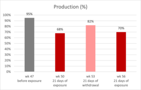
Giancarlo Bozzo’s team (39) reported and published a veterinary case regarding natural mycotoxin contamination in commercial egg production: up to week 47 of age, production parameters were on top of the genetic standards. However, a drop in egg production started at around week 47, and at week 50, egg production was only 68% (figure 1).

Figure
Figure 1: production of laying hens fed naturally contaminated feed with AFB1 and OTA
The house with the reduced performance received feed with linseed. In other houses of the same complex, which did not include linseed in the feed, production was unaffected. Therefore, this raw material was considered a possible cause of the issue. Linseed was removed from the formula, and three weeks after (53 weeks of age), egg production was at 84%. Afterward, linseed got back into the formulation, and the laying rate dropped again to 70% (week 56), this time accompanied by a significant increase in mortality.

Samples were collected at week 56, and AFB1 and OTA were detected in feed and the kidneys and livers of the hens consuming it (table 2). While the levels in the feed were not considered high risk, evidence from necropsy and histopathology suggested either a higher or a prolonged exposure; a synergistic effect of both mycotoxins on hen’s health and productivity can be inferred.

Table 2: mycotoxin analysis results for feed and organs

HPLC analysis results in samples of:
toxin Feed 1
(n=5)
Feed 2
(n=5)
Kidney

(n=10)

Liver

(n=10)

OTA 1.1 ± 0.1 ppb 31 ± 3 ppb 47 ± 3 ppb 24 ± 2 ppb
AFB1 ND 5.6 ± 0.3 ppb 1.4 ± 0.3 ppb 3.6 ± 0.4 ppb

The liver and kidneys were enlarged and showed signs of damage. Furthermore, urate crystals in the peritoneum and the abdominal air sac were observed, indicating renal failure. This limited the excretion of both toxins in the urine, increasing their half-life in the organism and enhancing the effects in target organs, contributing to the synergistic effect observed.

After using mycotoxin-free certified linseed, the problem receded. Though this is the best option to keep animals healthy and productive, it may not be practical in the long term due to the ubiquitous nature of the toxins and the cost and availability constraints of feed raw materials. Moreover, the mycotoxin levels present in the feed were relatively low and fell under recommended guidelines. For these reasons, in-feed toxin mitigation solutions must also be considered to reduce exposure for production animals.

In-feed intervention mitigates the effects of intermittent exposure to multiple mycotoxins

EW Nutrition conducted a study with Hy-Line W-36 layer-breeders intercalating three 10-day cycles of feed with 100ppb AFB1 + 100ppb OTA, with two 21-day cycles of non-challenged feed. An in-feed intervention (Solis Max 2.0, displayed as IFI) containing bentonite, yeast cell wall components, and a mixture of phytogenic components mitigated all effects.

Table 3: experimental groups and mycotoxin challenge

Treatment Group 100 ppb AFB1+ 100 ppb OTA IFI (2 kg/ton)
T-1 Control (C)
T-2 C+IFI X
T-3 Challenge (Ch) X
T-4 Ch+IFI X X

Trial design:

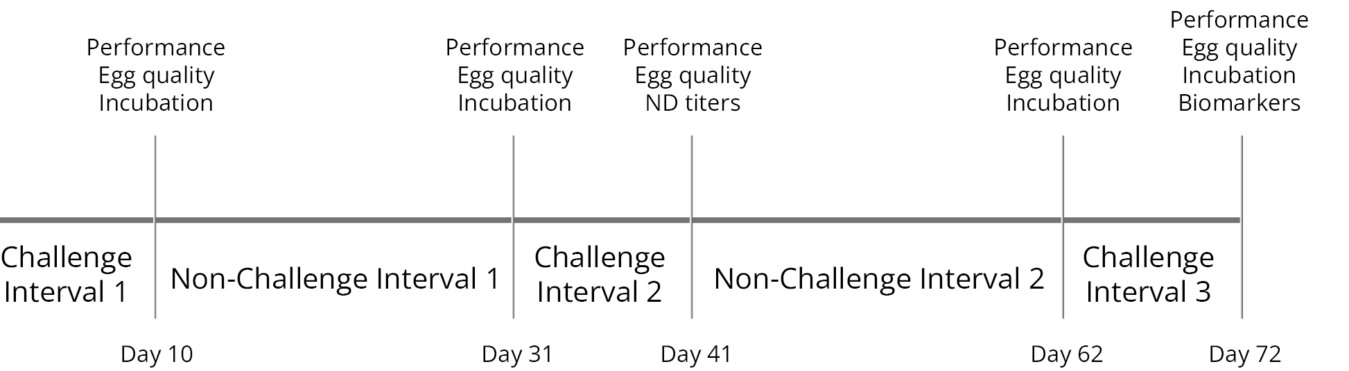
A total of 576 hens (18 replicates per diet, 8 hens each) and 58 roosters were randomly assigned to four diets at 28 weeks of age, as shown in Table 3. The 72-day experimental period included alternating 10-day challenge and 21-day non-challenge intervals (Figure 2). During the challenge intervals, the breeders in T-3 and T-4 were fed the mycotoxin-contaminated feed with and without the IFI.

FigureFigure 2: trial timeline showing challenge and non-challenge intervals and days of data collection and sampling.

Mitigated effects on egg production and egg quality

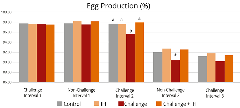
The challenge decreased overall egg production (Figure 3), egg mass, and shell thickness (Table 4). The first challenge interval did not affect production, but days later, from the first non-challenge period, all parameters were lower for the challenged group.

FigureDifferent letters indicate significant differences (p<0.05). Statistical tendencies (p<0.1) are indicated by (*).

Figure 3: Egg production of hens intermittently challenged with AFB1 and OTA, with and without in-feed Solis Max

The adverse effects on productivity and egg quality started after the first challenged feed was withdrawn and persisted through the following intervals until the end of the experiment. Similar effects in chronic mycotoxin challenges have been previously found (37, 39).

Table 4: Average egg quality parameters of hens intermittently challenged with AFB1+OTA, with and without an in-feed intervention (IFI)

Group Eggshell strength (N) Eggshell thickness (mm) Haugh Units
Control 21,02a 0,3661ab 70,88
IFI 21,16a 0,3702a 71,68
Challenge 20,05b 0,3630b   70,07*
Ch+IFI 21,06a 0,3698a 71,06

Different letters indicate significant differences (p<0.05). Statistical tendencies (p<0.1) are indicated by (*).

Mitigated effects on the progeny in incubation trials

Three incubation trials were performed: after the first challenge and non-challenge interval and at the end of the trial period after the third challenge interval. A significant decrease in fertility and hatchability was observed for the challenged group in all incubation trials. As mycotoxins affect egg quality (22-24) and can be transferred to the eggs (10, 14, 27), the effects were also shown in the case of hatchability and offspring performance. Fertility was affected from the first challenge interval onwards, continuing to be low for the challenge group until the end of the trial. However, the hatchability of fertile eggs dropped after the withdrawal of the contaminated feed and showed the lowest value during the third challenge interval.

The in-feed supplementation of Solis Max 2.0 (IFI) resulted in the consistent recovery of egg production and egg quality throughout the whole experimental period, achieving the same levels of productivity as the non-challenged control.

Figure
Letters indicate significant differences (p<0.05). Statistical tendencies (p<0.1), indicated by (*).

Figure 4: Hatchery parameters of eggs from breeders intermittently challenged with AFB1 and OTA, with and without an in-feed intervention (IFI).

Results in hatch of fertile can be related to egg quality, as the thickness of the eggshell influences the egg’s moisture loss and exchange with the environment during the incubation period. Thinner eggshells lead to higher embryo mortality (31, 32). The group having the challenge with Solis Max showed the same performance as the non-challenged control regarding hatchery performance.

Day-old chick weight was not affected. However, weight gain and mortality after ten days were hindered for the chicks from breeders taking the mycotoxin-contaminated feed (Table 5).

Table 5: Average day- and 10-day-old chick parameters from hens intermittently challenged with AFB1+OTA, with and without an in-feed intervention (IFI)

Parameter Control Challenge Ch + IFI
DOC body weight (g) 36,67 36,24 36,80
10-day body weight (g) 76,30a 75,94b 79,50a
10-day mortality (%) 0,94 1,26 0,97

Letters indicate significant differences (p<0.05). Statistical tendencies (p<0.1) indicated by (*)

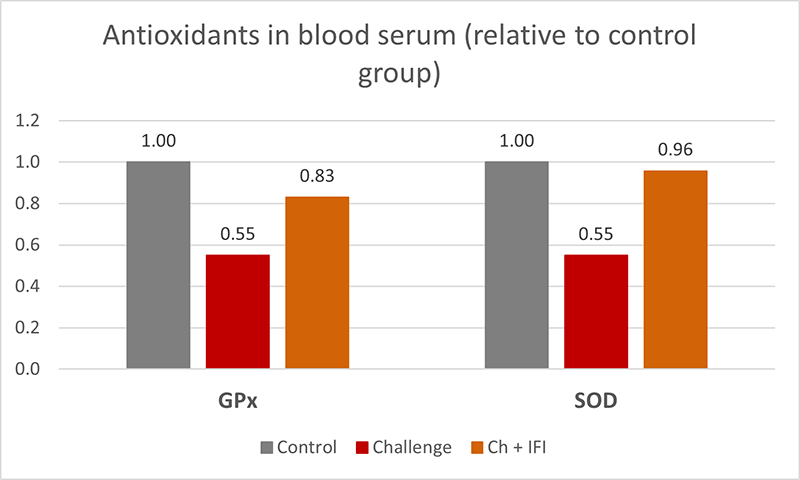
At the end of the experiment, oxidative stress biomarkers were measured in the blood serum of 15 hens per treatment, showing significantly lower GPx, and SOD (figure 5) in the challenged group, which indicates a depletion of the mechanisms to fight oxidative stress (40), the hens taking the in-feed product did not show this depletion.

FigureFigure 5: Antioxidants in blood serum, glutathione peroxidase (GPx), and superoxide dismutase (SOD) from breeders intermittently challenged with AFB1 and OTA, with and without an in-feed intervention (IFI).

Intermittent exposure to AFB1 and OTA negatively affected layer breeder productivity, egg quality, and hatchability and promoted oxidative stress in the birds. Intermittent mycotoxin challenges may affect animals even after the contamination is withdrawn. In-feed interventions showed effectiveness in mitigating these effects.

Climate changes bring new mycotoxin challenges – the right in-feed solutions can help

Today’s mycotoxin scenario shows increased frequency, quantity, and variety. Mainly long-living animals, such as laying hens and breeders, can be at more risk. Additionally, the contamination can be carried over to the eggs, potentially risking human health in the case of table eggs and hatchery performance and DOC quality in the case of breeders.

From case reports, we learn the consequences of real challenges and struggles in commercial production; from scientific trials based on possible commercial situations, we realize the advantages of interventions designed to tackle those challenges.

References

  1. Kos, Jovana, Mislav Anić, Bojana Radić, Manuela Zadravec, Elizabet Janić Hajnal, and Jelka Pleadin. “Climate Change—a Global Threat Resulting in Increasing Mycotoxin Occurrence.” Foods 12, no. 14 (July 14, 2023): 2704. https://doi.org/10.3390/foods12142704.
  2. Zingales, Veronica, Mercedes Taroncher, Piera Anna Martino, María-José Ruiz, and Francesca Caloni. “Climate Change and Effects on Molds and Mycotoxins.” Toxins 14, no. 7 (June 30, 2022): 445. https://doi.org/10.3390/toxins14070445.
  3. Loi, Martina, Antonio F. Logrieco, Tünde Pusztahelyi, Éva Leiter, László Hornok, and István Pócsi. “Advanced Mycotoxin Control and Decontamination Techniques in View of an Increased Aflatoxin Risk in Europe Due to Climate Change.” Frontiers in Microbiology 13 (January 10, 2023). https://doi.org/10.3389/fmicb.2022.1085891.
  4. Medina, Ángel, Jesús M González-Jartín, and María J Sainz. “Impact of Global Warming on Mycotoxins.” Current Opinion in Food Science 18 (December 2017): 76–81. https://doi.org/10.1016/j.cofs.2017.11.009.
  5. Grenier, Bertrand, and Todd Applegate. “Modulation of Intestinal Functions Following Mycotoxin Ingestion: Meta-Analysis of Published Experiments in Animals.” Toxins 5, no. 2 (February 21, 2013): 396–430. https://doi.org/10.3390/toxins5020396.
  6. Filazi, Ayhan, Begum Yurdakok-Dikmen, Ozgur Kuzukiran, and Ufuk Tansel Sireli. “Mycotoxins in Poultry.” Poultry Science, February 15, 2017. https://doi.org/10.5772/66302.
  7. Amirkhizi, Behzad, Seyed Rafie Arefhosseini, Masoud Ansarin, and Mahboob Nemati. “Aflatoxin B1in Eggs and Chicken Livers by Dispersive Liquid–Liquid Microextraction and HPLC.” Food Additives &amp; Contaminants: Part B, August 27, 2015, 1–5. https://doi.org/10.1080/19393210.2015.1067649.
  8. Emmanuel K, Tangni, Van Pamel Els, Huybrechts Bart, Delezie Evelyne, Van Hoeck Els, and Daeseleire Els. “Carry-over of Some Fusarium Mycotoxins in Tissues and Eggs of Chickens Fed Experimentally Mycotoxin-Contaminated Diets.” Food and Chemical Toxicology 145 (November 2020): 111715. https://doi.org/10.1016/j.fct.2020.111715.
  9. Ebrahem, Mohammad, Susanne Kersten, Hana Valenta, Gerhard Breves, and Sven Dänicke. “Residues of Deoxynivalenol (Don) and Its Metabolite de-Epoxy-Don in Eggs, Plasma and Bile of Laying Hens of Different Genetic Backgrounds.” Archives of Animal Nutrition 68, no. 5 (August 20, 2014): 412–22. https://doi.org/10.1080/1745039x.2014.949029.
  10. Salwa, A. Aly, and W. Anwer. “Effect of Naturally Contaminated Feed with Aflatoxins on Performance of Laying Hens and the Carryover of Aflatoxin B1 Residues in Table Eggs.” Pakistan Journal of Nutrition 8, no. 2 (January 15, 2009): 181–86. https://doi.org/10.3923/pjn.2009.181.186.
  11. Devreese, Mathias, Gunther Antonissen, Nathan Broekaert, Siegrid De Baere, Lynn Vanhaecke, Patrick De Backer, and Siska Croubels. “Comparative Toxicokinetics, Absolute Oral Bioavailability, and Biotransformation of Zearalenone in Different Poultry Species.” Journal of Agricultural and Food Chemistry 63, no. 20 (May 19, 2015): 5092–98. https://doi.org/10.1021/acs.jafc.5b01608.
  12. Galtier, P. “Biotransformation and Fate of Mycotoxins.” Toxin Reviews 18, no. 3 (August 1, 1999): 295–312. https://doi.org/10.3109/15569549909162648.
  13. Galtier, P., M. Alvinerie, and J.L. Charpenteau. “The Pharmacokinetic Profiles of Ochratoxin A in Pigs, Rabbits and Chickens.” Food and Cosmetics Toxicology 19 (January 1981): 735–38. https://doi.org/10.1016/0015-6264(81)90528-9.
  14. Hassan, Zahoor Ul, Muhammad Z Khan, Ahrar Khan, Ijaz Javed, and Zahid Hussain. “Effects of Individual and Combined Administration of Ochratoxin A and Aflatoxin B1 in Tissues and Eggs of White Leghorn Breeder Hens.” Journal of the Science of Food and Agriculture 92, no. 7 (December 16, 2011): 1540–44. https://doi.org/10.1002/jsfa.4740.
  15. Li, Shao-Ji, Guangzhi Zhang, Bin Xue, Qiaoling Ding, Lu Han, Jian-chu Huang, Fuhai Wu, Chonggao Li, and Chunmin Yang. “Toxicity and Detoxification of T-2 Toxin in Poultry.” Food and Chemical Toxicology 169 (November 2022): 113392. https://doi.org/10.1016/j.fct.2022.113392.
  16. Prelusky, D.B., R.M.G. Hamilton, and H.L. Trenholm. “Transmission of Residues to Eggs Following Long-Term Administration of 14 C-Labelled Deoxynivalenol to Laying Hens.” Poultry Science 68, no. 6 (June 1989): 744–48. https://doi.org/10.3382/ps.0680744.
  17. Ringot, Diana, Abalo Chango, Yves-Jacques Schneider, and Yvan Larondelle. “Toxicokinetics and Toxicodynamics of Ochratoxin A, an Update.” Chemico-Biological Interactions 159, no. 1 (January 2006): 18–46. https://doi.org/10.1016/j.cbi.2005.10.106.
  18. Osselaere, Ann, Mathias Devreese, Joline Goossens, Virginie Vandenbroucke, Siegrid De Baere, Patrick De Backer, and Siska Croubels. “Toxicokinetic Study and Absolute Oral Bioavailability of Deoxynivalenol, T-2 Toxin and Zearalenone in Broiler Chickens.” Food and Chemical Toxicology 51 (January 2013): 350–55. https://doi.org/10.1016/j.fct.2012.10.006.
  19. Sudhakar, BV. “A Study on Experimentally Induced Aflatoxicosis on the Carryover of Aflatoxin B1 into Eggs and Liver Tissue of White Leghorn Hens.” The Pharma Innovation Journal 11, no. 2S (2022): 213–17.
  20. Yiannikouris, Alexandros, and Jean-Pierre Jouany. “Mycotoxins in Feeds and Their Fate in Animals: A Review.” Animal Research 51, no. 2 (March 2002): 81–99. https://doi.org/10.1051/animres:2002012.
  21. Bouhet, Sandrine, and Isabelle P. Oswald. “The Intestine as a Possible Target for Fumonisin Toxicity.” Molecular Nutrition &amp; Food Research 51, no. 8 (August 2007): 925–31. https://doi.org/10.1002/mnfr.200600266.
  22. Fakhri, Yadolah, Mansour Sarafraz, Amene Nematollahi, Vahid Ranaei, Moussa Soleimani-Ahmadi, Van Nam Thai, and Amin Mousavi Khaneghah. “A Global Systematic Review and Meta-Analysis of Concentration and Prevalence of Mycotoxins in Birds’ Egg.” Environmental Science and Pollution Research 28, no. 42 (September 9, 2021): 59542–50. https://doi.org/10.1007/s11356-021-16136-y.
  23. Osaili, Tareq M., Akram R. Al-Abboodi, Mofleh AL. Awawdeh, and Samah Aref Jbour. “Assessment of Mycotoxins (Deoxynivalenol, Zearalenone, Aflatoxin B1 and Fumonisin B1) in Hen’s Eggs in Jordan.” Heliyon 8, no. 10 (October 2022). https://doi.org/10.1016/j.heliyon.2022.e11017.
  24. Wang, Lan, Qiaoyan Zhang, Zheng Yan, Yanglan Tan, Runyue Zhu, Dianzhen Yu, Hua Yang, and Aibo Wu. “Occurrence and Quantitative Risk Assessment of Twelve Mycotoxins in Eggs and Chicken Tissues in China.” Toxins 10, no. 11 (November 16, 2018): 477. https://doi.org/10.3390/toxins10110477.
  25. Tolosa, J., Y. Rodríguez-Carrasco, M.J. Ruiz, and P. Vila-Donat. “Multi-Mycotoxin Occurrence in Feed, Metabolism and Carry-over to Animal-Derived Food Products: A Review.” Food and Chemical Toxicology 158 (December 2021): 112661. https://doi.org/10.1016/j.fct.2021.112661.
  26. Völkel, Inger, Eva Schröer-Merker, and Claus-Peter Czerny. “The Carry-over of Mycotoxins in Products of Animal Origin with Special Regard to Its Implications for the European Food Safety Legislation.” Food and Nutrition Sciences 02, no. 08 (2011): 852–67. https://doi.org/10.4236/fns.2011.28117.
  27. Yuan, Tao, Junyi Li, Yanan Wang, Meiling Li, Ao Yang, Chenxi Ren, Desheng Qi, and Niya Zhang. “Effects of Zearalenone on Production Performance, Egg Quality, Ovarian Function and Gut Microbiota of Laying Hens.” Toxins 14, no. 10 (September 21, 2022): 653. https://doi.org/10.3390/toxins14100653.
  28. Song, Bin, Teng Ma, Damien P. Prévéraud, Keying Zhang, Jianping Wang, Xuemei Ding, Qiufeng Zeng, et al. “Research Note: Effects of Feeding Corn Naturally Contaminated with Aflatoxin B1, Deoxynivalenol, and Zearalenone on Reproductive Performance of Broiler Breeders and Growth Performance of Their Progeny Chicks.” Poultry Science 102, no. 11 (November 2023): 103024. https://doi.org/10.1016/j.psj.2023.103024.
  29. Qureshi, MA, J Brake, PB Hamilton, WM Hagler, and S Nesheim. “Dietary Exposure of Broiler Breeders to Aflatoxin Results in Immune Dysfunction in Progeny Chicks.” Poultry Science 77, no. 6 (June 1998): 812–19. https://doi.org/10.1093/ps/77.6.812.
  30. Ul-Hassan, Zahoor, Muhammad Zargham Khan, Ahrar Khan, and Ijaz Javed. “Immunological Status of the Progeny of Breeder Hens Kept on Ochratoxin a (OTA)- and Aflatoxin B1(Afb1)-Contaminated Feeds.” Journal of Immunotoxicology 9, no. 4 (April 24, 2012): 381–91. https://doi.org/10.3109/1547691x.2012.675365.
  31. Devegowda, G., and D. Ravikiran. “Mycotoxins and Eggshell Quality: Cracking the Problem.” World Mycotoxin Journal 1, no. 2 (May 1, 2008): 203–8. https://doi.org/10.3920/wmj2008.1037.
  32. Onagbesan, O., V. Bruggeman, L. De Smit, M. Debonne, A. Witters, K. Tona, N. Everaert, and E. Decuypere. “Gas Exchange during Storage and Incubation of Avian Eggs: Effects on Embryogenesis, Hatchability, Chick Quality and Post-Hatch Growth.” World’s Poultry Science Journal 63, no. 4 (December 1, 2007): 557–73. https://doi.org/10.1017/s0043933907001614.
  33. Ebrahem, Mohammad, Susanne Kersten, Hana Valenta, Gerhard Breves, Andreas Beineke, Kathrin Hermeyer, and Sven Dänicke. “Effects of Feeding Deoxynivalenol (Don)-Contaminated Wheat to Laying Hens and Roosters of Different Genetic Background on the Reproductive Performance and Health of the Newly Hatched Chicks.” Mycotoxin Research 30, no. 3 (April 11, 2014): 131–40. https://doi.org/10.1007/s12550-014-0197-z.
  34. Yegani, M., T.K. Smith, S. Leeson, and H.J. Boermans. “Effects of Feeding Grains Naturally Contaminated with Fusarium Mycotoxins on Performance and Metabolism of Broiler Breeders.” Poultry Science 85, no. 9 (September 2006): 1541–49. https://doi.org/10.1093/ps/85.9.1541.
  35. Calini, F, and F Sirri. “Breeder Nutrition and Offspring Performance.” Revista Brasileira de Ciência Avícola 9, no. 2 (June 2007): 77–83. https://doi.org/10.1590/s1516-635×2007000200001.
  36. Hassan, ZU, MZ Khan, A Khan, I Javed, U Sadique, and A Khatoon. “Ochratoxicosis in White Leghorn Breeder Hens: Production and Breeding Performance.” Vet. J. 32, no. 4 (2012): 557–61.
  37. Verma, J., T. S. Johri, and B. K. Swain. “Effect of Varying Levels of Aflatoxin, Ochratoxin and Their Combinations on the Performance and Egg Quality Characteristics in Laying Hens.” Asian-Australasian Journal of Animal Sciences 16, no. 7 (January 1, 2003): 1015–19. https://doi.org/10.5713/ajas.2003.1015.
  38. Johnson-Dahl, M.L., M.J. Zuidhof, and D.R. Korver. “The Effect of Maternal Canthaxanthin Supplementation and Hen Age on Breeder Performance, Early Chick Traits, and Indices of Innate Immune Function.” Poultry Science 96, no. 3 (March 2017): 634–46. https://doi.org/10.3382/ps/pew293.
  39. Bozzo, Giancarlo, Nicola Pugliese, Rossella Samarelli, Antonella Schiavone, Michela Maria Dimuccio, Elena Circella, Elisabetta Bonerba, Edmondo Ceci, and Antonio Camarda. “Ochratoxin A and Aflatoxin B1 Detection in Laying Hens for Omega 3-Enriched Eggs Production.” Agriculture 13, no. 1 (January 5, 2023): 138. https://doi.org/10.3390/agriculture13010138.
  40. Surai, Peter F., Ivan I. Kochish, Vladimir I. Fisinin, and Michael T. Kidd. “Antioxidant Defence Systems and Oxidative Stress in Poultry Biology: An Update.” Antioxidants 8, no. 7 (July 22, 2019): 235. https://doi.org/10.3390/antiox8070235.


Masked mycotoxins – particularly dangerous for dairy cows

Cows

By Si-Trung Tran, SEAP Regional Technical Manager, EW Nutrition

Technical Team and Dr. Inge Heinzl, Editor, EW Nutrition

Mycotoxins are secondary metabolites of fungi, commonly found as contaminants in agricultural products. In some cases, these compounds are used in medicine or industry, such as penicillin and patulin. In most cases, however, they are considered xenobiotics that are toxic to animals and humans, causing the disease collectively known as mycotoxicosis. The adverse effects of mycotoxins on human and animal health have been documented in many publications. Aflatoxins (AFs) and deoxynivalenol (DON, vomitoxin) are amongst the most critical mycotoxins affecting milk production and -quality.

Aflatoxins do not only affect cows

Aflatoxins (AFs) are highly oxygenated, heterocyclic difuranocoumarin compounds produced by Aspergillus flavus and Aspergillus parasiticus. They colonize crops, including many staple foods and feed ingredients. Within a group of over 20 AFs and derivatives, aflatoxin B1 (AFB1), B2, G1, and G2 are the most important naturally occurring compounds.

Among the aflatoxins, AFB1 is the most widespread and most toxic to humans and animals. Concern about mycotoxin contamination in dairy products began in the 1960s with the first reported cases of contamination by aflatoxin M1 (AFM1), a metabolite of AFB1 formed in the liver of animals and excreted in the milk.

There is ample evidence that lactating cows exhibit a significant reduction in feed efficiency and milk yield within a few days of consuming aflatoxin-contaminated feed. At the cellular level, aflatoxins cause degranulation of endoplasmic membranes, loss of ribosomes from the endoplasmic reticulum, loss of nuclear chromatin material, and altered nuclear shapes. The liver, as the organ mainly dealing with the decontamination of the organism, gets damaged, and performance drops. Immune cells are also affected, reducing immune competence and vaccination success (Arnold and Gaskill, 2023).

DON reduces cows’ performance

Another mycotoxin that can also reduce milk quality and affect metabolic parameters, as well as the immune function of dairy cows, is DON. DON is produced by different fungi of the Fusarium genus that infect plants. DON synthesis is associated with rainy weather from crop flowering to harvest. Whitlow and co-workers (1994) reported the association between DON and poor performance in dairy herds and showed decreased milk production in dairy cows fed 2.5 mg DON/kg. However, in cows fed 6 to 12 mg DON/kg dry matter for 10 weeks, no DON or its metabolite DOM-1 residues were detected in milk.

Masked mycotoxins hide themselves during analysis

Plants suffering from fungal infestations and thus confronted with mycotoxins convert the harmful forms of mycotoxins into less harmful or harmless ones for themselves by conjugation to sulfates, organic acids, or sugars. Conjugated mycotoxins cannot always be detected by standard analytical methods. However, in animals, these forms can be released and transformed into parent compounds by enzymes and microorganisms in the gastrointestinal tract. Thus, the feed may show a concentration of mycotoxins that is still below the limit value, but in the animal, this concentration is suddenly much higher. In dairy cows, the release of free mycotoxins from conjugates during digestion may play an important role in understanding the silent effects of mycotoxins.

Fusarium toxins, in particular, frequently occur in this “masked form”. They represent a serious health risk for animals and humans.

Aflatoxins first show up in the milk

Masked aflatoxins may also play a role in total aflatoxin contamination of feed materials. Research has harvested little information on masked aflatoxins that may be present in TMR ingredients. So far, metabolites such as Aflatoxin M2 have been identified (Righetti, 2021), which may reappear later in milk as AFM1.

DON-related symptoms without DON?

Sometimes, animals show DON-related symptoms, with low levels detected in the feed or raw materials. Besides sampling errors, this enigma could be due to conjugated or masked DON, which is structurally altered DON bound to various compounds such as glucose, fatty acids, and amino acids. These compounds escape conventional feed analysis techniques because of their modified chemical properties but can be released as their toxic precursors after acid hydrolysis.

Masked DON was first described in 1984 by Young and co-workers, who found that the DON content of yeast-fermented foods was higher than that of the contaminated wheat flour used in their production. The most plausible reason for this apparent increase was that the toxin from the wheat had been converted to a compound other than DON, which could be converted back to DON under certain conditions. Since this report, there has been much interest in conjugated or masked DON.

Silage: masked DON is a challenge for dairy producers

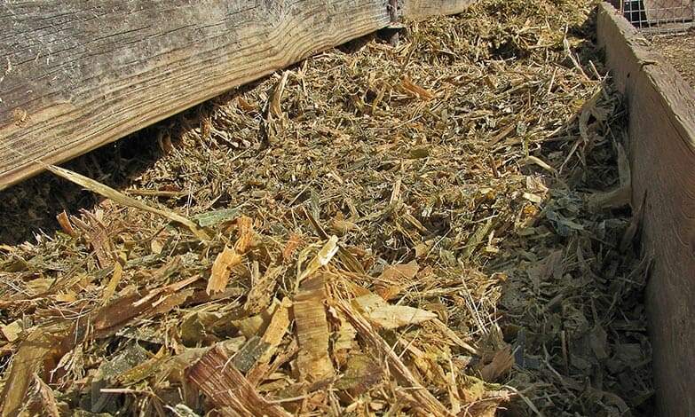
Silage is an essential feed for dairy cows, supporting milk production. Most silage is made from corn and other grains. The whole green plant is used, which can be infected by fungi. Since infection of corn with Fusarium spp. and subsequent DON contamination is usually a major problem in the field worldwide, a relatively high occurrence of this toxin in silage must be expected. The ensiling process may reduce the amount of Fusarium fungi, but the DON formed before ensiling is very stable.

Corn Silage

Silage samples show DON levels of concern

It is reasonable to assume that the DON biosynthesized by the fungi was metabolized by the plants to a new compound and thus masked DON. Under ensiling conditions, masked DON can be hydrolyzed, producing free DON again. Therefore, the level of free DON in the silage may not reflect the concentration measured in the plants before ensiling.

A study analyzed 50 silage samples from different farms in Ontario, Canada. Free DON was found in all samples, with levels ranging from 0.38 to 1.72 µg/g silage (unpublished data). Eighty-six percent of the samples contained DON at concentrations higher than 0.5 µg/g. Together with masked DON, it poses a potential threat to dairy cattle.

Specific hydrolysis conditions allow detection

However, in the natural ensiling process, the conditions for hydrolysis of masked DON are not optimal. The conditions that allow improved analysis of masked DON were recently described. This method detected masked DON in 32 of 50 silage samples (64%) along with free DON, increasing DON concentration by 23% in some cases (unpublished data).

Mycotoxins impact humans and animals

Aflatoxins, as well as DON, have adverse effects. In the case of DON, the impact on the animal is significant; in the case of aflatoxin, the possible long-term effects on humans are of higher relevance.

DON has more adverse effects on the animal and its performance

Unlike AFs, DON may be found in milk at low or trace concentrations. It is more associated with negative effects in the animal, altered rumen fermentation, and reduced flow of usable protein into the duodenum. For example, milk fat content was significantly reduced when cows were fed 6 µg DON/kg. However, the presence of DON also indicates that the feed probably contains other mycotoxins, such as zearalenone (ZEA) (estrogenic mycotoxin) and fusaric acid (pharmacologically active compound). All these mycotoxins may interact to cause symptoms that are different or more severe than expected, considering their individual effects. DON and related compounds also have immunosuppressive effects, resulting in increased somatic cell counts in milk. The U.S. FDA has established an action level for DON in wheat and wheat-derived products intended for cows, which is 5µg DON/g feed and the contaminated ingredient must not exceed 40% of the ration.

Aflatoxins decrease milk quality and pose a risk to humans

Aflatoxins are poorly degraded in the rumen, with aflatoxicol being the main metabolite that can be reconverted to AFB1. Most AFs are absorbed and extensively metabolized/hydrolyzed by enzymes found mainly in the liver. This results in the formation of AFM1, a part of which is conjugated to glucuronic acid and subsequently excreted in the bile. The other part enters the systemic circulation. It is either excreted in urine or milk. AFM1 appears within 12-48 hours after ingestion in cow’s milk. The excreted amount of AFM1 in milk from dairy cows usually ranges from 0.17% to 3% of the ingested AFB1. However, this carryover rate may vary from day to day and from one milking to the next in individual animals, as it is influenced by various factors, such as feeding regime, health status, individual biotransformation capacity, and, of course, by actual milk production. Carryover rates of up to 6.2% have been reported in high-yielding dairy cows producing up to 40 liters of milk per day.

In various experiments, AFM1 showed both carcinogenic and immunosuppressive effects. Accordingly, the International Agency for Research on Cancer (IARC) classified AFM1 as being in Group 2B and, thus, possibly carcinogenic in humans. The action level of 0.50 ppb and 0.05 ppb for AFM1 in milk is strictly adhered to by the U.S. Food and Drug Administration (FDA) and the European Food Safety Authority (EFSA), respectively.

Trials show the high adsorption capacity of Solis Max

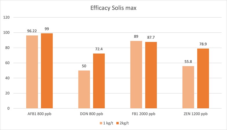
A trial was conducted at an independent laboratory located in Spain. The evaluation of the performance of Solis Max was executed with the following inclusion levels:

  • 0.10% equivalent to 1.0 kg of Solis Max per ton of feed
  • 0.20% equivalent to 2.0 kg of Solis Max per ton of feed

A phosphate buffer solution at pH 7 was prepared for the trial to simulate rumen conditions. Each mycotoxin was tested separately, preparing solutions with known contamination (final concentration described in the table below). The contaminated solutions were divided into 3 parts: A positive control, 0.10% Solis Max and 0.20% Solis Max. All samples were incubated at 41°C for 1 hour, centrifuged, and the supernatant was analyzed for the mycotoxin added to determine the binding efficacy. All analyses were carried out by high-performance liquid chromatography (HPLC) with standard detectors.

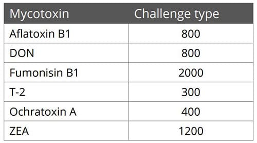
Mycotoxin Contamination Level (ppb)
Aflatoxin B1 800
DON 800
Fumonisin B1 2000
ZEA 1200

Results:
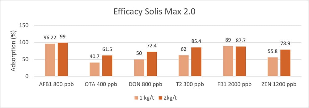
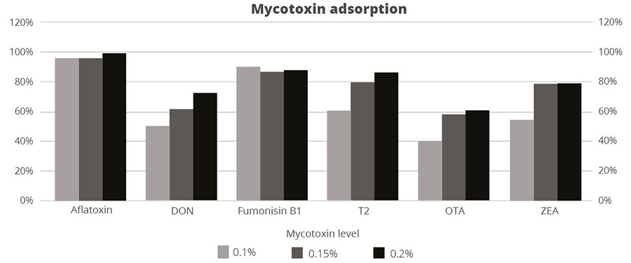
The higher concentration of Solis max showed a higher adsorption rate for most mycotoxins. The high dose of Solis Max adsorbed 99% of the AFB1 contamination. In the case of DON, more than 70% was bound. For fumonisin B1 and zearalenone, Solis max showed excellent binding rates of 87.7% and 78.9%, respectively (Figure 1).

FigureFigure 1: Solis Max showed a high binding capacity for the most relevant mycotoxins

Another trial was conducted at an independent laboratory serving the food and feed industry and located in Valladolid, Spain.

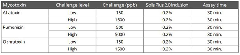
All tests were carried out as duplicates and using a standard liquid chromatography/mass spectrometry (LC/MS/MS) quantification. Interpretation and data analysis were carried out with the corresponding software. The used pH was 3.0, toxin concentrations and anti-mycotoxin agent application rates were set as follows (Table 1):

TableTable 1: Trial set-up testing the binding capacity of Solis Plus 2.0 for several mycotoxins in different contamination levels

Results:

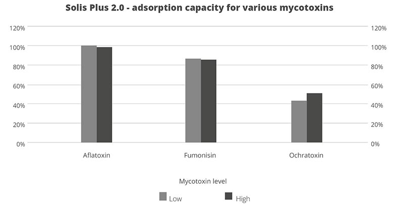
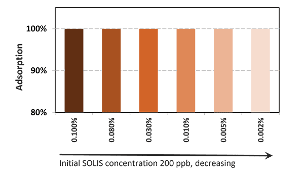
Under acidic conditions (pH3), Solis Plus 2.0 effectively adsorbs the three tested mycotoxins at low and high levels. 100% binding of aflatoxin was achieved at a level of 150ppb and 98% at 1500ppb.In the case of fumonisin, 87% adsorption could be reached at 500ppb and 86 for a challenge with 5000ppb. 43% ochratoxin was adsorbed at the contamination level of 150ppb and 52% at 1500ppb.

FigureFigure 2: The adsorption capacity of Solis Plus 2.0 for three different mycotoxins at two challenge levels

Mycotoxins – Effective risk management is of paramount importance

Although the rumen microflora may be responsible for conferring some mycotoxin resistance to ruminants compared to monogastric animals, there are still effects of mycotoxins on rumen fermentation and milk quality. In addition, masked mycotoxins in feed present an additional challenge for dairy farms because they are not readily detectable by standard analyses.

Feeding dairy cows with feed contaminated with mycotoxins can lead to a reduction in milk production. Milk quality may also deteriorate due to an adverse change in milk composition and mycotoxin residues, threatening the innocuousness of dairy products. Dairy farmers should therefore have feed tested regularly, consider masked mycotoxins, and take action. EW Nutrition’s MasterRisk tool provides a risk evaluation and corresponding recommendations for the use of products that mitigate the effects of mycotoxin contamination and, in the end, guarantee the safety of all of us.


Toxin Mitigation 101: Essentials for Animal Production

Fusarium Mycotoxins

By Monish Raj, Assistant Manager-Technical Services, EW Nutrition
Inge Heinzl, Editor, EW Nutrition  

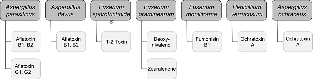
Mycotoxins, toxic secondary metabolites produced by fungi, are a constant and severe threat to animal production. They can contaminate grains used for animal feed and are highly stable, invisible, and resistant to high temperatures and normal feed manufacturing processes. Mycotoxin-producing fungi can be found during plant growth and in stored grains; the prevalence of fungi species depends on environmental conditions, though in grains, we find mainly three genera: Aspergillus, Penicillium, and Fusarium. The most critical mycotoxins for poultry production and the fungi that produce them are detailed in Fig 1.

FigureFigure 1: Fungi species and their mycotoxins of worldwide importance for poultry production (adapted from Bryden, 2012).

The effects of mycotoxins on the animal are manifold

When, usually, more than one mycotoxin enters the animal, they “cooperate” with each other, which means that they combine their effects in different ways. Also, not all mycotoxins have the same targets.

The synergistic effect: When 1+1 ≥3

Even at low concentrations, mycotoxins can display synergistic effects, which means that the toxicological consequences of two or more mycotoxins present in the same sample will be higher than the sum of the toxicological effects of the individual mycotoxins. So, disregarded mycotoxins can suddenly get important due to their additive or synergistic effect.

Table 1: Synergistic effects of mycotoxins in poultry

Synergistic interactions
DON ZEN T-2 DAS
FUM * * *
NIV * * *
AFL * *

Table 2: Additive effects of mycotoxins in poultry

Additive interactions
AFL T2 DAS MON
FUM + + + +
DON + +
OTA + +

Recognize the effects of mycotoxins in animals is not easy

The mode of action of mycotoxins in animals is complex and has many implications. Research so far could identify the main target organs and effects of high levels of individual mycotoxins. However, the impact of low contamination levels and interactions are not entirely understood, as they are subtle, and their identification requires diverse analytical methods and closer observation.

With regard to the gastrointestinal tract, mycotoxins can inhibit the absorption of nutrients vital for maintaining health, growth, productivity, and reproduction. The nutrients affected include amino acids, lipid-soluble vitamins (vitamins A, D, E, and K), and minerals, especially Ca and P (Devegowda and Murthy, 2005). As a result of improper absorption of nutrients, egg production, eggshell formation, fertility, and hatchability are also negatively influenced.

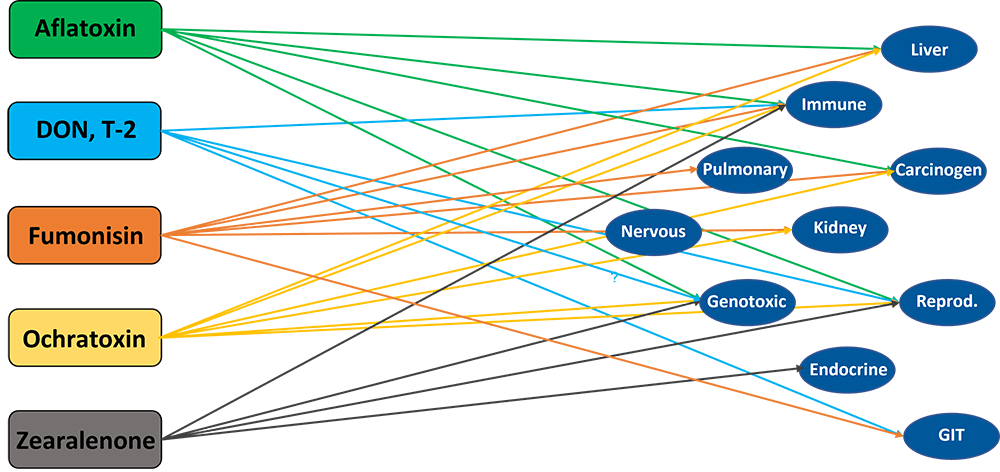
Most mycotoxins also have a negative impact on the immune system, causing a higher susceptibility to disease and compromising the success of vaccinations. Besides that, organs like kidneys, the liver, and lungs, but also reproduction, endocrine, and nervous systems get battered.

Mycotoxins have specific targets

Aflatoxins, fumonisins, and ochratoxin impair the liver and thus the physiological processes modulated and performed by it:

  • lipid and carbohydrate metabolism and storage
  • synthesis of functional proteins such as hormones, enzymes, and nutrient transporters
  • metabolism of proteins, vitamins, and minerals.

For trichothecenes, the gastrointestinal tract is the main target. There, they hamper digestion, absorption, and intestinal integrity. T-2 can even produce necrosis in the oral cavity and esophagus.

Figure Main Targets Of Important MycotoxinsFigure 2: Main target organs of important mycotoxins

How to reduce mycotoxicosis?

There are two main paths of action, depending on whether you are placed along the crop production, feed production, or animal production cycle. Essentially, you can either prevent the formation of mycotoxins on the plant on the field during harvest and storage or, if placed at a further point along the chain, mitigate their impact.

Preventing mycotoxin production means preventing mold growth

To minimize the production of mycotoxins, the development of molds must be inhibited already during the cultivation of the plants and later on throughout storage. For this purpose, different measures can be taken:

Selection of the suitable crop variety, good practices, and optimal harvesting conditions are half of the battle

Already before and during the production of the grains, actions can be taken to minimize mold growth as far as possible:

  • Choose varieties of grain that are area-specific and resistant to insects and fungal attacks.
  • Practice crop rotation
  • Harvest proper and timely
  • Avoid damage to kernels by maintaining the proper condition of harvesting equipment.

Optimal moisture of the grains and the best hygienic conditions are essential

The next step is storage. Here too, try to provide the best conditions.

  • Dry properly: grains should be stored at <13% of moisture
  • Control moisture: minimize chances of moisture to increase due to condensation, and rain-water leakage
  • Biosecurity: clean the bins and silos routinely.
  • Prevent mold growth: organic acids can help prevent mold growth and increase storage life.

Mold production does not mean that the war is lost

Even if molds and, therefore, mycotoxins occur, there is still the possibility to change tack with several actions. There are measures to improve feed and support the animal when it has already ingested the contaminated feed.

1.    Feed can sometimes be decontaminated

If a high level of mycotoxin contamination is detected, removing, replacing, or diluting contaminated raw materials is possible. However, this is not very practical, economically costly, and not always very effective, as many molds cannot be seen. Also, heat treatment does not have the desired effect, as mycotoxins are highly heat stable.

2.    Effects of mycotoxins can be mitigated

Even when mycotoxins are already present in raw materials or finished feed, you still can act. Adding products adsorbing the mycotoxins or mitigating the effects of mycotoxins in the organism has been considered a highly-effective measure to protect the animals (Galvano et al., 2001).

This type of mycotoxin mitigation happens at the animal production stage and consists of suppressing or reducing the absorption of mycotoxins in the animal. Suppose the mycotoxins get absorbed in the animal to a certain degree. In that case, mycotoxin mitigation agents help by promoting the excretion of mycotoxins, modifying their mode of action, or reducing their effects. As toxin-mitigating agents, the following are very common:

Aluminosilicates: inorganic compounds widely found in nature that are the most common agents used to mitigate the impact of mycotoxins in animals. Their layered (phyllosilicates) or porous (tectosilicates) structure helps “trap” mycotoxins and adsorbs them.

  • Bentonite / Montmorillonite: classified as phyllosilicate, originated from volcanic ash. This absorbent clay is known to bind multiple toxins in vivo. Incidentally, its name derives from the Benton Shale in the USA, where large formations were discovered 150 years ago.
    Bentonite mainly consists of smectite minerals, especially montmorillonite (a layered silicate with a larger surface area and laminar structure).
  • Zeolites: porous crystalline tectosilicates, consisting of aluminum, oxygen, and silicon. They have a framework structure with channels that fit cations and small molecules. The name “zeolite” means “boiling stone” in Greek, alluding to the steam this type of mineral can give off in the heat). The large pores of this material help to trap toxins.

Activated charcoal: the charcoal is “activated” when heated at very high temperatures together with gas. Afterward, it is submitted to chemical processes to remove impurities and expand the surface area. This porous, powdered, non-soluble organic compound is sometimes used as a binder, including in cases of treating acute poisoning with certain substances.

Yeast cell wall: derived from Saccharomyces cerevisiae. Yeast cell walls are widely used as adsorbing agents. Esterified glucomannan polymer extracted from the yeast cell wall was shown to bind to aflatoxin, ochratoxin, and T-2 toxin, individually and combined (Raju and Devegowda 2000).

Bacteria: In some studies, Lactic Acid Bacteria (LAB), particularly Lactobacillus rhamnosus, were found to have the ability to reduce mycotoxin contamination.

Which characteristics are crucial for an effective toxin-mitigating solution

If you are looking for an effective solution to mitigate the adverse effects of mycotoxins, you should keep some essential requirements:

  1. The product must be safe to use:
    1. safe for the feed-mill workers.
    2. does not have any adverse effect on the animal
    3. does not leave residues in the animal
    4. does not bind with nutrients in the feed.
  2. It must show the following effects:
    1. effectively adsorbs the toxins relevant to your operation.
    2. helps the animals to cope with the consequences of non-bound toxins.
  3. It must be practical to use:
    1. cost-effective
    2. easy to store and add to the feed.

Depending on

  • the challenge (one mycotoxin or several, aflatoxin or another mycotoxin),
  • the animals (short-cycle or long-living animals), and
  • the economical resources that can be invested,

different solutions are available on the market. The more cost-effective solutions mainly contain clay to adsorb the toxins. Higher-in-price products often additionally contain substances such as phytogenics supporting the animal to cope with the consequences of non-bound mycotoxins.

Solis – the cost-effective solution

In the case of contamination with only aflatoxin, the cost-effective solution Solis is recommended. Solis consists of well-selected superior silicates with high surface area due to its layered structure. Solis shows high adsorption of aflatoxin B1, which was proven in a trial:

FigureFigure 3: Binding capacity of Solis for Aflatoxin

Even at a low inclusion rate, Solis effectively binds the tested mycotoxin at a very high rate of nearly 100%. It is a high-efficient, cost-effective solution for aflatoxin contamination.

Solis Max 2.0: The effective mycotoxin solution for sustainable profitability

Solis Max 2.0 has a synergistic combination of ingredients that acts by chemi- and physisorption to prevent toxic fungal metabolites from damaging the animal’s gastrointestinal tract and entering the bloodstream.

Figure

Figure 4: Composition and effects of Solis Max 2.0

Solis Max 2.0 is suitable for more complex challenges and longer-living animals: in addition to the pure mycotoxin adsorption, Solis Max 2.0 also effectively supports the liver and, thus, the animal in its fight against mycotoxins.

In an in vitro trial, the adsorption capacity of Solis Max 2.0 for the most relevant mycotoxins was tested. For the test, the concentrations of Solis Max 2.0 in the test solutions equated to 1kg/t and 2kg/t of feed.

FigureFigure 5: Efficacy of Solis Max 2.0 against different mycotoxins relevant in poultry production

The test showed a high adsorption capacity: between 80% and 90% for Aflatoxin B1, T-2 Toxin (2kg/t), and Fumonisin B1. For OTA, DON, and Zearalenone, adsorption rates between 40% and 80% could be achieved at both concentrations (Figure 5). This test demonstrated that Solis Max 2.0 could be considered a valuable tool to mitigate the effects of mycotoxins in poultry.

Broiler trial shows improved performance in broilers

Protected and, therefore, healthier animals can use their resources for growing/laying eggs. A trial showed improved liver health and performance in broilers challenged with two different mycotoxins but supported with Solis Max 2.0.

For the trial, 480 Ross-308 broilers were divided into three groups of 160 birds each. Each group was placed in 8 pens of 20 birds in a single house. Nutrition and management were the same for all groups. If the birds were challenged, they received feed contaminated with 30 ppb of Aflatoxin B1 (AFB1) and 500 ppb of Ochratoxin Alpha (OTA).

Negative control: no challenge no mycotoxin-mitigating product
Challenged group: challenge no mycotoxin-mitigating product
Challenge + Solis Max 2.0 challenge Solis Max 2.0, 1kg/t

The body weight and FCR performance parameters were measured, as well as the blood parameters of alanine aminotransferase and aspartate aminotransferase, both related to liver damage when increased.

Concerning performance as well as liver health, the trial showed partly even better results for the challenged group fed with Solis Max 2.0 than for the negative, unchallenged control (Figures 6 and 7):

  • 6% higher body weight than the negative control and 18.5% higher body weight than the challenged group
  • 12 points and 49 points better FCR than the negative control and the challenged group, respectively
  • Lower levels of AST and ALT compared to the challenged group, showing a better liver health

The values for body weight, FCR, and AST, even better than the negative control, may be owed to the content of different gut and liver health-supporting phytomolecules.

FigureFigure 6: Better performance data due to the addition of Solis Max 2.0

FigureFigure 7: Healthier liver shown by lower values of AST and ALT

Effective toxin risk management: staying power is required

Mycotoxin mitigation requires many different approaches. Mycotoxin mitigation starts with sewing the appropriate plants and continues up to the post-ingestion moment. From various studies and field experience, we find that besides the right decisions about grain crops, storage management, and hygiene, the use of effective products which mitigate the adverse effects of mycotoxins is the most practical and effective way to maintain animals healthy and well-performing. According to Eskola and co-workers (2020), the worldwide contamination of crops with mycotoxins can be up to 80% due to the impact of climate change and the availability of sensitive technologies for analysis and detection. Using a proper mycotoxin mitigation program as a precautionary measure is, therefore, always recommended in animal production.

Toxin Risk ManagementFigure

EW Nutrition’s Toxin Risk Management Program supports farmers by offering a tool (MasterRisk) that helps identify and evaluate the risk and gives recommendations concerning using toxin solutions.


Price hikes = more cereal byproducts in animal feed. What about mycotoxin risk?

animal feed

By Technical Team, EW Nutrition

Most grains used in feed are susceptible to mycotoxin contamination, causing severe economic losses all along feed value chains. As skyrocketing raw material prices force producers to include a higher proportion of economical cereal byproducts in the feed, the risks of mycotoxin contamination likely increase. In this article, we review why mycotoxins cause the damage they do – and how effective toxin-mitigating solutions prevent this damage.

Mycotoxin contamination of cereal byproducts requires solutions

Cereal byproducts may become more important feed ingredients as grain prices increase. But also from a sustainability point of view and considering population growth, using cereal byproducts in animal feed makes  a lot of sense. Dried distiller’s grains with solubles (DDGS) are a good example of how byproducts from food processing industries can become high-quality animal feed.

Figure 1: Byproducts are a crucial protein source (data from FEFAC Feed & Food 2021 report)

Still, research on what happens to mycotoxins during food processing shows that mycotoxins are concentrated into fractions that are commonly used as animal feed (cf. Pinotti et al., 2016 + link to article IH+MC ). To safeguard animal health and performance when feeding lower-quality cereals, it is essential to monitor mycotoxin risks through regular testing and to use toxin-mitigating solutions.

Problematic effects of mycotoxins on the intestinal epithelium

Most mycotoxins are absorbed in the proximal part of the gastrointestinal tract. This absorption can be high, as in the case of aflatoxins (ca. 90%), but also very limited, as in the case of fumonisins (< 1%); moreover, it depends on the species. Importantly, a significant portion of unabsorbed toxins remains within the lumen of the gastrointestinal tract.

Importantly, studies based on realistic mycotoxin challenges (e.g., Burel et al., 2013) show that the mycotoxin levels necessary to trigger damaging processes are lower than the levels reported as safe by EFSA, the Food Safety Agency of the European Union. The ultimate consequences range from diminished nutrient absorption to inflammatory responses and pathogenic disorders in the animal (Figure 2).

Figure 2: Mycotoxins’ impact on the GIT and consequences for monogastric animals

  1.  Alteration of the intestinal barrier‘s morphology and functionality

    Several studies indicate that mycotoxins such as aflatoxin B1, DON, fumonisin B1, ochratoxin A, and T2, can increase the permeability of the intestinal epithelium of poultry and swine (e.g. Pinton & Oswald, 2014). This is mostly a consequence of the inhibition of protein synthesis.

    As a result, there is an increase in the passage of antigens into the bloodstream (e.g., bacteria, viruses, and toxins). This increases the animal’s susceptibility to infectious enteric diseases. Moreover, the damage that mycotoxins cause to the intestinal barrier entails that they are also being absorbed at a higher rate.

  2. Impaired immune function in the intestine

    The intestine is a very active immune site, where several immuno-regulatory mechanisms simultaneously defend the body from harmful agents. Immune cells are affected by mycotoxins through the initiation of apoptosis, the inhibition or stimulation of cytokines, and the induction of oxidative stress.

    For poultry production, one of the most severe enteric problems of bacterial origin is necrotic enteritis, which is caused by Clostridium perfringens toxins. Any agent capable of disrupting the gastrointestinal epithelium – e.g. mycotoxins such as DON, T2, and ochratoxin – promotes the development of necrotic enteritis.

  3. Alteration of the intestinal microflora

    Recent studies on the effect of various mycotoxins on the intestinal microbiota show that DON and other trichothecenes favor the colonization of coliform bacteria in pigs. DON and ochratoxin A also induce a greater invasion of Salmonella and their translocation to the bloodstream and vital organs in birds and pigs – even at non-cytotoxic concentrations.

    It is known that fumonisin B1 may induce changes in the balance of sphingolipids at the cellular level, including for gastrointestinal cells. This facilitates the adhesion of pathogenic bacteria, increases in their populations, and prolongs infections, as has been shown for the case of E. coli. The colonization of the intestine of food-producing animals by pathogenic strains of E. coli and Salmonella also poses a risk for human health.

  4. Interaction with bacterial toxins

    When mycotoxins induce changes in the intestinal microbiota, this can lead to an increase in the endotoxin concentration in the intestinal lumen. Endotoxins promote the release of several cytokines that induce an enhanced immune response, causing inflammation, thus reducing feed consumption and animal performance, damage to vital organs, sepsis, and death of the animals in some cases.

    The synergy between mycotoxins and endotoxins can result in an overstimulation of the immune system. The interaction between endotoxins and estrogenic agents such as zearalenone, for example, generates chronic inflammation and autoimmune disorders because immune cells have estrogen receptors, which are stimulated by the mycotoxin.

Increased mycotoxin risks through byproducts? Invest in mitigation solutions

To prevent the detrimental consequences of mycotoxins on animal health and performance, proactive solutions are needed that support the intestinal epithelium’s digestive and immune functionality and help maintain a balanced microbiome in the GIT. As the current market conditions will likely engender a long-term shift towards the inclusion of more cereal byproducts in animal diets, this becomes even more important.

Trial data shows that EW Nutrition’s toxin-mitigating solution SOLIS MAX provides effective protection against feedborne mycotoxins. The synergistic combination of ingredients in SOLIS MAX mycotoxins from damaging the animals’ gastrointestinal tract and entering the blood stream:

In-vitro study shows SOLIS MAX’ strong mitigation effects against wide range of mycotoxins

Animal feed is often contaminated with two or more mycotoxins, making it important for an anti-mycotoxin agent to be effective against a wide range of different mycotoxins. A dose response evaluation of SOLIS MAX was conducted a at an independent laboratory in Spain, for inclusion levels of 0.10%, 0.15%, and 0.20% (equivalent to 1 kg, 1.5 kb, and 2 kg per ton of feed). A phosphate buffer solution at pH 7 was prepared to simulate intestinal conditions in which a portion of the mycotoxins may be released from the binder (desorption).

Each mycotoxin was tested separately by adding a challenge to buffer solutions, incubating for one hour at 41°C, to establish the base line (see table). At the same time a solution with the toxin challenge and SOLIS MAX was prepared, incubated, and analyzed for the residual mycotoxin. All analyses were carried out by high performance liquid chromatography (HPLC) with standard detectors.

Figure 3: SOLIS MAX adsorption capacity against different mycotoxins (%)

The results demonstrate that SOLIS MAX is a very effective solution against the most common mycotoxins found in raw materials and animal feed, showing clear dose-response effects.

Mycotoxin risk management for better animal feed

A healthy gastrointestinal tract is crucial to animals’ overall health: it ensures that nutrients are optimally absorbed, it provides effective protection against pathogens through its immune function, and it is key to maintaining a well-balanced microflora. Even at levels considered safe by the European Union, mycotoxins can compromise different intestinal functions, resulting in lower productivity and susceptibility to disease.

The globalized feed trade, which spreads mycotoxins beyond their geographical origin, climate change and raw material market pressures only escalates the problem. On top of rigorous testing, producers should mitigate unavoidable mycotoxin exposures through the use of solutions such as SOLIS MAX – for stronger animal health, welfare, and productivity.

References

Antonissen, Gunther, An Martel, Frank Pasmans, Richard Ducatelle, Elin Verbrugghe, Virginie Vandenbroucke, Shaoji Li, Freddy Haesebrouck, Filip Van Immerseel, and Siska Croubels. “The Impact of Fusarium Mycotoxins on Human and Animal Host Susceptibility to Infectious Diseases.” Toxins 6, no. 2 (January 28, 2014): 430–52. https://doi.org/10.3390/toxins6020430.

Burel, Christine, Mael Tanguy, Philippe Guerre, Eric Boilletot, Roland Cariolet, Marilyne Queguiner, Gilbert Postollec, et al. “Effect of Low Dose of Fumonisins on Pig Health: Immune Status, Intestinal Microbiota and Sensitivity to Salmonella.” Toxins 5, no. 4 (April 23, 2013): 841–64. https://doi.org/10.3390/toxins5040841.

Burton, Emily J., Dawn V. Scholey, and Peter E. Williams. “Use of Cereal Crops for Food and Fuel – Characterization of a Novel Bioethanol Coproduct for Use in Meat Poultry Diets.” Food and Energy Security 2, no. 3 (September 19, 2013): 197–206. https://doi.org/10.1002/fes3.30.

Ghareeb, Khaled, Wageha A. Awad, Josef Böhm, and Qendrim Zebeli. “Impacts of the Feed Contaminant Deoxynivalenol on the Intestine of Monogastric Animals: Poultry and Swine.” Journal of Applied Toxicology 35, no. 4 (October 28, 2014): 327–37. https://doi.org/10.1002/jat.3083.

Mani, V., T. E. Weber, L. H. Baumgard, and N. K. Gabler. “Growth and Development Symposium: Endotoxin, Inflammation, and Intestinal Function in livestock1,2.” Journal of Animal Science 90, no. 5 (May 1, 2012): 1452–65. https://doi.org/10.2527/jas.2011-4627.

Obremski, K. “The Effect of in Vivo Exposure to Zearalenone on Cytokine Secretion by Th1 and Th2 Lymphocytes in Porcine Peyer’s Patches after in Vitro Stimulation with LPS.” Polish Journal of Veterinary Sciences 17, no. 4 (2014): 625–32. https://doi.org/10.2478/pjvs-2014-0093.

Oswald, I. P., C. Desautels, J. Laffitte, S. Fournout, S. Y. Peres, M. Odin, P. Le Bars, J. Le Bars, and J. M. Fairbrother. “Mycotoxin Fumonisin B1 Increases Intestinal Colonization by Pathogenic Escherichia Coli in Pigs.” Applied and Environmental Microbiology 69, no. 10 (2003): 5870–74. https://doi.org/10.1128/aem.69.10.5870-5874.2003.

Pinotti, Luciano, Matteo Ottoboni, Carlotta Giromini, Vittorio Dell’Orto, and Federica Cheli. “Mycotoxin Contamination in the EU Feed Supply Chain: A Focus on Cereal Byproducts.” Toxins 8, no. 2 (February 15, 2016): 45. https://doi.org/10.3390/toxins8020045.

Pinton, Philippe, and Isabelle Oswald. “Effect of Deoxynivalenol and Other Type B Trichothecenes on the Intestine: A Review.” Toxins 6, no. 5 (May 21, 2014): 1615–43. https://doi.org/10.3390/toxins6051615.