EU guidance values for key mycotoxins in animal feeds

Shutterstock

By Marie Gallissot, Global Manager Feed Quality Solutions, EW Nutrition

EU guidance values for key mycotoxins in animal feeds

After years of discussions, and the publication by EFSA of several scientific opinions in the past 10 years, the new EC recommendation on the presence of deoxynivalenol (DON), zearalenone (ZEA), ochratoxin A (OTA), T-2 and HT-2 toxins (T-2/HT-2) and fumonisins (FB1+2) in feed is out!
EW Nutrition dissected the new recommendation – see below the summary of the key points.

2006 : first recommendation of mycotoxin levels in raw materials and feed destined to animal feed is published (2006/576/EC).
• Toxicity for animals is recognized, risk for humans (consuming animal products) is considered marginal
• T-2/HT-2 not considered, by lack of data and reliable analytical method. The EC is amended in 2013 (2013/637/EU) to provide guidance value for cat feed (considered most at risk)
• The need for more analysis (occurrence data), especially simultaneous analysis (for co-occurrence estimation) is stressed
• Values for cereals and their products are set as upper values, considering most tolerant species, and should be used carefully.

2026: 20 years of research have enriched the knowledge and risk understanding, so now much more complete recommendations are out.
• Switch from considering “toxic effects” to “animal health” > better consideration for sub-clinical toxicity
• Risk for humans no longer considered marginal
• Need for continuous monitoring still up to date
• New EFSA opinions fed the European commission for this new recommendation (mainly from 2017 to 2023, depending on the mycotoxin)
• Very new: the EC acknowledges the use of mitigation measures when the guidance values cannot be respected!
• Recommendation coming into effect July 1, 2027 (October 1, 2027 for some raw materials).
• Many more categories included: recommendations for aqua species, equines, rabbits; inclusion of soybean and oilseed feedstuffs, forages…

TABLES ART ALL New EFSA Feed Guidance

How can EW Nutrition help you?

  • We provide EU-approved proven mitigation strategies to secure your feed and animal performance, in a profitable way. Contact us to find the adequate solution for your situation.
  • With our team of experts, we support you in measuring, understanding and managing mycotoxin risk, for sustainable operations and production.


Mycotoxins and Gut Integrity: Strengthening the Intestinal Barrier to Secure Performance

Mycotoxins and Gut Integrity

By Elise Nacer-Khodja, Toxin Solution Product Manager EW Nutrition

The gut under siege: understanding the direct assault on epithelial integrity

The gastrointestinal tract (GIT) is the primary site of interaction between animals and ingested mycotoxins, playing a pivotal role in the absorption and oral bioavailability of these contaminants. While high-dose clinical mycotoxicosis is rare in modern production, the chronic ingestion of low to moderate levels triggers a cascade of metabolic, physiological, and immunological disorders. The intestinal epithelium, a single layer of cells, is the animal’s most critical interface, functioning simultaneously as a nutrient harvester and a frontline barrier against pathogens and toxins.

Mycotoxins, specifically trichothecenes like deoxynivalenol (DON), and fumonisins (FB1), but also aflatoxins (AFLA) and ochratoxins (OTA) directly sabotage this barrier. They downregulate the mRNA expression of tight junction proteins, compromise cell viability, and degrade the protective mucus layer. Beyond this structural damage, mycotoxins induce a pro-inflammatory cytokine response and disrupt the gut microbiota. These alterations do more than just damage the gut; they increase susceptibility to secondary infections such as coccidiosis, necrotic enteritis, salmonellosis and many others.

Protecting the gastrointestinal tract from mycotoxins becomes an essential pillar for health and performance because GIT is not just an organ for digestion; it is the largest immune organ in the body. When its integrity is compromised, the animal’s entire biological priority shifts from growth to defense, leading to hidden performance losses that are often only noticed at the end of the production cycle.

Fig Impact Of Mycotoxins On Different Functions Of The GIT
Figure 01 – Impact of mycotoxins on different functions of the GIT

Beyond physical damage: the catalyst for antibiotic resistance

Recent research highlights a critical link between mycotoxins and the global rise of antimicrobial resistance (AMR). While the misuse of drugs is the primary driver of AMR, toxins such as deoxynivalenol (DON) act as potent environmental catalysts. DON significantly disrupts the microbial balance of the gut, providing a survival advantage to bacteria carrying resistance genes. Furthermore, mycotoxins have been shown to activate specific bacterial resistance genes and accelerate horizontal gene transfer, allowing resistant strains to spread more rapidly through the microbiota. Bacteria employ molecular defense mechanisms against mycotoxins (such as efflux pumps and detoxification enzymes) that are similar to those used against antibiotics. This cross-resistance not only weakens therapeutic effectiveness but also creates a systemic “One Health” challenge.

From gut porosity to hepatic stress

In a study led by EW Nutrition in a research center in 2025, the oral exposure to 2 ppm of DON and 5 ppm of Fumonisin B1 from day 11 to 42 of 480 broiler chickens (Ross 308) acted as a direct assault on their intestinal and hepatic functions. Specifically:

  • Intestinal porosity was increased: a significant downregulation of the tight junction protein ZO-1 (p<0.001) expression was observed, compromising gut integrity.

  • Systemic leakage was revealed: an increased level of serum E. coli lipopolysaccharide (LPS), indicated that pathogens bypassed the degraded epithelial barrier.

  • Hepatic damage was observed: severe hepatocellular necrosis, fibrosis, and a massive upregulation of IL-6 (inflammatory interleukin) and NOX-4 (marker of oxidative stress) was measured in the liver.

The liver is the primary metabolic hub for birds. By forcing the liver to deal with an influx of intestinal pathogens and oxidative damage, mycotoxins divert energy away from muscle protein synthesis. This redirection of resources is a primary driver of poor feed conversion rates, even when the animals do not show obvious signs of illness.

Economic consequences: the true cost of a compromised barrier

The biological sabotage detailed in the EW Nutrition trial translates directly into technical failure and heavy economic losses. The exposure to DON and FB1 significantly hindered performance during the growing-finishing period:

  • Feed efficiency: The Feed Conversion Ratio (FCR) increased by 5 points (3%, p<0.01) from 11 to 42 days,

  • Growth inhibition: At 42 days, challenged birds weighed 67g less (2.5%) than the control group,

  • Productivity drop: The European Production Efficiency Factor (EPEF) decreased by 7% (p<0.01),

  • Mortality: mortality rates more than doubled, jumping from 2.50% in the control group to 6.67% in the challenged group.

For the producer, this resulted in an average loss of 0.18€ per head. In a large-scale commercial operation, these “sub-clinical” losses can represent tens of thousands of euros in lost revenue per house, largely driven by the indirect effects of gut leakage and liver stress.

Research led by Kolawole (2025) suggests that poultry producers lose $0.30 per broiler chicken due to subclinical mycotoxin exposure. By damaging gut health and weakening immune responses, these toxins reduce feed efficiency and trigger “hidden” financial leaks. Even when contamination appears low, the cumulative impact on profitability remains severe.

Securing gut barrier: a shield for profitability

To counteract these effects, the trial evaluated the capacity of EW Nutrition toxin risk solution to mitigate these mycotoxin-induced damage. The results showed that the inclusion of this solution acted as a definitive shield for the animals:

  • Restored gut integrity: EW Nutrition solution significantly improved the gut condition, reducing inflammation and restoring the intestinal barrier,
  • Reduced lesions: mucosal ulceration and lesion scores were greatly reduced compared to the challenged group,
  • Liver protection: supplementation returned the hepatic markers IL-6 and NOX-4 to control levels, effectively neutralizing the metabolic burden and oxidative stress on the liver.

Most importantly, this biological protection translated into a full recovery of animal performance. Birds receiving the supplementation reached higher body weights (2,782g vs 2,686g in the challenge group) and mortality was halved. Overall, groups treated with EW Nutrition toxin risk solution showed the highest productivity, with an EPEF 5% to 11% higher than their respective controls.

Conclusion

Effective mycotoxin management requires a multi-layered approach. While general biosecurity measures and raw material monitoring are essential to reduce initial exposure, they are rarely enough to eliminate the risk entirely in commercial environments. This study demonstrates that even moderate levels of toxins can trigger systemic metabolic stress and gut failure. Therefore, in addition to standard preventive measures, the use of EW Nutrition’s advanced solutions, such as Solis Max 2.0, represents a highly effective lever.

With a Return on Investment (ROI) of 5:1, EW Nutrition’s approach proves that protecting the intestinal epithelium and the liver is a fundamental technical and economic requirement. By ensuring nutrients are used for growth rather than inflammation, producers can secure the profitability and health of the broiler cycle, even under significant mycotoxin challenges.

References available upon request.


Environmental Stress and Mycotoxins in Breeders: Hidden Losses in Fertility, Egg Quality & Chick Output

Hatched Chick

Author: Dr. Vaibhav Gawande, Feed Safety and Toxin Control Specialist – South Asia, EW Nutrition

In Tropical countries like India, Bangladesh & Sri Lanka, high temperatures and humidity significantly increase the risk of mycotoxin contamination in animal feed. FAO surveys report mycotoxin contamination in over 70% of cereals and oilseeds used for animal feed, making them a major threat to poultry health and productivity.

The problem becomes particularly severe during the summer & monsoon when:

  • The new maize crop with high moisture enters the market
  • Drying is often inadequate
  • Feed mill humidity remains high

The Science of Mycotoxins & Environmental Risk

  • Summer temperatures (28–38°C) and high humidity promote fungal growth and mycotoxin production in feed ingredients.
  • Freshly harvested “new maize” often enters feed mills with unsafe moisture levels (13–15%), exceeding the safe storage limit of 11–12%.
  • High-moisture grain undergoes self-heating during storage, creating ideal conditions for fungal proliferation.
  • Poor aeration, condensation, insect damage, and humid storage environments further accelerate contamination.
  • Common summer mycotoxins include Aflatoxins, Ochratoxins, and T-2 toxins, which may develop during both pre- and post-harvest stages.
  • Although pelleting and heat treatment may destroy molds, mycotoxins are generally heat-stable and remain toxic to poultry.

Regional Case Studies: South Asia

India

Coastal Humidity Crisis – Andhra Pradesh, Telangana, Tamil Nadu, Odisha & West Bengal
  • Pre-monsoon humidity promotes Aspergillus flavus growth in stored maize.
  • Aflatoxin contamination commonly reduces shell quality and egg production.
  • Fatty Liver Hemorrhagic Syndrome (FLHS) is frequently observed.
Short-Storage Trap – Punjab & Haryana
  • Wet maize stored during summer heat retains internal moisture, favoring T-2 toxin and Ochratoxin formation.
  • Broilers commonly show oral lesions, feed refusal, and poor FCR.
Mixed Toxin Challenge – Maharashtra
  • Co-contamination with Aflatoxin and Fumonisin is common.
  • Combined toxicity causes immunosuppression, poor vaccine response, and increased mortality during disease outbreaks.

Bangladesh

High Humidity Feed Risk
  • Persistent humidity and post-flood harvesting increase moisture retention in maize and rice by-products.
  • Aflatoxin frequently co-occurs with Ochratoxin A.
  • Causes immunosuppression, uneven flock uniformity, and poor hatchability in breeders.

Sri Lanka

Tropical Storage Challenge
  • Tropical coastal humidity and prolonged ingredient storage favor fungal proliferation.
  • Aflatoxin and Fumonisin commonly develop during humid transit and storage.
  • Causes thin shells, liver damage, and poor FCR.

Nepal

Mountain Moisture Variability
  • Humid Terai grains stored in cool hill regions favor mixed mycotoxin contamination.
  • Aflatoxins commonly co-occur with DON and Zearalenone.
  • DON causes feed refusal, while Zearalenone induces prolapse and false layers.

Mycotoxins and High Temperature Humidity Index (THI): Synergistic effects on poultry health, immunity & productivity

The Immunological “Blackout”

  • Aflatoxins, Trichothecenes, and Ochratoxins inhibit protein synthesis, reducing the formation of antibodies and immune cells.
  • Mycotoxins cause atrophy of immune organs (bursa of Fabricius, thymus, and spleen)
  • Macrophage activity and phagocytosis are reduced, weakening bacterial clearance.
  • Cytokine signaling is disrupted, delaying immune activation against infections.
  • Enhanced oxidative stress: Mycotoxins increase the occurrence of reactive oxygen species (ROS), and heat stress weakens antioxidant defenses, resulting in severe cellular and liver damage.
  • Oxidative stress caused by aflatoxins and trichothecenes leads to immune cell apoptosis and tissue damage.

Gut Health & Barrier Damage

The gut is the first line of defense. Mycotoxins and heat stress act like a “chemical and physical abrasive” on the intestinal lining.

  • Villi Destruction: T-2 and Aflatoxins cause necrosis (cell death) of the intestinal villi. This reduces the surface area for nutrient absorption, leading to poor FCR.
  • The “Leaky Gut” Phenomenon: Heat stress causes blood to be diverted from the internal organs to the skin for cooling (vasodilation). As a result, the gut receives less oxygen, causing the tight junctions (the “glue” between intestinal cells) to break down and become permeable. Mycotoxins also have a direct effect, inhibiting tight junctions proteins.
  • Pathogen Entry: Mycotoxins further erode the protective mucus layer. With the “gates” (tight junctions) open and the “walls” (mucus) gone, bacteria can freely enter the bloodstream.

Disease Susceptibility

Because the immune system is “blind” and the gut is “leaky,” the bird becomes a target for opportunistic infections.

  • Secondary Bacterial Infections: Normal gut bacteria like E. coli and Salmonella transition from harmless to fatal, causing systemic septicemia.
  • Viral Synergism: Small viral loads, such as Inclusion Body Hepatitis (IBH) that a healthy bird would normally survive, become highly fatal.
  • Coccidiosis Flare-ups: Damaged gut linings are more easily colonized by Eimeria, making standard anti-coccidial programs less effective.

Vaccine Failure:

  • Mycotoxins suppress B-cell and T-cell maturation, reducing vaccine effectiveness.
  • Low immunoglobulin (IgG, IgA, IgM) production results in poor antibody titers.
  • Memory immune cells fail to develop properly, causing weak long-term immunity.
  • Common field outcomes:
    • Poor seroconversion
    • Breakthrough infections
    • Uneven flock protection
    • Failure of ND/IBD/IBV vaccination programs
  • Maternal toxin exposure reduces immunity transfer to chicks, increasing early-age disease vulnerability.

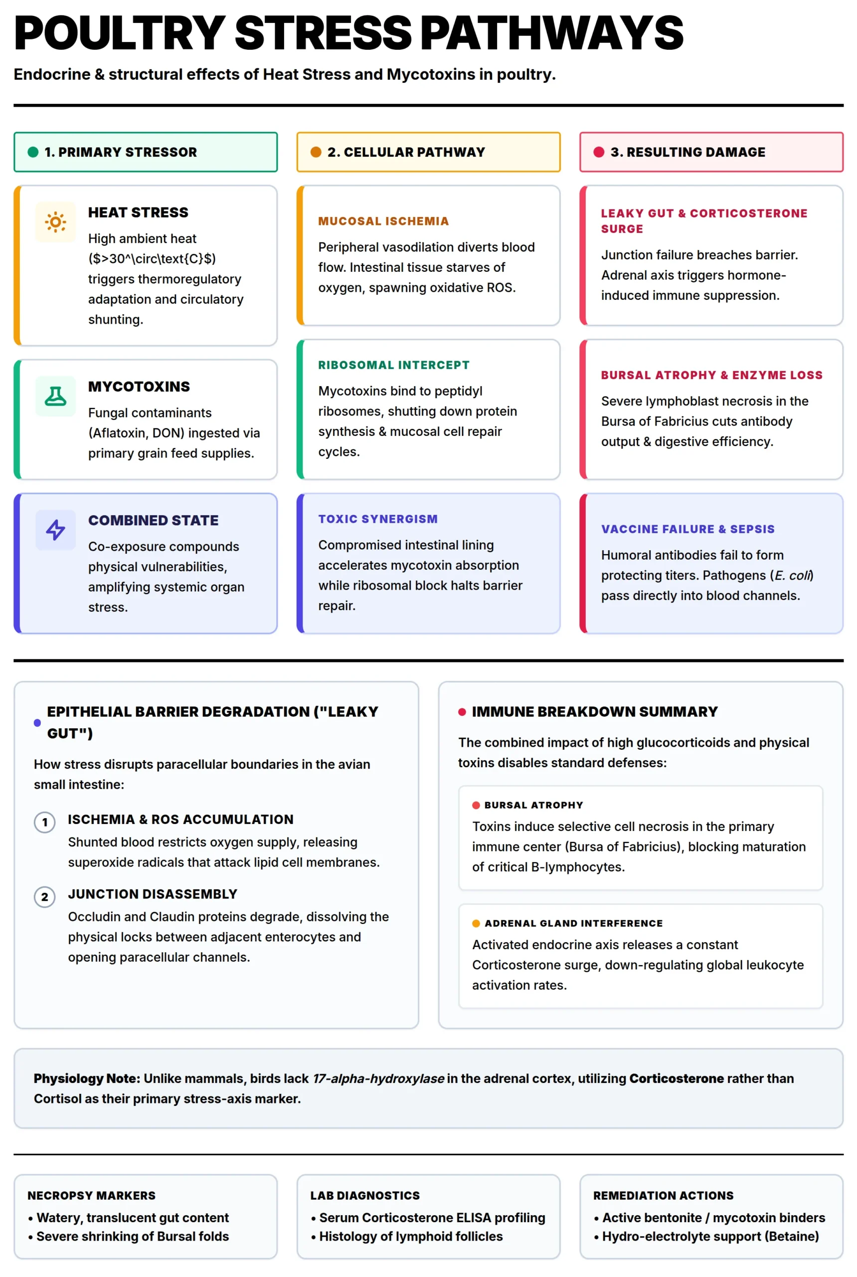
POULTRY STRESS PATHWAYS
Figure 1: How mycotoxins and heat stress cause damage in poultry

Breeder Reproductive Dysfunction and Transgenerational Effects of Mycotoxins

In breeder operations, mycotoxins represent a catastrophic economic threat because they are vertically transmitted. Unlike commercial layers, where the loss is limited to the individual bird’s production, breeder contamination compromises the viability of the entire next generation.

1. Impact on the Reproductive Systems (Male & Female)

Mycotoxins hit both sides of the fertility equation, often exacerbated by summer heat.

Female Reproductive System
  • Mode of action: Mycotoxins (especially Zearalenone) mimic estrogen. This disrupts the hypothalamic-pituitary-ovarian axis.
  • Impact: inflammation of the oviduct, cystic ovaries, and reduced synthesis of yolk precursors in the liver, resulting in a sharp drop in egg production and poor internal egg quality.
Male Fertility
  • Mode of action: Toxins like T-2 and Aflatoxin induce oxidative stress that damages the phospholipid membrane of sperm cells.
  • Impact: Under heat stress, rooster semen quality already declines; mycotoxins accelerate this by reducing sperm motility, concentration, and increasing morphological abnormalities. This leads to a massive spike in infertility rates.

2. Hatchability & Embryonic Mortality

For breeders, mycotoxins represent a “generational loss” via vertical transmission.

  • Mode of action (the yolk bridge): Some mycotoxins are highly lipophilic. As the liver assembles the yolk, it deposits toxins directly into the egg.
  • The “three-wave” mortality:
    1. Early (Days 1–7): Toxins interfere with mitosis (cell division), leading to early deaths often mistaken for “infertility.”
    2. Mid-Term (Days 8–18): As the embryo begins intensive absorption of the toxic yolk, its developing liver and kidneys are compromised. This is the classic “Toxin Fingerprint.”
    3. Late (Days 19–21): Ochratoxins impair the embryo’s ability to mobilize calcium from the eggshell. As a result, the chick becomes too weak to pip and dies fully developed inside the shell (“dead-in-shell”).

3. Chick Quality, Grading, and Settability

The “Chick Quality” starts in the breeder’s gut and kidney health.

  • Mode of action (nutrient malabsorption): Mycotoxins reduce pancreatic lipase and bile salts. This prevents the mother from absorbing fat-soluble vitamins (A, D, E, K) and pigments. They also lead to lower intestinal adsorption due to a reduced absorption area and lower transporter efficacy.
  • Impact on chick quality:
    • “Pale Bird Syndrome”: Chicks lack vital carotenoids for early-stage defense.
    • Skeletal weakness: Interference with Vitamin D3 metabolism results in weak legs and “rubbery beaks” in Day-Old-Chicks (DOCs).
    • High first week mortality (FWM): Chicks hatch immunosuppressed, leading to high mortality during the first week.
  • Impact on egg grading & settability: Mycotoxins (Ochratoxin) are nephrotoxic, damaging the kidneys and disrupting the blood calcium-carbonate balance. This leads to “Sandpaper” shells, misshapen eggs, and a 5–10% drop in the number of settable eggs fit for the incubator.

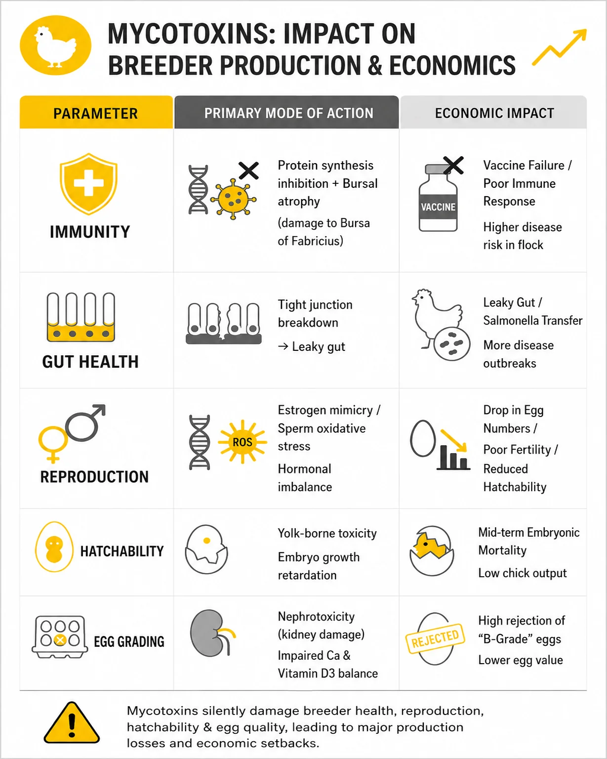
The impact of mycotoxins on breeder production and economics
Figure 2: The impact of mycotoxins on breeder production and economics


Pro tip for breeders
: In summers, high-moisture new maize triggers a mycotoxin surge that synergistically destroys the breeder’s kidneys and shell gland, crippling egg settability. A 5% spike in “Dead-in-Shell” embryos during breakout analysis is a definitive indicator of feed toxicity rather than incubator failure.

Integrated Mycotoxin Mitigation Strategies for Poultry Production

To pursue an effective mycotoxin mitigation strategy, it is essential first to identify which mycotoxins are relevant to a given region before implementing measures.

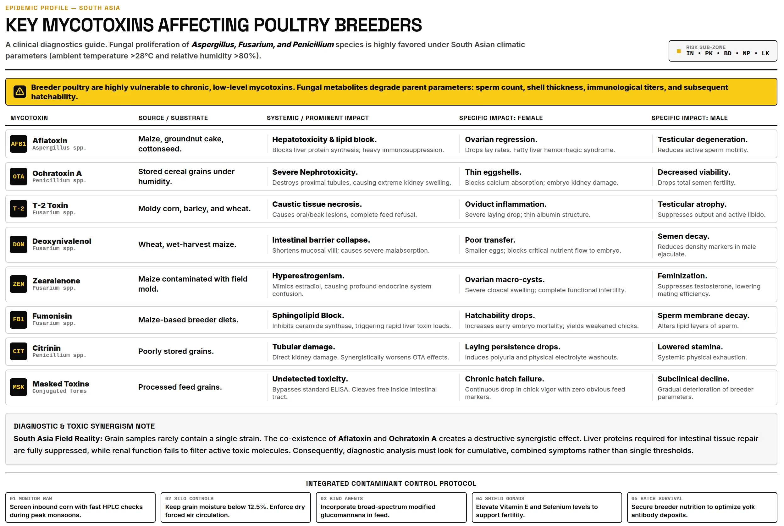
Key mycotoxins affecting poultry breeders in South Asia
Figure 3: Key mycotoxins affecting poultry breeders in South Asia

1. Raw Material Management

  • Strict moisture control: Reject any maize arriving with >14% moisture.
  • Rapid screening: Perform rapid mycotoxin screening before unloading raw materials.
  • Mechanical grain driers: To maintain a safe storage moisture level (<12%)

2. Feed Plant & Storage Hygiene

  • First-In, First-Out (FIFO): Ensure strict inventory rotation to prevent “pockets” of old, moldy feed from contaminating new batches.
  • Frequently clean silos and elevators: High temperature and humidity cause moisture condensation on silo walls, leading to localized mold growth.
  • Antifungal Treatment: Use buffered organic acids (propionic and formic acid) to limit mold proliferation in feed.

3. Broad-Spectrum binders:

Bentonites (for Aflatoxins) and Yeast Cell Walls: These components help bind pathogenic bacteria like E. coli that capitalize on the “leaky gut” caused by toxins and heat stress.

4. Physiological & Gut Health Support

  • Water acidification: Lower the drinking water pH to 4.5–5.5. This prevents bacterial blooms in the water lines when birds increase water intake by 3 times during heat stress.
  • Liver & kidney tonics: Supplemental hepatic (milk thistle/silymarin) and renal support to help the bird metabolize and export toxins more efficiently.
  • Metabolite supplementation: Use 25-hydroxyvitamin D3 in breeder diets to bypass the liver/kidney damage and ensure shell quality remains intact.
  • Antioxidant boost: Increase levels of Vitamin E, C, and Selenium to counter the oxidative stress caused by the heat-toxin synergy.

5. Monitoring & Diagnostics

  • Hatchery breakout analysis: Monitor “dead-in-shell” embryos. A spike in mid-term mortality is an immediate indicator that the breeder feed toxin binder needs a dosage increase.
  • Frequent lab testing: Mycotoxin testing at least weekly during the new maize transition to identify the specific toxin profile.

Solutions are available to support toxin risk management

In the challenging climate where high-moisture “new maize” and summer humidity create a complex cocktail of mycotoxins, endotoxins, and pesticide residues, traditional, single-ingredient binders often fall short. Modern poultry production requires a proactive solution that does more than just “bind”; it must protect the bird’s internal integrity.

Solis Max – The effective myco- and endotoxin solution for sustained profitability

Solis Max is engineered to meet customers’ demand for an effective solution, offering a multi-pronged defense mechanism that targets the root causes of performance collapse. Solis Max uses a synergistic blend of five key components to ensure the flock’s safety.

Trials prove the effectiveness of Solis Max

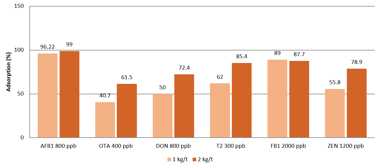
Solis Max shows dose-dependent adsorbing capacity against multiple mycotoxins:

Figure : Mycotoxin Binding Capacity Of Solis Max
Figure 4: Mycotoxin Binding Capacity Of Solis Max

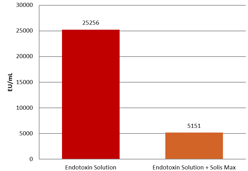
SOLIS MAX shows endotoxin adsorbing capacity – 1mg of SOLIS MAX absorbs 20 endotoxin units (EU) of E. coli endotoxin (80% adsorption rate):

Figure : Endotoxin Binding Capacity Of Solis Max
Figure 5: Endotoxin Binding Capacity Of Solis Max

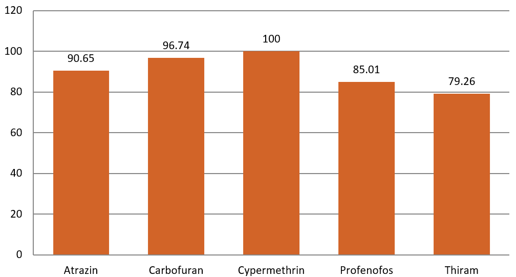
Solis Max demonstrates high pesticide binding efficiency across multiple compounds:

Figure : Pesticide Net Binding Capacity Of Solis Max (%)
Figure 6: Pesticide Net Binding Capacity Of Solis Max (%)

Conclusion:

The convergence of a high Temperature–Humidity Index (THI) and mycotoxicosis represents a critical, multisystem challenge in poultry production, precipitating severe pathology across the hepatic, renal, and gastrointestinal systems. In breeding operations, this crisis exhibits a transgenerational impact: lipophilic mycotoxins are vertically transmitted to the yolk, inducing mid-term embryonic mortality and compromising post-hatch progeny immunity.

Mitigation demands stringent control of raw material moisture alongside advanced, broad-spectrum interventions. Utilizing an advanced multi-pronged solution like Solis Max counters this synergy by providing physicochemical adsorption and targeted organ protection. By neutralizing the concurrent threats of mycotoxins, endotoxins, and pesticides, it preserves cellular integrity, mitigates systemic pathology, and maintains optimal performance under extreme environmental stress.

References available upon request.


Mycotoxins & Poultry Egg Quality in Southeast Asia

Mycotoxins & Poultry Egg Quality In Southeast Asia

Tran Si Trung, PhD
EWN SEAP – Regional Technical Manager for Toxin Risk Management

1. Introduction

The global egg market is experiencing steady and robust growth, playing a vital role in food security and animal protein nutrition. According to reports from RaboResearch and the World Egg Organization, global egg production has more than doubled, rising from 46 million tonnes in 1995 to approximately 99 million tonnes in 2025. By 2035, the market is projected to expand by a further 22%, with an annual growth rate of approximately 2.0%. Asia leads with more than 64% of global output, with China and India being the largest producing countries. The global egg market value was estimated at USD 352 billion in 2025 and is expected to reach USD 585 billion by 2033, representing a CAGR of approximately 6.6%. This growth is driven by urbanization, rising incomes, demand for high-quality protein, and the widespread use of eggs in the processed food industry.

In Vietnam, the poultry industry has expanded rapidly and become one of the pillars of the agricultural sector. In 2023, poultry egg production reached approximately 19.22 billion eggs. In 2024, this figure exceeded 20 billion eggs, with chicken eggs accounting for the dominant share. Per-capita egg consumption rose from 108 eggs per year in 2017 to approximately 185–190 eggs per year in 2024, though this remains below the average of many other countries (300–350 eggs). In the near term, despite limited exports, domestic consumption is fairly stable, and the sector has the potential to achieve a value of USD 3 billion. Alongside these opportunities, the industry also faces challenges such as occasional local oversupply, price volatility, disease outbreaks, and food quality and safety issues. Among these, mycotoxins can be regarded as one of the most silent yet serious threats to egg quality and consumer health.

The principal mycotoxins include aflatoxins (AFs), ochratoxin A (OTA), zearalenone (ZEN), deoxynivalenol (DON), fumonisins (FBs), and T-2 toxin. These compounds form in feed raw materials — including maize, wheat, soybean, groundnut, and other oil seeds — under field stress conditions or during storage under inadequate conditions. In the tropical humid climates of Vietnam and much of Southeast Asia, the risk of natural contamination is particularly elevated. Mycotoxins not only reduce livestock productivity (organ damage, immune suppression, etc.) but also directly affect egg quality, nutritional value, and toxin residue levels in eggs, thereby impacting both the economic value of the product and consumer health.

2. Key Aspects of Egg Quality

Egg quality is typically assessed across multiple dimensions: external appearance (clean, intact shell, uniform shape and color), internal quality (albumen height, yolk color, Haugh unit score), nutritional value (high protein ~12–13%, lipids, vitamins A/D/E, carotenoids, choline, lutein), and food safety (freedom from microbiological contamination, antibiotic residues, mycotoxin residues, and heavy metals).

Key technical parameters include: egg weight and grade (AA, A, B); shell thickness and strength; Haugh unit (reflecting albumen freshness); yolk color (Roche scale or DSM Yolk Fan); air cell size; and absence of blood spots (meat spots). High quality ensures commercial value, shelf life, and nutritional benefit to the consumer.

Each of these quality dimensions (shell integrity, albumen height, yolk color, and residue status) is, to varying degrees, susceptible to mycotoxin insult, as the following section demonstrates.

3. Adverse Effects of Mycotoxins on Egg Quality and Value

Mycotoxins cause harm through multiple mechanisms: hepato-renal toxicity, hormonal disruption, oxidative stress, intestinal damage (reduced nutrient absorption), immune and enzyme suppression. Effects are often evident at relatively low concentrations (20–500 ppb depending on the toxin type) and are amplified when multiple mycotoxins are present simultaneously. Field surveys across Asia consistently demonstrate that co-contamination, the presence of two or more mycotoxins in a single feed ingredient or complete diet, is the norm rather than the exception, particularly in maize-based diets during wet-season harvests. Effective risk management must therefore address the full toxin spectrum rather than individual contaminants in isolation.

Mycotoxin Main Feed
Substrates
Primary
Mechanism(s)
Key Effects on Egg Quality
Aflatoxins
(AFs/AFB1)
Maize, groundnut,
soybean, cottonseed
Hepatotoxicity;
oxidative stress;
Ca & Zn absorption
inhibition
↓ Laying rate; ↓ shell quality; ↓ yolk carotenoids & color; residues (AFB1, AFM1) in eggs
Ochratoxin A
(OTA)
Wheat, barley,
maize, sorghum
Nephrotoxicity;
immunosuppression
↓ Laying rate; ↑ cracked/thin/
misshapen shells; residues in eggs
Zearalenone
(ZEN)
Maize, wheat,
barley
Estrogenic receptor
disruption
(HPG axis)
↓ FSH/LH/progesterone; ↓ ovarian
function; ↓ fertility & hatchability in breeders (roosters + hens)
Deoxynivalenol
(DON)
Wheat, maize,
barley, oats
Intestinal inflammation;
protein synthesis
inhibition
↓ Feed intake; ↓ shell breaking
strength (10–15%); ↓ Haugh unit;
↓ yolk color
Fumonisins
(FBs/FB1)
Maize and
maize by-products
Sphingolipid synthesis
inhibition; liver damage
↓ Laying performance; ↓ nutrient
absorption; ↓ albumen & yolk quality
T-2 Toxin Cereal grains
(wheat, barley, oats)
Mucosal necrosis;
immunosuppression;
ribotoxic effect
↓ Feed intake (oral lesions); ↓ Haugh unit (especially combined with DON); ↓ eggshell quality

Table 1. Overview of principal mycotoxins, their main feed substrates, primary mechanisms of action, and key effects on poultry egg quality.

3.1. Reduced Laying Performance

AFs and OTA can reduce laying rate by 5–10% at field-relevant dietary concentrations, with greater reductions reported under conditions of more severe or prolonged contamination. ZEN, acting as a potent estrogen mimic, disrupts the hypothalamic-pituitary-gonadal (HPG) axis, reducing FSH, LH, and progesterone levels, thereby impairing follicular development and ovarian function. DON and FBs cause intestinal inflammation and reduced nutrient absorption. T-2 toxin can cause ulcerative lesions of the oral mucosa or gizzard, thereby reducing feed intake or impairing gizzard motility and feed digestion.

3.2. Impact on Eggshell Quality

Mycotoxins in general can reduce eggshell thickness and strength by inhibiting calcium absorption, vitamin D3 utilization, and carbonic anhydrase activity (zinc-dependent). In particular, AFs may induce secondary zinc deficiency through liver damage. OTA has been associated with increased incidence of cracked, thin, misshapen, and urate-spotted eggs. Experimental studies have shown that DON can reduce eggshell breaking strength by 10–15% under controlled conditions.

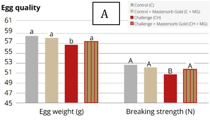
Figure A
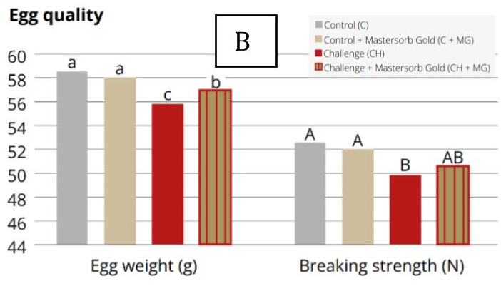
Figure B
Figure 1. Egg quality parameters of laying hens challenged with (A) 100 ppb AFB1 & 9,000 ppb fumonisins and (B) 1,400 ppb DON & 300 ppb T-2 toxin, with and without in-feed Mastersorb Gold. Significant differences (p<0.05) indicated by lowercase letters; statistical tendencies (p<0.1) by uppercase letters.

3.3. Impact on Internal Egg Quality

Research conducted at Kasetsart University (Thailand) demonstrates that DON and T-2 toxin can reduce albumen height and Haugh unit scores at relatively low dietary concentrations (Tables 2 and 3). Additionally, experimental data indicate that DON can impair yolk carotenoid content and yolk color score, diminishing both antioxidant value and visual appeal, at dietary concentrations as low as 2,500 ppb.

Table
Table 2. Egg quality parameters of laying hens challenged with 100 ppb AFB1 & 9,000 ppb FB1, with and without in-feed Mastersorb Gold.
Table
Table 3. Egg quality parameters of laying hens challenged with 1,400 ppb DON & 300 ppb T-2 toxin, with and without in-feed Mastersorb Gold.

3.4. Residues and Food Safety

After mycotoxins are absorbed, the host begins detoxification and excretion processes, while organ damage simultaneously occurs. Detoxification is primarily carried out by the liver, and accumulation occurs mainly in the liver and kidneys. However, accumulation in other tissues, including meat and eggs, has also been documented. AFB1 and its liver-derived metabolites, including AFM1, have been detected in eggs at transfer rates of approximately 0.05% of the dietary AFB1 intake; OTA transfers at ~0.15%; T-2 at ~0.10%; while DON, FB1, and ZEN transfer at lower rates.

3.5. Economic and Indirect Impacts

While ZEN is considered to have limited impact on commercial broiler performance, the situation is markedly different for breeder flocks. Acting primarily through its active hepatic metabolite α-zearalenol (α-ZOL), which has a higher affinity for estrogen receptors than the parent compound, ZEN may reduce fertility (impaired semen quality in roosters) and hatchability (increased embryo mortality, reduced chick quality at hatch). More broadly, mycotoxins negatively affect animal health, growth performance, and egg quality, leading to increased culling and veterinary costs, as well as lower selling prices for substandard eggs. As a concrete example, with Vietnam producing more than 20 billion eggs per year, even a 1–2% reduction in productivity or egg quality could translate into losses of tens of millions of USD annually – a scale of impact applicable across every major egg-producing nation in the region.

4. Key Considerations for Mycotoxin Risk Management

Managing mycotoxin risks requires an increasingly comprehensive and integrated approach. The “3F – from Feedmill, Farm to Fork” process is an integrated management framework developed by EW Nutrition in the region to prevent, trace, and mitigate mycotoxin-related risks for poultry producers and egg manufacturers.

4.1. Prevention at Source (Feedmill)

  • Upon raw material intake: conduct sensory inspection, then perform proper sampling and test for mycotoxins using rapid test strips or ELISA.
  • Storage: pay close attention to ambient relative humidity and temperature in warehouses/silos, as these two factors directly influence the moisture content and water activity (Aw) of stored materials, creating favorable conditions for the growth of Aspergillus spp. and/or Penicillium spp. (mold species capable of producing mycotoxins such as AFs, OTA, citrinin, patulin, etc. during storage). As practical targets: keep grain moisture below 14% for maize and wheat (below 10% for groundnut meal); Aspergillus spp. can proliferate at Aw ≥ 0.80, while Penicillium spp. remain active at Aw ≥ 0.78; maintaining Aw below these thresholds is the single most effective storage intervention.
  • Finished feed samples from each batch must be properly collected and analyzed for multiple mycotoxins using ELISA or chromatographic methods (HPLC, LC-MS/MS, etc.). Retained samples should be stored under cool, dry conditions for a minimum of two weeks to enable analysis and traceability in the event of a subsequent incident.
  • Periodically inspect hygiene of storage facilities and equipment (e.g., mixer, cooler, feed transport trucks from feedmill to farm).
  • Develop preventive strategies against the adverse effects of mycotoxins on the health and performance of commercial laying hens, including supplementation with broad-spectrum solutions (Mastersorb Gold, etc.) adsorbing a diverse range of toxins and providing antioxidant support to mitigate oxidative stress.

4.2. Prevention at Farm Level

  • Establish a routine hygiene monitoring program for housing facilities, particularly feed storage areas/silos and associated equipment (e.g., automated feeders, egg and manure conveyors, etc.).
  • Feed samples from each batch at the farm level should also be properly collected and retained (under cool, dry conditions) for a minimum of two weeks for analysis and traceability should any subsequent issue arise.
  • When animal health or performance issues arise and mycotoxicosis is suspected, in complement to analyzing retained feed samples, the analysis of mycotoxin residues in Dried Blood Spots (DBS), a technique developed by EW Nutrition and its partner, can be a valuable complementary measurement to make a diagnosis.

4.3. Food Safety at the Table (Fork)

Vietnamese Standard TCVN 1858:2018 stipulates that commercial chicken eggs must be clean, uncracked, undistorted, and free from spoilage; air cell depth must not exceed 6 mm (depending on grade); yolk must not be visibly off-center; and no off-odors are permitted. Eggs are graded based on both external and internal quality criteria. Additionally, Circular 34/2012/TT-BNNPTNT and food safety and hygiene regulations require traceability, Salmonella control, and monitoring of specific residues. The national technical regulation QCVN 01-190:2020/BNNPTNT on animal feed sets maximum limits for mycotoxins in feed raw materials. While Vietnam serves as a concrete example, analogous frameworks are in place across Southeast and South Asia, with many producers also referencing the Codex Alimentarius maximum levels for aflatoxins in food (4 µg/kg total AFs; 2 µg/kg AFB1) and EU feed maximum limits as de facto benchmarks for export-oriented operations.

According to the International Agency for Research on Cancer (IARC), AFB1 is classified as a Group 1 carcinogen (carcinogenic to humans), while OTA, AFM1, and FB1 are classified as Group 2B (possibly carcinogenic to humans). It is of particular concern that these mycotoxins are also highly heat-stable, they are not destroyed by cooking or standard food processing temperatures. Consequently, their residues in eggs represent an important aspect to be monitored and controlled before eggs reach the consumer’s table.

5. Conclusion

Mycotoxins are a critical factor affecting egg quality across all dimensions – external appearance, internal quality, food safety, and economic value. In the context of Vietnam’s egg industry, and the broader dynamic growth of egg production across Southeast and South Asia, moving toward modernization and export competitiveness, mycotoxin control is not merely a loss-reduction measure, but a strategy for sustainable competitive advantage. The sector requires close collaboration among feed manufacturers, poultry and egg producers, regulatory authorities, and scientific researchers to turn these challenges into opportunities for development.

In line with this broader direction, EW Nutrition has developed and is actively supporting the implementation of the integrated 3F Management Process (from Feedmill, Farm to Fork), grounded in scientific evidence and technology, to help protect poultry flocks, enhance egg quality, and ensure consumer safety across the region.

About the Author

Dr. Tran Si Trung holds PhD degrees in Food Safety & Quality in France and serves as Regional Technical Manager for Toxin Risk Management at EW Nutrition, Southeast Asia and Pacific. He specializes in mycotoxin risk assessment and feed quality management. For further information or technical inquiries, please contact EW Nutrition Vietnam.


Mycotoxins as contributors to antibiotic resistance?

Mycotoxins as contributors to antibiotic resistance?

By Dr. Inge Heinzl, Editor EW Nutrition and
Marie Gallissot, Global Manager Feed Quality Solutions EW Nutrition

Antibiotic resistance is a growing global health concern, making infections more complicated to treat and increasing the risk of disease spread, severe illness, and death. While overuse and misuse of antibiotics are the primary causes, recent research has uncovered another unexpected contributor: mycotoxins. Among these, deoxynivalenol (DON), a toxin commonly found in contaminated grains, has been shown to significantly alter gut microbiota and promote antibiotic resistance. This article examines how DON impacts gut bacteria, influences antibiotic resistance, and highlights why this issue warrants urgent attention.

Mycotoxins – originators of antimicrobial resistance?

Actually, it would be logical…

Alexander Fleming discovered Penicillin when he returned after the summer holidays and saw that a mold had grown on the agar plate he had prepared. Around the mold, Staphylococcus was unable to proliferate. The reason was a substance produced by the mold – penicillin, which, like other toxins produced by molds, is a mycotoxin. In his article about the origin of antibiotics and mycotoxins, Shier (2011) stated that antibiotics and mycotoxins share considerable similarities in structure, metabolic roles, and biosynthesis.

A short excursus to antimicrobial resistance

In general, the primary mechanisms of resistance involve the prevention or limitation of the antimicrobial substance’s uptake, modifying the drug target, inactivating the drug, or facilitating its discharge with efflux pumps.

There are two types of resistance: natural resistance, which is further divided into intrinsic and induced resistance, and acquired resistance.

Intrinsic resistance is a “characteristic” of a bacterial species and is not dependent on antibiotic exposure. An example is the reduced permeability of the outer membrane of gram-negative bacteria, which prevents certain antibiotics from entering.

Induced resistance, however, needs to be initiated by antibiotics. Here, multidrug-efflux pumps can be mentioned.

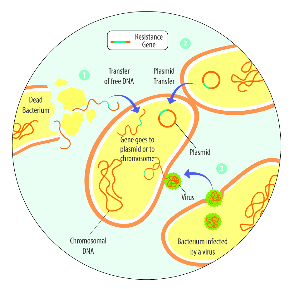
The third one, acquired resistance, refers to the process by which bacteria acquire genetic material, the resistance genes, from other bacteria that are resistant. The mechanisms include vertical transfer to daughter cells and horizontal transfer, such as the transfer from dead bacteria to living ones, by viruses, or the transfer of plasmids (Reygaert, 2018).

Different possibilities of transfer of resistance genes
Figure 1: Different possibilities of transfer of resistance genes

Deoxynivalenol (DON) promotes resistance in gut microbiota

A Chinese group of researchers (Deng et al., 2025) examined for the first time the influence of DON on the intestinal microbiota of chickens. One of the most alarming findings is DON’s ability to enhance antibiotic resistance. It contributes to this issue in several ways:

  1. Encouraging resistant bacteria – By disrupting microbial balance, DON provides a survival advantage to bacteria that carry resistance genes.
  2. Activating resistance genes – Studies suggest that DON can increase the expression of genes that help bacteria withstand antibiotics.
  3. Enhancing gene transfer – Bacteria can share resistance genes through horizontal gene transfer. DON appears to promote this process, making antibiotic-resistant strains spread more rapidly.
  4. Weakening antibiotic effectiveness – DON-induced changes in the gut environment can reduce the effectiveness of antibiotics, making treatments less successful.

A further indication that mycotoxins can enhance resistance is the significant overlap in the geographical distribution of antimicrobial-resistant bacteria and genes with that of mycotoxins, as noted by Deng et al.

Which protection mechanisms do bacteria have against mycotoxins?

In the case of mycotoxins, bacteria employ similar molecular mechanisms to those used against antibiotics. In an in vitro experiment, Hassan et al. (2019) challenged Devosia mutans, a gram-negative bacterium, with DON in the growth medium. DON inhibits protein synthesis, induces oxidative stress, and compromises cell membrane integrity in eucaryotic cells. Hassan et al. asserted three adaptive mechanisms as the response to the challenge:

  1. Activation of cellular membrane proteins (adenosine 5’-triphosphate-binding cassette -ABC- transporters) responsible for the unidirectional transport of substrates, either outward or inward. These ABC transporters can work as drug efflux pumps.
  2. Production of DON-specific deactivation enzymes, thereby engaging a toxin-specific pyrroloquinoline quinone-dependent detoxification pathway. This enables the bacterial isolate to transform DON to a non-toxic stereoisomer.
  3. Upregulation of auxiliary coping proteins, such as porins (transmembrane proteins involved in metabolite exchange), glutathione S-transferases, and phosphotransferases, both of which are likely involved in the detoxification of xenobiotics.

Public health implications and preventive measures

Given the widespread presence of DON in food and animal feed, its potential role in antibiotic resistance poses a serious threat. The combination of increased bacterial resistance and weakened antibiotic efficacy could lead to more difficult-to-treat infections. This is particularly concerning in hospital settings, where antibiotic-resistant infections already cause high mortality rates.

To address the issue, several strategies can be implemented:

  1. Reducing DON contamination: Implementing improved agricultural practices, such as crop rotation, the use of fungal-resistant crop varieties, and maintaining proper storage conditions, can help limit fungal growth and DON production.
  2. Monitoring food and feed supply – Strict regulations and testing for DON contamination in grains and animal feed are essential to minimize human and animal exposure.
  3. Effective mycotoxin risk management at feed mill and farm levels: Using tools such as MasterRisk and effective products combatting mycotoxins.
  4. Maintaining gut health: A healthy diet rich in fiber, probiotics, and gut health-supporting feed supplements, such as Ventar D or products from the Activo line, may help counteract some of the adverse effects of DON on gut microbiota.
  5. Developing new treatments: Research into alternative therapies and new antibiotics is crucial to combat the rise of antibiotic resistance.

Antimicrobial resistance: Be aware of the mycotoxins!

The connection between mycotoxins, such as DON, and antibiotic resistance underscores the need for a broader perspective on public health and food safety and once again brings the “One Health Concept” into focus. While antibiotic overuse remains the primary driver of resistance, environmental factors, such as exposure to mycotoxins, should not be overlooked. By increasing awareness, enhancing food safety regulations, and investing in research, we can take steps to mitigate this emerging threat and safeguard the effectiveness of antibiotics for future generations.

References:

Deng, Fengru, Chuying Yao, Linyu Ke, Meichan Chen, Mi Huang, Jikai Wen, Qingmei Chen, Jun Jiang, and Yiqun Deng. “Emerging Threat to Antibiotic Resistance: Impact of Mycotoxin Deoxynivalenol on Gut Microbiota and Clonal Expansion of Extensively Drug-Resistant Enterococci.” Environment International 197 (March 2025): 109353.
https://doi.org/10.1016/j.envint.2025.109353.

Hassan, Yousef I., Jian Wei He, Dion Lepp, and Ting Zhou. “Understanding the Bacterial Response to Mycotoxins: The Transcriptomic Analysis of Deoxynivalenol-Induced Changes in Devosia Mutans 17-2-E-8.” Frontiers in Pharmacology 10 (November 14, 2019).
https://doi.org/10.3389/fphar.2019.01098.

Reygaert, Wanda C. “An Overview of the Antimicrobial Resistance Mechanisms of Bacteria.” AIMS Microbiology 4, no. 3 (2018): 482–501.
https://doi.org/10.3934/microbiol.2018.3.482.

Shier, W. Thomas. “On the Origin of Antibiotics and Mycotoxins.” Toxin Reviews 30, no. 1 (January 28, 2011): 6–30.
https://doi.org/10.3109/15569543.2011.550862.

Smith, William P., Benjamin R. Wucher, Carey D. Nadell, and Kevin R. Foster. “Bacterial Defences: Mechanisms, Evolution and Antimicrobial Resistance.” Nature Reviews Microbiology 21, no. 8 (April 24, 2023): 519–34.
https://doi.org/10.1038/s41579-023-00877-3.


Sustainability will push more by-products into pig feed – Keep track of mycotoxins!

Header DSC

Mycotoxin Team EW Nutrition

Most grains used in feed are susceptible to mycotoxin contamination, causing severe economic losses all along feed value chains. As skyrocketing raw material prices force producers to include a higher proportion of economical cereal by-products in the feed, the risks of mycotoxin contamination likely increase. This article reviews why mycotoxins cause the damage they do – and how effective toxin-mitigating solutions prevent this damage.

Mycotoxin contamination of cereal by-products requires solutions

Cereal by-products may become more important feed ingredients as grain prices increase. However, from a sustainability point of view and considering population growth, using cereal by-products in animal feed makes much sense. Distiller’s dried grains with solubles (DDGS) are a good example of how by-products from food processing industries can become high-quality animal feed.

Fefac Stats
Figure 1: By-products are a crucial protein source (data from FEFAC Feed&Food 2021 report)


Still, research on what happens to mycotoxins during food processing shows that mycotoxins are concentrated into fractions that are commonly used as animal feed (cf. Pinotti et al., 2016; Caballero and Heinzl, 2022). To safeguard animal health and performance when feeding lower-quality cereals, monitoring mycotoxin risks through regular testing and using toxin-mitigating solutions is essentialy.

Problematic effects of mycotoxins on the intestinal epithelium

Most mycotoxins are absorbed in the proximal part of the gastrointestinal tract. This absorption can be high, as in the case of aflatoxins (ca. 90%), but also very limited, as in the case of fumonisins (< 1%); moreover, it depends on the species. Notably, a significant portion of unabsorbed toxins remains within the lumen of the gastrointestinal tract.

Importantly, studies based on realistic mycotoxin challenges (e.g., Burel et al., 2013) show that the mycotoxin levels necessary to trigger damaging processes are lower than the levels reported as safe by EFSA, the Food Safety Agency of the European Union. The ultimate consequences range from diminished nutrient absorption to inflammatory responses and pathogenic disorders in the animal (Figure 2).

Figure Scheme
Figure 2: Mycotoxins’ impact on the GIT and consequences for monogastric animals

1. Alteration of the intestinal barrier‘s morphology and functionality

Several studies indicate that mycotoxins such as aflatoxin B1, DON, fumonisin B1, ochratoxin A, and T2, can increase the permeability of the intestinal epithelium of poultry and swine (e.g., Pinton & Oswald, 2014). This is primarily a consequence of the inhibition of protein synthesis.

As a result, there is an increase in the passage of antigens into the bloodstream (e.g., bacteria, viruses, and toxins). This increases the animal’s susceptibility to infectious enteric diseases. Moreover, the damage that mycotoxins cause to the intestinal barrier entails that they are also being absorbed at a higher rate.

2. Impaired immune function in the intestine

The intestine is a very active immune site, where several immuno-regulatory mechanisms simultaneously defend the body from harmful agents. Immune cells are affected by mycotoxins through the initiation of apoptosis, the inhibition or stimulation of cytokines, and the induction of oxidative stress.

3. Alteration of the intestinal microflora

Piglets ART

Recent studies on the effect of various mycotoxins on the intestinal microbiota show that DON and other trichothecenes favor the colonization of coliform bacteria in pigs. DON and ochratoxin A also induce a greater invasion of Salmonella and their translocation to the bloodstream and vital organs in birds and pigs – even at non-cytotoxic concentrations.

It is known that fumonisin B1 may induce changes in the balance of sphingolipids at the cellular level, including for gastrointestinal cells. This facilitates the adhesion of pathogenic bacteria, increases in their populations, and prolongs infections, as has been shown in the case of E. coli. The colonization of the intestine of food-producing animals by pathogenic strains of E. coli and Salmonella also poses a risk to human health.

4. Interaction with bacterial toxins

When mycotoxins induce changes in the intestinal microbiota, this can increase the endotoxin concentration in the intestinal lumen. Endotoxins promote the release of several cytokines that induce an enhanced immune response, causing inflammation, thus reducing feed consumption and animal performance, damage to vital organs, sepsis, and death of the animals in some cases.

The synergy between mycotoxins and endotoxins can result in an overstimulation of the immune system. The interaction between endotoxins and estrogenic agents such as zearalenone, for example, generates chronic inflammation and autoimmune disorders because immune cells have estrogen receptors, which are stimulated by the mycotoxin.

Increased mycotoxin risks through by-products? Invest in mitigation solutions

To prevent the detrimental consequences of mycotoxins on animal health and performance, proactive solutions are needed that support the intestinal epithelium’s digestive and immune functionality and help maintain a balanced microbiome in the GIT. This becomes even more important as the current market conditions will likely engender a long-term shift towards including more cereal by-products in animal diets.

Trial data shows that EW Nutrition’s toxin-mitigating solution SOLIS MAX 2.0 provides adequate protection against feedborne mycotoxins. The synergistic combination of ingredients in SOLIS MAX 2.0 prevents mycotoxins from damaging the animals’ gastrointestinal tract and entering the bloodstream and additionally acts as antioxidant and liver-protecting:

Figure MOA Solis Max
Figure 3: Moa of Solis Max 2.0

In-vitro study shows strong mitigation effects of SOLIS MAX 2.0 against a wide range of mycotoxins

Animal feed is often contaminated with two or more mycotoxins, making it essential for an anti-mycotoxin agent to be effective against a wide range of different mycotoxins. A trial with SOLIS MAX 2.0 was conducted at an independent laboratory in Spain with an inclusion level of the product of 0.10% (equivalent to 1 kg per ton of feed). A phosphate buffer solution at pH 7 was prepared to simulate intestinal conditions in which a portion of the mycotoxins may be released from the binder (desorption). The following mycotoxins were evaluated in the test (see Table 1):

Table 1: Mycotoxin challenges

Table Efficacy Solis Max Table

Each mycotoxin was tested separately by adding a challenge to buffer solutions, incubating for one hour at 41°C, to establish the baseline (table). At the same time, a solution with the toxin challenge and Solis Max 2.0 was prepared, incubated, and analyzed for the residual mycotoxin to find the binding efficacy. All analyses were carried out using high-performance liquid chromatography (HPLC) with standard detectors.

Figure Efficacy Solis Max Mycotoxins
Figure 4: SOLIS MAX 2.0 (1 kg/t of feed) adsorption capacity against different mycotoxins (%)

The results (Figure 4) demonstrate that SOLIS MAX 2.0 is a highly effective solution against the most common mycotoxins in raw materials and animal feed.

Mycotoxin risk management for better animal feed

A healthy gastrointestinal tract is crucial to animals’ overall health: it ensures that nutrients are optimally absorbed, provides adequate protection against pathogens through its immune function, and is key to maintaining a well-balanced microflora. Even at levels considered safe by the European Union, mycotoxins can compromise different intestinal functions, resulting in lower productivity and susceptibility to disease.

The globalized feed trade, which spreads mycotoxins beyond their geographical origin, climate change, and raw material market pressures additionally escalate the problem. On top of rigorous testing, producers should mitigate unavoidable mycotoxin exposures by using solutions such as SOLIS MAX 2.0 – for stronger animal health, welfare, and productivity.

References

Antonissen, Gunther, An Martel, Frank Pasmans, Richard Ducatelle, Elin Verbrugghe, Virginie Vandenbroucke, Shaoji Li, Freddy Haesebrouck, Filip Van Immerseel, and Siska Croubels. “The Impact of Fusarium Mycotoxins on Human and Animal Host Susceptibility to Infectious Diseases.” Toxins 6, no. 2 (January 28, 2014): 430–52. https://doi.org/10.3390/toxins6020430.

Burel, Christine, Mael Tanguy, Philippe Guerre, Eric Boilletot, Roland Cariolet, Marilyne Queguiner, Gilbert Postollec, et al. “Effect of Low Dose of Fumonisins on Pig Health: Immune Status, Intestinal Microbiota and Sensitivity to Salmonella.” Toxins 5, no. 4 (April 23, 2013): 841–64. https://doi.org/10.3390/toxins5040841.

Burton, Emily J., Dawn V. Scholey, and Peter E. Williams. “Use of Cereal Crops for Food and Fuel – Characterization of a Novel Bioethanol Coproduct for Use in Meat Poultry Diets.” Food and Energy Security 2, no. 3 (September 19, 2013): 197–206. https://doi.org/10.1002/fes3.30.

Ghareeb, Khaled, Wageha A. Awad, Josef Böhm, and Qendrim Zebeli. “Impacts of the Feed Contaminant Deoxynivalenol on the Intestine of Monogastric Animals: Poultry and Swine.” Journal of Applied Toxicology 35, no. 4 (October 28, 2014): 327–37. https://doi.org/10.1002/jat.3083.

Mani, V., T. E. Weber, L. H. Baumgard, and N. K. Gabler. “Growth and Development Symposium: Endotoxin, Inflammation, and Intestinal Function in livestock1,2.” Journal of Animal Science 90, no. 5 (May 1, 2012): 1452–65. https://doi.org/10.2527/jas.2011-4627.

Obremski, K. “The Effect of in Vivo Exposure to Zearalenone on Cytokine Secretion by Th1 and Th2 Lymphocytes in Porcine Peyer’s Patches after in Vitro Stimulation with LPS.” Polish Journal of Veterinary Sciences 17, no. 4 (2014): 625–32. https://doi.org/10.2478/pjvs-2014-0093.

Oswald, I. P., C. Desautels, J. Laffitte, S. Fournout, S. Y. Peres, M. Odin, P. Le Bars, J. Le Bars, and J. M. Fairbrother. “Mycotoxin Fumonisin B1 Increases Intestinal Colonization by Pathogenic Escherichia Coli in Pigs.” Applied and Environmental Microbiology 69, no. 10 (2003): 5870–74. https://doi.org/10.1128/aem.69.10.5870-5874.2003.

Pinotti, Luciano, Matteo Ottoboni, Carlotta Giromini, Vittorio Dell’Orto, and Federica Cheli. “Mycotoxin Contamination in the EU Feed Supply Chain: A Focus on Cereal Byproducts.” Toxins 8, no. 2 (February 15, 2016): 45. https://doi.org/10.3390/toxins8020045.

Pinton, Philippe, and Isabelle Oswald. “Effect of Deoxynivalenol and Other Type B Trichothecenes on the Intestine: A Review.” Toxins 6, no. 5 (May 21, 2014): 1615–43. https://doi.org/10.3390/toxins6051615.


Building and boosting the immunity shield of pigs

ART IMAGE

Conference report

A well-functioning immune system is vital for the survival and performance of animals. It helps piglets cope with challenging periods, such as their first days of life or weaning. Measures can be taken around farrowing to support the piglets during their first days by enhancing the quality and quantity of colostrum and helping them develop their own immune system as fast as possible.

Adequate feeding of the sow before and around farrowing

Feeding of both the sow and the piglet has an important influence on farrowing, the health of the sow, colostrum and milk production, piglets’ development of immunity, and their later performance. A well-functioning immune system is crucial for the piglets to withstand upcoming challenges such as weaning.

Colostrum quality can be influenced by feeding

Newborn piglets have no functioning immunity system. They rely entirely on immunoglobulin G (IgG) absorption from colostrum within the first few hours after birth to establish their immunity shield. Dr. Megan Edwards, Animal Nutrition Consultant from Integral Nutrition (S) Pte Ltd, highlighted the payback of adequate colostrum quality and intake: Adequate colostrum intake can positively affect whole-of-life immunity and, ultimately, growth performance. The contained IgG is essential for providing passive immunity to piglets, protecting them from infections during their early days of life when their immune systems are still developing. There is a positive correlation between the amount of IgG they absorb from colostrum and their performance. This benefit of colostrum intake is independent of birth weight.

We have a 3-week window to influence colostrogenesis. However, the fat content of colostrum is determined in the last 48 hours before farrowing. According to Dr. Edwards, influencing colostrum quality is generally easier than affecting quantity. She identified several compounds that can serve as immunomodulators, such as MCFAs, yeast extracts, and butyrate. However, by moving IgG to colostrum and milk in late gestation and lactation, the sow compromises her immunity status by depleting her own reserves for about two weeks.

Feeding at farrowing

Sow body condition has been shown to have more impact on colostrum yield than feeding level. The highest colostrum yield was achieved when sows entered the farrowing unit with a moderate body condition (3-3.25 – the ribs, spine, and hip bones can only be felt with firm pressure but are not visibly prominent). Overfeeding should be avoided to prevent sows from becoming excessively fat pre-farrowing.

Sows experience increased energy demands during farrowing due to the physical demands of parturition and the physiological changes occurring in their bodies. Dr. Edwards does not encourage withholding feed on the day of farrowing and suggests offering up to 3kg if the sow has the appetite. Feeding just below the energy requirement helps the sow to mobilize her own body fat.

Many producers mistakenly withhold feed on the day of the farrow to reduce the incidence of constipation. Feeding, however, stimulates gut motility. Withholding feed can slow down gut transit time and actually increase the likelihood of constipation.

Piglet feeding for developing intestinal tract and immune system

In piglet feeding, two strategies are decisive: the early intake of immunoglobulins via colostrum to protect the piglets against pathogens during their first days of life and the offering of creep feed to stimulate their intestinal development.

High-quality colostrum as much and as soon as possible

When the piglets are born, it is of the highest importance that they ingest colostrum as much and as soon as possible. The piglet can only absorb intact large IgG molecules, the primary source of passive immunity, before gut closure, which begins about 6–12 hours after birth and progresses rapidly to completion in about 24 hours. In any case, the sow will start producing milk by this time and no more colostrum. The concentration of colostrum IgG decreases by 50% within 6 hours after the birth of the first piglet. The target is for piglets to consume 250 g of colostrum within the first 24 hours, ideally within the first 6 hours. However, about 30% of sows produce insufficient colostrum.

Figure 1: Mortality of piglets until 42 days of age
Figure 1: Mortality of piglets until 42 days of age according to intervals of birthweight and colostrum intake
(Hasan et al. 2019; the numbers of piglets are shown in parenthesis)

Split suckling jump-starts weak piglets

Split suckling is an effective management strategy to improve piglets’ access to colostrum and milk, particularly in increasingly common situations where sows give birth to large litters. This involves temporarily separating the more vigorous piglets from the sow to allow smaller or weaker piglets better access to the teats. This method helps ensure that all piglets receive adequate nutrition during the critical early hours after birth.

Large litters provoke energy deficiency in piglets

Piglets are born with limited energy reserves (glycogen and brown fat tissue). Ingestion of colostrum is associated with a considerable increase in the metabolic rate, contributing to maintaining body temperature. About 70% of the piglets’ energy requirement in the first 72 hours is provided by colostrum. “Most piglets that die within this period do so primarily due to energy deficiencies rather than immune-related issues. The trend towards larger litter sizes has exacerbated the issue of energy deficiency,” stated Dr. Edwards.

Creep feeding

The primary role of creep feed is to accelerate the development of the piglets, their digestive and immune systems, and their gut microbiome, not for weight gain. Creep feeding helps evolve digestive enzymes and acid secretion necessary for breaking down complex carbohydrates and proteins. This early feeding supports piglets in adapting to solid diets, mitigating stress during weaning.
Creep feeding also helps piglets develop an oral tolerance to avoid transient hypersensitivity due to various dietary ingredients. This process is essential for preventing allergic reactions and hypersensitivity, which can occur when the immune system mistakenly identifies harmless substances as threats. It takes about two weeks for the piglet to recognize an ingredient as a nutrient, not a pathogen. To facilitate this process, she recommends that creep diets contain a broad range of ingredients at low doses. This approach gradually exposes piglets to various nutrients, allowing their immune systems to adapt without overwhelming them with high concentrations of any single ingredient.

Mycotoxins must be managed – even in piglets

The significance of mycotoxins in piglets is often underestimated due to their relatively small feed intake. However, there is substantial evidence that mycotoxins can be transferred from sows to piglets through colostrum and milk, which can have profound health implications.

Dr. Edwards is convinced that managing mycotoxins is managing immunity. Mycotoxins are transferrable via the placenta, colostrum, and milk. There is a positive correlation between the mycotoxin levels in feed and colostrum. For example, adverse effects seen in piglets consuming colostrum with low doses of deoxynivalenol (DON) include:
• Decreased villus height
• Reduced mucosal integrity
• Increased inflammation
• Alternated immune response
The bottom line is that mycotoxins are a real and everyday risk to the immune quality of your piglets.

Nutrition influences piglets’ immune development

Dr. Edwards summarized that adequate nutrition is fundamental for developing a strong immune system in pigs, which is the basis for high performance. By focusing on the appropriate nutrition of the sow, ensuring an adequate intake of high-quality colostrum intake in piglets, and implementing creep feeding strategies, producers can significantly enhance the lifetime health and productivity of their piglets from an early age.

EW Nutrition’s Swine Academy took place in Ho Chi Minh City and Bangkok in October 2024. Dr. Megan Edwards, an Australian animal nutrition consultant with global research and praxis experience and a keen interest in immuno-nutrition and functional nutrients, was an esteemed guest speaker at this event.


Mycotoxins in poultry – External signs can give a hint

Header

Part 4: Paleness

By Dr. Inge Heinzl, Editor and Technical Team, EW Nutrition

We already showed bad feathering, mouth and beak lesions, bone issues, and foot pad lesions as signs of mycotoxin contamination in the feed, but there is another indicator: paleness. Paleness can signify a low count of red blood cells resulting from blood loss or inadequate production of these cells. Other possibilities are higher bilirubin levels in the blood due to an impaired liver, leading to jaundice or missing pigmentation.

Hen With Pale Comb And Wattles Large
Hen with pale comb and wattles (adapted from Bozzo et al., 2023)

The mycotoxins mainly causing anemia are Aflatoxins, Ochratoxin, DON, and T-2 toxin

Anemia can be diagnosed using parameters such as red blood cell count, hemoglobin levels, and hematocrit/packed cell volume (PCV). Numerous studies have examined the impact of mycotoxins on hematological parameters. They reveal their propensity to affect red blood cell production by impairing the function of the spleen and inducing hematological alterations. On the other hand, anemia can be caused by blood loss. Due to affecting coagulation factors, mycotoxins can lead to internal hemorrhages. The gut wall damage, probably due to secondary infections such as coccidiosis and necrotic enteritis, can entail bloody diarrhea in various animal species.

Impact on the production of blood cells

Low values of blood parameters such as red blood cells, hemoglobin, and hematocrit can result from inadequate production due to impacted production organs. The World Health Organization (WHO, 1990) and European Commission (European Commission, 2001) have identified hematopoietic tissues as targets for necrosis caused by T-2 toxin. Chu (2003) even stated that “the major lesion of T-2 toxin is its devastating effect on the hematopoietic system in many mammals, including humans”. Pande et al. (2006) suggested that reduced hemoglobin values result from decreased protein synthesis due to mycotoxin contamination, a notion supported by Pronk et al. (2002), who described trichothecenes as potent inhibitors of protein, DNA, and RNA synthesis, particularly affecting tissues with high cell division rates. Additionally, the European Commission (2001) highlighted the sensitivity of red blood cell progenitor cells (in this trial, the cells of mice, rats, and humans) to the toxic effects of T-2 and HT-toxins. DAS also seems to attack the hematopoietic system, as shown in humans (WHO, 1990). A further cause for anemia might be low feed intake or nutrient absorption, which inhibits adequate iron absorption and leads to iron deficiency. In their case report, Bozzo et al. (2023) assumed that renal failure and a resulting impaired excretion capacity caused by OTA might even increase the half-life of the toxins. This would enhance their effects on their target organs, such as the liver and bone marrow, and lead to anemia.

Several studies utilizing different animal species and mycotoxin dosages have been conducted to assess the effects of Aflatoxins, Ochratoxin, and T-2 Toxin on hematological parameters. The following table provides a summary of some of these studies.

Animal species Dosage Impact Reference
T-2 Toxin and other Trichothecenes
Broilers T-2 – 0, 1, 2, and 4 mg T-2 toxin/kg

n=30 per group

Significant reduction in hemoglobin at 1, 2, and 4 ppm; PCV significantly reduced at 4 ppm Pande et al., 2006
Broilers T-2 – 0 and 4 mg/kg diet

n=60 per group

Decrease in hemoglobin, mean corpuscular volume, and mean corpuscular hemoglobin concentration Kubena et al., 1989a
Broilers 4, 16, 50, 100, 300 ppm for seven days

n=5-20 chickens per group

Anemia; significant reduction of hematocrit (50 and 100 ppm); survivors had atrophied lymphoid organs and were anemic Hoerr et al., 1982
Yangzhou goslings 0, 0.2, 0.4, 0.6, 0.8, 1.0, 2.0 mg/kg; n=6 per group Red blood cell count decreased in the 2.0 mg/kg group along with an increase in mean corpuscular hemoglobin (p<0.05) and reduced mean platelet volume (P<0.05) Gu et al., 2023
Broilers 2 ppm; 32 birds per group Anemia, as indicated by significantly (P<0.05) lower total erythrocyte count (TEC) values, lower hemoglobin levels, and packed cell volume; additional thrombocytopenia could be the cause of bleeding Yohannes et al., 2013
DON
Broilers 5 and 15 mg/kg of feed for 42 days Decrease in erythrocytes, mean corpuscular volume (MCV), and mean corpuscular hemoglobin concentration (MCHC) at 15 mg/kg; decrease in hematocrit and hemoglobin at both levels of DON.


Riahi, 2021
Piglets 0.6 mg/kg and 2.0 mg/kg Significant decrease in mean corpuscular volume Modrá et al., 2013
Broilers 16 mg/kg diet

n=60 per group

Significant decrease in mean corpuscular volume Kubena et al., 1989c
Ochratoxin
Broilers 2 mg/kg diet singly or combined with

DAS 6 mg/kg

Reduced mean corpuscular hemoglobin values Kubena et al., 1994
Broilers 2 mg/kg diet Significant decrease in hemoglobin, hematocrit, mean corpuscular volume and mean corpuscular hemoglobin concentration Kubena et al., 1989b
Aflatoxins
Broilers 2.5 µg/g Decrease in red blood cell count Huff et al., 1988
Broilers ≥1.25 µg/g Significant decrease in hemoglobin and erythrocyte count Tung et al., 1975
AFB1 + OTA
Laying hens Natural feed contamination OTA – 31 ± 3.08 µg/kg and

AFB1 – 5.6 ± 0.33 µg/kg dry weight

Anemia signs (pale appearance of combs and wattles), evidenced by the discoloration of the content of the femoral medullary cavity.


Bozzo et al., 2023


Table 1: The effects of different mycotoxins on hematological parameters – hematopoiesis

In their meta-analysis, Andretta et al. (2012) reported that the presence of mycotoxins in broiler diets decreased the hematocrit and the hemoglobin concentration by 5% and 15%, and aflatoxin alone decreased the parameters by 6% and 20%.

It should be evident that a simultaneous occurrence of several mycotoxins even aggravates the situation. In an experiment involving Sprague Dawley rats, administering T-2, DON, NIV, ZEA, NEO, and OTB decreased hematocrit and red blood cell counts across all mycotoxins. However, for DON, NIV, ZEN, and OTB, red blood cell values showed partial recovery after 24 hours (Chattopadhyay, 2013). Perhaps the organism learns to cope with the mycotoxins.

The examples show that Trichothecenes, such as T-2 toxin, DON, and others, as well as Ochratoxins and Aflatoxins, impact blood parameters such as hematocrit, hemoglobin, red blood cell count, and mean corpuscular volume. All these changes might lead to paleness of the skin and birds’ feet and combs.

Blood loss caused by bleeding or destruction of erythrocytes

The second possibility for anemia is blood loss due to injuries or lesions. In addition to directly causing hemorrhages, mycotoxins can promote secondary infections such as coccidiosis, which damages the gut and may produce bloody feces.

Parent-Massin (2004) e.g. reports on rapidly progressing coagulation problems after the ingestion of trichothecenes leading to septicemia and massive hemorrhages. Table 2 shows more examples of mycotoxins causing paleness due to blood loss.

Animal species Dosage Impact Reference
T-2 Toxin and other Trichothecenes
Cats T-2 toxin – 0.06-0.1 mg/kg body weight/day Bloody feces, hemorrhages Lutsky et al., 1978
Cats T-2 toxin – 0.08 mg/kg BW every 48 h until death Bloody feces Lutzky and Mor, 1981
Pigeon DAS in oat, sifting Emesis and bloody stools Szathmary (1983)
Calves 0.08, 0.16, 0.32, or 0.6 mg/kg BW per day for 30 days; 1 calf per treatment Bloody feces at doses ≥0.32 mg/kg BW per day Pier et al., 1976
Ochratoxin
Rats Single dosages of 0, 17, or 22 mg/kg BW in 0.1 Mol/L NaHCO3, gavage Multifocal hemorrhages in many organs Albassam et al., 1987
 
DON
Broilers 0, 35, 70, 140, 280, 560, and 1120 mg/kg body weight Ecchymotic hemorrhages throughout the intestinal tract, liver, and musculature; relationship to hemorrhagic anemia syndrome seems warranted Huff et al., 1981
Sterigmatocystin (ST)
10-12-day old chicks (93-101 g) 10 and 14 mg/kg BW intraperitoneal Hemorrhages and foci of necrosis in the liver Sreemannarayana et al., 1987
Aflatoxins
Broiler chickens 100 µg/kg feed Hemorrhages in the liver Abdel-Sattar, 2019
Turkeys 500 and 1000 ppb in the diet Bloody diarrhea, spleens with hemorrhages, petechial hemorrhages in the small intestine Giambrone et al., 1984
Broilers 0, 0.625, 1.25, 2.5, 5.0, and 10.0 mg/kg of diet combined with Infectious Bursal Disease Slight hemorrhages in the skeletal muscles; decreased hematocrit and hemoglobin due to hemolytic anemia. Chang and Hamilton, 1981
Broilers 0, 1, and 2 mg AFB1/kg of diet Downregulation of the genes involved in blood coagulation (coagulation factor IX and X) and upregulation of anticoagulant protein C precursor, an inactivator of coagulation factors Va and VIIIa, and antithrombin-III precursor with 2 mg/kg Yarru, 2009
Pigs 1-4 mg/kg, 4 weeks

0.4-0.8 mg/kg, 10 weeks

Hemorrhages Henry et al., 2001

Table 2: The effects of different mycotoxins on hematological parameters – blood loss

Poor pigmentation

The fourth reason for paleness can be inadequate pigmentation. According to Hy Line (2021), the so-called pale bird syndrome is characterized by poor skin and egg yolk pigmentation and is caused by reduced absorption of fat and carotenoid pigments in compromised birds. This is also the case when the diets contain pigment supplements. Tyczkowski and Hamilton (1986) observed in their experiment with chickens exposed to doses of 1-8 µg of Aflatoxins/g of diet for three weeks that aflatoxins can cause poor pigmentation in chickens, probably by impairing carotenoids absorption but also transport and deposition. Osborne et al. (1982) asserted that carotenoids were significantly (P<0.05) depressed by 2 ppm ochratoxin as well as by 2.5 ppm aflatoxin in the diet.

Another possibility is oxidative stress due to the mycotoxin challenge. As pigments also serve as antioxidants, they may be expended for this purpose and are no longer available for pigmentation.

Paleness in poultry – a reason to think about mycotoxins

Paleness can have different causes, some of which are influenced by mycotoxins. If your chickens or hens are pale, checking the feed concerning mycotoxins is always recommended. A feed analysis can give information about possible contamination (see our tool MasterRisk).

In the case of contamination, effective products binding the mycotoxins and mitigating the adverse effects of these harmful substances can help protect your birds. As paleness is usually not the only effect of mycotoxins but also a decrease in growth, toxin binders can help maintain the performance of your animals.

References:

Abdel-Sattar, Ward Masoud, Kadry Mohamed Sadek, Ahmed Ragab Elbestawy, and Disouky Mohamed Mourad. “The Protective Role of Date Palm (Phoenix Dactylifera Seeds) against Aflatoxicosis in Broiler Chickens Regarding Carcass Characterstics, Hepatic and Renal Biochemical Function Tests and Histopathology.” Journal of World’s Poultry Research 9, no. 2 (June 25, 2019): 59–69. https://doi.org/10.36380/scil.2019.wvj9.

Albassam, M. A., S. I. Yong, R. Bhatnagar, A. K. Sharma, and M. G. Prior. “Histopathologic and Electron Microscopic Studies on the Acute Toxicity of Ochratoxin a in Rats.” Veterinary Pathology 24, no. 5 (September 1987): 427–35. https://doi.org/10.1177/030098588702400510.

Andretta, I., M. Kipper, C.R. Lehnen, and P.A. Lovatto. “Meta-Analysis of the Relationship of Mycotoxins with Biochemical and Hematological Parameters in Broilers.” Poultry Science 91, no. 2 (February 2012): 376–82. https://doi.org/10.3382/ps.2011-01813.

Bhat, RameshV, Y Ramakrishna, SashidharR Beedu, and K.L Munshi. “Outbreak of Trichothecene Mycotoxicosis Associated with Consumption of Mould-Damaged Wheat Products in Kashmir Valley, India.” The Lancet 333, no. 8628 (January 1989): 35–37. https://doi.org/10.1016/s0140-6736(89)91684-x.

Bozzo, Giancarlo, Nicola Pugliese, Rossella Samarelli, Antonella Schiavone, Michela Maria Dimuccio, Elena Circella, Elisabetta Bonerba, Edmondo Ceci, and Antonio Camarda. “Ochratoxin A and Aflatoxin B1 Detection in Laying Hens for Omega 3-Enriched Eggs Production.” Agriculture 13, no. 1 (January 5, 2023): 138. https://doi.org/10.3390/agriculture13010138.

Chang, Chao-Fu, and Pat B. Hamilton. “Increased Severity and New Symptoms of Infectious Bursal Disease during Aflatoxicosis in Broiler Chickens.” Poultry Science 61, no. 6 (June 1982): 1061–68. https://doi.org/10.3382/ps.0611061.

Chattopadhyay, Pronobesh, Amit Agnihotri, Danswerang Ghoyary, Aadesh Upadhyay, Sanjeev Karmakar, and Vijay Veer. “Comparative Hematoxicity of Fusarium Mycotoxin in Experimental Sprague-Dawley Rats.” Toxicology International 20, no. 1 (2013): 25. https://doi.org/10.4103/0971-6580.111552.

European Commission. “Opinion of the Scientific Committee on Food on Fusarium Toxins Part 5: T-2 Toxin and HT-2 Toxin.” Food.ec.europa. Accessed May 30, 2001. https://food.ec.europa.eu/document/download/a859c348-a38e-404c-a2af-c3e29a3a8777_en?filename=sci-com_scf_out88_en.pdf.

Giambrone, J.J., U.L. Diener, N.D. Davis, V.S. Panangala, and F.J. Hoerr. “Effect of Purified Aflatoxin on Turkeys.” Poultry Science 64, no. 5 (May 1985): 859–65. https://doi.org/10.3382/ps.0640859.

Gu, Wang, Qiang Bao, Kaiqi Weng, Jinlu Liu, Shuwen Luo, Jianzhou Chen, Zheng Li, et al. “Effects of T-2 Toxin on Growth Performance, Feather Quality, Tibia Development and Blood Parameters in Yangzhou Goslings.” Poultry Science 102, no. 2 (February 2023): 102382. https://doi.org/10.1016/j.psj.2022.102382.

Henry, H., T. Whitaker, I. Rabban, J. Bowers, D. Park, W. Price, F.X. Bosch, et al. “Aflatoxin M1.” Aflatoxin M1 (JECFA 47, 2001). Accessed July 29, 2024. https://inchem.org/documents/jecfa/jecmono/v47je02.htm.

Hoerr, F., W. Carlton, B. Yagen, and A. Joffe. “Mycotoxicosis Caused by Either T-2 Toxin or Diacetoxyscirpenol in the Diet of Broiler Chickens.” Fundamental and Applied Toxicology 2, no. 3 (May 1982): 121–24. https://doi.org/10.1016/s0272-0590(82)80092-4.

Huff, W.E., J.A. Doerr, P.B. Hamilton, and R.F. Vesonder. “Acute Toxicity of Vomitoxin (Deoxynivalenol) in Broiler Chickens,” Poultry Science 60, no. 7 (July 1981): 1412–14. https://doi.org/10.3382/ps.0601412.

Huff, W.E., R.B. Harvey, L.F. Kubena, and G.E. Rottinghaus. “Toxic Synergism between Aflatoxin and T-2 Toxin in Broiler Chickens.” Poultry Science 67, no. 10 (October 1988): 1418–23. https://doi.org/10.3382/ps.0671418.

Hy-Line. “Mycotoxins: How to deal with the threat of mycotoxicosis.” Hy-Line International. Accessed July 29, 2024. https://www.hyline.com/.

Klein, P. J., T. R. Vleet, J. O. Hall, and R. A. Coulombe. “Dietary Butylated Hydroxytoluene Protects against Aflatoxicosis in Turkey.” Poisonous plants and related toxins, November 24, 2003, 478–83. https://doi.org/10.1079/9780851996141.0478.

Kubena, L.F., R.B. Harvey, T.S. Edrington, and G.E. Rottinghaus. “Influence of Ochratoxin A and Diacetoxyscirpenol Singly and in Combination on Broiler Chickens.” Poultry Science 73, no. 3 (March 1994): 408–15. https://doi.org/10.3382/ps.0730408.

Kubena, L.F., R.B. Harvey, W.E. Huff, D.E. Corrier, T.D. Philipps, and G.E. Rottinghaus. “Influence of Ochratoxin A and T-2 Toxin Singly and in Combination on Broiler Chickens.” Poultry Science 68, no. 7 (July 1989): 867–72. https://doi.org/10.3382/ps.0680867.

Kubena, L.F., R.B. Harvey, W.E. Huff, D.E. Corrier, T.D. Phillips, and G.E. Rottinghaus. “Influence of Ochratoxin A and T-2 Toxin Singly and in Combination on Broiler Chickens.” Poultry Science 68, no. 7 (July 1989): 867–72. https://doi.org/10.3382/ps.0680867.

Kubena, L.F., W.E. Huff, R.B. Harvey, T.D. Phillips, and G.E. Rottinghaus. “Individual and Combined Toxicity of Deoxynivalenol and T-2 Toxin in Broiler Chicks.” Poultry Science 68, no. 5 (May 1989): 622–26. https://doi.org/10.3382/ps.0680622.

Lutsky, I.I., and N. Mor. “Alimentary Toxic Aleukia (Septic Angina, Endemic Panmyelotoxicosis, Alimentary Hemorrhagic Aleukia): T-2 Toxin-Induced Intoxication of Cats.” The American journal of pathology, 1980. https://pubmed.ncbi.nlm.nih.gov/6973281/.

Lutsky, Irving, Natan Mor, Boris Yagen, and Avraham Z. Joffe. “The Role of T-2 Toxin in Experimental Alimentary Toxic Aleukia: A Toxicity Study in Cats.” Toxicology and Applied Pharmacology 43, no. 1 (January 1978): 111–24. https://doi.org/10.1016/s0041-008x(78)80036-2.

MEJ, Pronk, Schothorst RC, and H.P. van Egmond. “Toxicology and Occurrence of Nivalenol, Fusarenon X, Diacetoxyscirpenol, Neosolaniol and 3- and 15- Acetyldeoxynivalenol; a Review of Six Trichothecenes.” Home – Web-based Archive of RIVM Publications, November 7, 2002. https://rivm.openrepository.com/handle/10029/9184.

Modra, Helena, Jana Blahova, Petr Marsalek, Tomas Banoch, Petr Fictum, and Martin Svoboda. “The Effects of Mycotoxin Deoxynivalenol (DON) on Haematological and Biochemical Parameters and Selected Parameters of Oxidative Stress in Piglets.” Neuro Endocrinol Lett. 34, no. Suppl 2 (2013): 84–89.

Osborne, D.J., W.E. Huff, P.B. Hamilton, and H.R. Burmeister. “Comparison of Ochratoxin, Aflatoxin, and T-2 Toxin for Their Effects on Selected Parameters Related to Digestion and Evidence for Specific Metabolism of Carotenoids in Chickens,” Poultry Science 61, no. 8 (August 1982): 1646–52. https://doi.org/10.3382/ps.0611646.

Pande, Vivek, Nitin Kurkure, and A.G. Bhandarkar. “Effect of T-2 Toxin on Growth, Performance and Haematobiochemical Alterations in Broilers .” Indian Journal of Experimental Biology 44, no. 1 (February 2006): 86–88.

Pier , A.C., S.J. Cysewski, J.L. Richard , A.L. Baetz, and L. Mitchell. “Experimental Mycotoxicoses in Calves with Aflatoxin, Ochratoxin, Rubratoxin, and T-2 Toxin.” Proceedings, annual meeting of the United States Animal Health Association, 1976. https://pubmed.ncbi.nlm.nih.gov/1078072/.

Resanovic, Radmila, Ksenija Nesic, Vladimir Nesic, Todor Palic, and Vesna Jacevic. “Mycotoxins in Poultry Production.” Zbornik Matice srpske za prirodne nauke, no. 116 (2009): 7–14. https://doi.org/10.2298/zmspn0916007r.

Riahi, Insaf, Virginie Marquis, Anna Maria Pérez-Vendrell, Joaquim Brufau, Enric Esteve-Garcia, and Antonio J. Ramos. “Effects of Deoxynivalenol-Contaminated Diets on Metabolic and Immunological Parameters in Broiler Chickens.” Animals 11, no. 1 (January 11, 2021): 147. https://doi.org/10.3390/ani11010147.

Sreemannarayana, O., A. A. Frohlich, and R. R. Marquardt. “Acute Toxicity of Sterigmatocystin to Chicks.” Mycopathologia 97, no. 1 (January 1987): 51–59. https://doi.org/10.1007/bf00437331.

Stack, Jim, and Mike Carlson. “Fumonisins in Corn.” DigitalCommons@University of Nebraska – Lincoln, 2003. https://core.ac.uk/download/pdf/188054556.pdf.

Szathmary, C.I. “Trichothecene Toxicoses and Natural Occurrence in Hungary.” Essay. In Ueno, Y: Developments in Food Science IV. Trichothecenes, 229–50. New York: Elsevier, 1983.

Tung, Hsi-Tang, F.W. Cook, R.D. Wyatt, and P.B. Hamilton. “The Anemia Caused by Aflatoxin.” Poultry Science 54, no. 6 (November 1975): 1962–69. https://doi.org/10.3382/ps.0541962.

Tyczkowski, Juliusz K., and Pat B. Hamilton. “Altered Metabolism of Carotenoids during Aflatoxicosis in Young Chickens,” Poultry Science 66, no. 7 (July 1987): 1184–88. https://doi.org/10.3382/ps.0661184.

WHO. “Selected Mycotoxins : Ochratoxins, Trichothecenes, Ergot / Published under the Joint Sponsorship of the United Nations Environment Programme, the International Labour Organisation and the World Health Organization.” World Health Organization, January 1, 1990. https://apps.who.int/iris/handle/10665/39552.

Yohannes, T., A. K. Sharma, S. D. Singh, and V. Sumi. “Experimental Haematobiochemical Alterations in Broiler Chickens Fed with T-2 Toxin and Co-Infected with IBV.” Open Journal of Veterinary Medicine 03, no. 05 (2013): 252–58. https://doi.org/10.4236/ojvm.2013.35040.


Mycotoxins in poultry – External signs can give a hint

DSC

Part 3: Bone disorders and foot pad lesions

By Dr. Inge Heinzl, Editor, and Marisabel Caballero, Global Technical Manager Poultry


Bone health is essential for animals and humans. Besides giving structural support, allowing movement, and protecting vital organs, the bones release hormones that are crucial for mineral homeostasis and acid balance and serve as reservoirs of energy and minerals (Guntur & Rosen, 2012; Rath, N.C. & Durairaj, 2022; Suchacki et al., 2017).

Bone disorders and foot pad lesions are considerable challenges in poultry production, especially for fast-growing birds with high final weights. Due to pain, the animals do not move, and dominant, healthy birds may restrict lame birds’ access to feed and water. In consequence, these birds are often culled. Moreover, processing these birds is problematic, and often, they must be discarded or downgraded.

Foot pad lesions, another common issue in poultry production, can also have significant economic implications. On the one hand, pain restricts birds from eating and drinking and reduces weight gain. On the other hand, for many producers, chicken feet constitute a substantial part of the economic value of the bird; therefore, discarding them represents a significant financial loss. Additionally, to push poultry production in the right direction concerning animal health and welfare, a foot pad scoring system at the processing plant is in place in European countries.

Mycotoxins affect bones in different ways

Mycotoxins, depending on their target organs, can have diverse effects on the skeleton of birds. For example, mycotoxins that target the liver can disrupt calcium metabolism, which in turn affects the mineralization of the bones (rickets) and the impairment of chondrocytes can slow down bone growth (e.g., tibial dyschondroplasia). When the kidneys are impacted, urate clearance decreases, plasma uric acid consequently increases, and urate crystals form in the synovial fluid and tendon sheaths of various joints, particularly the hock joints. These examples highlight the complex and varied ways mycotoxins can impact poultry bone health.

Inadequate bone mineralization and strength – Rickets and layer cage fatigue

Sufficient bone mineralization is essential for the stability of the skeleton. Calcium (Ca), Vitamin D, and Phosphorous (P) deficiency leads to inadequate mineralization, weakens the bone, and can cause soft and bent bones or, in the case of layers, cage fatigue – a collapse of the spinal bone- and paralysis. Inadequate bone mineralization can be caused in different ways, among them:

  1. Decrease in the availability of the nutrients necessary for mineralization. This can occur if the digestibility of these nutrients deteriorates
  2. Impact on the Ca/P ratio—A ratio of 1 – 2:1 is vital for adequate bone development (Loughrill et al., 2016). Mycotoxins can alter absorption and transporters for one or both elements, altering their ratio.
  3. Impact on the Vitamin D receptor, affecting its expression or the transporters for Ca and P.

Aflatoxins can impair bone mineralization by different modes of action. An important one is the impairment of the digestibility of Ca and P: Kermanshahi et al. (2007) fed broilers diets with high levels of aflatoxins (0.8 to 1.2 mg AFB1/kg feed) for three weeks, which resulted in a significant reduction of Ca and P digestibility. Other researchers, however, did not find an effect on Ca and P digestibility with lower aflatoxin levels:  Bai et al. (2014) feeding diets contaminated with 96 (starter) and 157 µg Aflatoxins (grower) per kg of feed to broilers and Han et al. (2008) saw no impact on cherry valley ducks with levels of 20 and 40 µg AFB1/kg diet.

Indirectly, a decrease in the availability of Ca and P due to aflatoxin-contaminated feed can be shown by blood or tibia levels of these minerals, as demonstrated by  Zhao et al. (2010): They conducted a trial with broilers, resulting in blood serum levels of Ca and P levels significantly (P<0.05) dropped with feed contaminated with 2 mg/kg of AFB1. Another trial conducted by Bai et al. (2014) showed decreased Ca in the tibia and reduced tibial break strength.

To get more information about the effect of mycotoxins on bone mineralization and the utilization of Ca, P, and Vit. D in animal organisms, Costanzo et al. (2015) challenged osteosarcoma cells with 5 and 50 ppb of aflatoxin B1. They asserted a significant down-modulation of the expression of the Vitamin D receptor. Furthermore, they assumed an interference of AFB1 with the actions of vitamin D on calcium-binding gene expression in the kidney and intestine.  Paneru et al. (2024) could confirm this downregulation of the Vit D receptor and additionally of the Ca and P transporters in broilers with levels of ≥75 ppb AFB1. They also saw a significant reduction in tibial bone ash content at AFB1 levels >230 ppb, a decreased trabecular bone mineral content and density at AFB1 520 ppb, and a reduced bone volume and tissue volume of the cortical bone of the femur at the level of 230 ppb (see Figure 1). They concluded that AFB1 levels of already 230 ppb contribute to bone health issues in broilers.
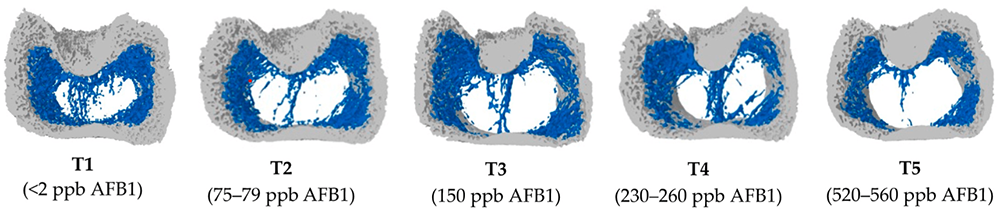
Figure
Figure 1: Increasing doses of AFB1 (<2 ppb – 560 ppb) deteriorate bone quality (Paneru, 2024): Cross-sectional images of femoral metaphysis with increasing AFB1 levels (left to right). The outer cortical bone is shown in light grey, and the inner trabecular bone in blue. Higher levels of AFB1 (T4 and T5) show a disruption of the trabecular bone pattern (less dense blue pattern with thinner and more fragmented bone strands and with wide spaces between the trabecular bone) (shown in white).

All experiments strongly suggest that aflatoxins harm bone homeostasis. Additional liver damage, oxidative stress, and impaired cellular processes can exacerbate bone health issues.

Trichothecenes also negatively impact bone mineralization. Depending on the mycotoxin, they may affect the gut, decreasing the absorption of Ca and P and probably provoking an imbalance in the Ca/P ratio.

For instance, when T-2 toxin was fed to Yangzhou goslings at 0.4, 0.6, and 0.8 mg/kg of diet, it decreased the Ca levels (halved at 0.8 mg/kg) and increased the P levels in the blood serum, so the Ca/P ratio decreased from the adequate ratio of 1 – 2 to 0.85, 0.66, and 0.59 (P<0.05) (Gu et al., 2023). The alterations of the Ca and P levels, the resulting decreasing Ca/P ratio, and an additional increase in alkaline phosphatase (ALP) suggest that T-2 toxin negatively impacts Ca absorption, increases ALP, and, therefore, disturbs calcification and bone development.

Other studies show that serum P levels decreased in broilers fed DON-contaminated feed with levels of only 2.5 mg/kg (Keçi et al., 2019). One reason for the lower P level is probably the lower dry matter intake, affecting Ca and P intake. Ca serum level is not typically reduced, which can be explained by the fact that Ca plays many critical physiological roles (e.g., nerve communication, blood coagulation, hormonal regulation), so the body keeps the blood levels by reducing bone mineralization. Another explanation is delivered by Li et al. (2020): After their trial with broilers, they stated that dietary P deficiency is more critical for bone development than Ca deficiency or Ca & P deficiency. The results of the trial conducted by Keçi et al. with DON (see above) were reduced bone mineralization, affected bone density, ash content, and ash density in the femur and tibiotarsus with a stronger impact on the tibiotarsus than on the femur.

In line with trichothecenes effects in Ca and P absorption, Ledoux et al. (1992) suppose that diarrhea caused by intake of fumonisins leads to malabsorption or maldigestion of vitamin D, calcium and phosphorus, having birds with rickets as a secondary effect.

Ochratoxin A (OTA) impairs kidney function, negatively affects vitamin D metabolism, reduces Ca absorption, and contributes to deteriorated bone strength (Devegowda and Ravikiran, 2009). Indications from Huff et al. (1980) show decreased tibia strength after feeding chickens OTA levels of 2, 4, and 8 µ/g, and Duff et al. (1987) report similar results also in turkey poults.

A further mycotoxin possibly contributing to leg weakness is cyclopiazonic acid produced by Aspergillus and Penicillium. This mycotoxin is known for leading to eggs with thin or visibly racked shells, indicating an impairment of calcium metabolism (Devegowda and Ravikiran, 2009). Tran et al. (2023) also showed this fact with multiple mycotoxins.

The co-occurrence of different mycotoxins in the feed – the standard in praxis – increases the risk of leg issues. A trial with broiler chickens conducted by Raju and Devegowda (2000) showed a bone ash-decreasing effect of AFB1 (300 µg/kg), OTA (2 mg/kg), and T-2 toxin (3 mg/kg), fed individually but an incomparable higher effect when fed in combination.

Impairment of bone growth – tibial dyschondroplasia (TD)

In TD, the development of long bones is impaired, and abnormal cartilage development occurs. It is frequent in broilers, with a higher incidence in males than females. It happens when the bone grows, as the soft cartilage tissue is not adequately replaced by hard bone tissue. Some mycotoxins have been related to this condition: According to Sokolović et al. (2008), actively dividing cells such as bone marrow are susceptible to T-2 toxin, including the tibial growth plates, which regulate chondrocyte formation, maturation, and turnover.

T-2 toxin: In a study with primary cultures of chicken tibial growth plate chondrocytes (GPCs) and three different concentrations of T-2 toxin (5, 50, and 500 nM), He et al. (2011) found that T-2 toxin decreased cell viability, alkaline phosphatase activity, and glutathione content (P < 0.05). Additionally, it increased the level of reactive oxygen species and malondialdehyde in a dose-dependent way, which could be partly recompensated by adding an antioxidant (N-acetyl-cysteine). They concluded that T-2 toxin inhibits the proliferation and differentiation of GPCs and contributes, therefore, to the development of TD, altering cellular homeostasis. Antioxidants may help to reduce these effects.

Gu et al. (2023) investigated the closely bodyweight-related shank length and the tibia development in Yangzhou goslings fed feed with six different levels (0 to 2.0 mg/kg) of T-2 toxin for 21 days. They determined a clear dose-dependent slowed tibial length and weight growth (p<0.05), as well as abnormal morphological structures in the tibial growth plate. As tibial growth and shank length are closely related to weight gain (Gu et al., 2023; Gao et al., 2010; Ukwu et al., 2014; Yu et al., 2022), their slowdown indicates lower growth performance.

Fumonisin B1 is also a potential cause of this kind of leg issue. Feeding 100 and 200 mg/kg to day-old turkey poults for 21 days led to the development of TD (Weibking et al., 1993). Possible explanations are the reduced viability of chondrocytes, as found by Chu et al. (1995) after 48 h of exposure, or the toxicity of FB1 to splenocytes and chondrocytes, which was shown in different primary cell cultures from chicken (Wu et al., 1995).

Bacterial chondronecrosis with osteomyelitis lameness (BCO) can be triggered by DON and FUM

BCO presents a highly critical health and welfare issue in broiler production worldwide, and it is estimated that 1-2 % of condemnations in birds at the marketing age result from this disease. What is the reason? Today’s fast-growing broilers are susceptible to stress. This enables pathogenic bacteria to compromise epithelial barriers, translocate from the gastrointestinal tract or the pulmonary system into the bloodstream, and colonize osteochondrotic microfractures in the growth plate of the long bone. This can lead to bone necrosis and subsequent lameness.

In their experiment with DON and FUM in broilers, Alharbi et al. (2024) showed that these mycotoxins reduce the gut’s barrier strength and trigger immunosuppressive effects. They used contaminations of 0.76, 1.04, 0.94, and 0.93 mg DON/kg of feed and 2.40, 3.40, 3.20, and 3.50 mg FUM/kg diet in the starter, grower, finisher, and withdrawal phases, respectively. The team observed lameness on day 35; the mycotoxin groups always showed a significantly (P<0.05) higher incidence of cumulative lameness.

The increase in uric acid leads to gout

In general, mycotoxins, which damage the kidneys and, therefore, impact the renal excretion of uric acid, are potentially a factor for gout appearance.

One of these mycotoxins is T-2 toxin. With the trial mentioned before (Yangzhou goslings, 21 days of exposure), Gu et al. (2023) showed that the highest dosage of the toxin (2.0 mg/kg) significantly increased uric acid in the blood (P<0.05), possibly leading to the deposit of uric acid crystals in the joints and to gout.

Huff et al. (1975) applied Ochratoxin to chicks at 0, 0.5, 1.0, 2.0, 4.0, and 8.0 µg/g of feed during the first three weeks of life. They found ochratoxin A as a severe nephrotoxin in young broilers as it caused damage to the kidneys with doses of 1.0 µg/g and higher. At 4.0 and 8.0 µg/g doses, uric acid increased by 38 and 48%, respectively (see Figure 2). Page et al. (1980) also reported increased uric acid after feeding 0.5 or 1.0 mg/kg of Ochratoxin A to adult white Leghorn chickens.

FigureFigure 2: Effect of Ochratoxin A on plasma uric acid (mg/100 ml) (according to Huff et al., 1975)

Foot pad lesions – a further hint of mycotoxicosis

Foot pad lesions often result from wet litter, originating from diarrhea due to harmed gut integrity. Frequently, mycotoxins impact the intestinal tract and create ideal conditions for the proliferation of diarrhea-causing microorganisms and, therefore, secondary infections. Some also negatively impact the immune defense system, allowing pathogens to settle down or aggravate existing bacterial or viral parasitic diseases. In general, mycotoxins affect the physical (intestinal cell proliferation, cell viability, cell apoptosis), chemical (mucins, AMPs), immunological, and microbial barriers of the gut, as reported by Gao et al. (2020). Here are some examples of the adverse effects of mycotoxins leading to intestinal disorders and diarrhea:

  • Mycotoxins can modulate intestinal epithelial integrity and the renewal and repair of epithelial cells, negatively impacting the intestinal barrier’s intrinsic components; for instance, DON can significantly reduce the transepithelial electrical resistance (TEER)(Grenier and Applegate, 2013). A higher permeability of the epithelium and a decreased absorption of dietary proteins can lead to higher protein in the digesta in the small intestine, which serves as a nutrient for pathogens including perfringens (Antonissen et al., 2014; Antonissen et al., 2015).
  • The application of Ochratoxin A (3 mg/kg) increased the number of S. typhimurium in the duodenum and ceca of White Leghorn chickens (Fukata et al., 1996). Another trial with broiler chicks at a concentration of 2 mg/kg aggravated the symptoms due to an infection by S. gallinarum (Gupta et al., 2005).
  • In a trial by Grenier et al., 2016, feed contaminated with DON (1.5 mg/kg), Fumonisin B (20 mg/kg), or both mycotoxins aggravated lesions caused by coccidia.
  • DON impacts the mucus layer composition by downregulating the expression of the gene coding for MUC2, as shown in a trial with human goblet cells (Pinton et al., 2015). The mucus layer prevents pathogenic bacteria in the intestinal lumen from contacting the intestinal epithelium (McGuckin et al., 2011).
  • Furthermore, DON and other mycotoxins decrease the populations of lactic acid-producing bacteria, indicating a shift in the microbial balance (Antonissen et al., 2016).
  • FB1 causes intestinal disturbances such as diarrhea, although it is poorly absorbed in the intestine. According to Bouhet and Oswald (2007), the main toxicological effect ascertained in vivo and in vitro is the accumulation of sphingoid bases associated with the depletion of complex sphingolipids. This negative impact on the sphingolipid biosynthesis pathway could explain other adverse effects, such as reduced intestinal epithelial cell viability and proliferation, modification of cytokine production, and impairment of intestinal physical barrier function.
  • T-2 toxin can disrupt the immune response, enhance the proliferation of coli in the gut, and increase its efflux (Zhang et al., 2022).

All these mycotoxins can cause foot pad lesions by impacting gut integrity or damaging the gut mucosa. They promote pathogenic organisms and, thus, provoke diarrhea and wet litter.

Mitigating the negative impact of mycotoxins on bones and feet is crucial for performance

Healthy bones and feet are essential for animal welfare and performance. Mycotoxins can be obstructive. Consequently, the first step to protecting your animals is to monitor their feed. If the analyses show the occurrence of mycotoxins at risky levels, proactive measures must be taken to mitigate the issues and ensure the health and productivity of your poultry.

References

Alharbi, Khawla, Nnamdi Ekesi, Amer Hasan, Andi Asnayanti, Jundi Liu, Raj Murugesan, Shelby Ramirez, Samuel Rochell, Michael T. Kidd, and Adnan Alrubaye. “Deoxynivalenol and Fumonisin Predispose Broilers to Bacterial Chondronecrosis with Osteomyelitis Lameness.” Poultry Science 103, no. 5 (May 2024): 103598. https://doi.org/10.1016/j.psj.2024.103598.

Antonissen, Gunther, Filip Van Immerseel, Frank Pasmans, Richard Ducatelle, Freddy Haesebrouck, Leen Timbermont, Marc Verlinden, et al. “The Mycotoxin Deoxynivalenol Predisposes for the Development of Clostridium Perfringens-Induced Necrotic Enteritis in Broiler Chickens.” PLoS ONE 9, no. 9 (September 30, 2014). https://doi.org/10.1371/journal.pone.0108775.

Antonissen, Gunther, Filip Van Immerseel, Frank Pasmans, Richard Ducatelle, Geert P. Janssens, Siegrid De Baere, Konstantinos C. Mountzouris, et al. “Mycotoxins Deoxynivalenol and Fumonisins Alter the Extrinsic Component of Intestinal Barrier in Broiler Chickens.” Journal of Agricultural and Food Chemistry 63, no. 50 (December 10, 2015): 10846–55. https://doi.org/10.1021/acs.jafc.5b04119.

Antonissen, Gunther, Venessa Eeckhaut, Karolien Van Driessche, Lonneke Onrust, Freddy Haesebrouck, Richard Ducatelle, Robert J Moore, and Filip Van Immerseel. “Microbial Shifts Associated with Necrotic Enteritis.” Avian Pathology 45, no. 3 (May 3, 2016): 308–12. https://doi.org/10.1080/03079457.2016.1152625.

Bai, Shiping, Leilei Wang, Yuheng Luo, Xumei Ding, Jun Yang, Jie Bai, Keying Zhang, and Jianping Wang. “Effects of Corn Naturally Contaminated with Aflatoxins on Performance, Calcium and Phosphorus Metabolism, and Bone Mineralization of Broiler Chicks.” The Journal of Poultry Science 51, no. 2 (2014): 157–64. https://doi.org/10.2141/jpsa.0130053.

Bouhet, Sandrine, and Isabelle P. Oswald. “The Intestine as a Possible Target for Fumonisin Toxicity.” Molecular Nutrition &amp; Food Research 51, no. 8 (August 2007): 925–31. https://doi.org/10.1002/mnfr.200600266.

Chi, M.S., C.J. Mirocha, H.J. Kurtz, G. Weaver, F. Bates, W. Shimoda, and H.R. Burmeister. “Acute Toxicity of T-2 Toxin in Broiler Chicks and Laying Hens ,.” Poultry Science 56, no. 1 (January 1977): 103–16. https://doi.org/10.3382/ps.0560103.

Chu, Qili, Weidong Wu, Mark E. Cook, and Eugene B. Smalley. “Induction of Tibial Dyschondroplasia and Suppression of Cell-Mediated Immunity in Chickens by Fusarium Oxysporum Grown on Sterile Corn.” Avian Diseases 39, no. 1 (January 1995): 100. https://doi.org/10.2307/1591988.

Costanzo, Paola, Antonello Santini, Luigi Fattore, Ettore Novellino, and Alberto Ritieni. “Toxicity of Aflatoxin B1 towards the Vitamin D Receptor (VDR).” Food and Chemical Toxicology 76 (February 2015): 77–79. https://doi.org/10.1016/j.fct.2014.11.025.

Costanzo, Paola, Antonello Santini, Luigi Fattore, Ettore Novellino, and Alberto Ritieni. “Toxicity of Aflatoxin B1 towards the Vitamin D Receptor (VDR).” Food and Chemical Toxicology 76 (February 2015): 77–79. https://doi.org/10.1016/j.fct.2014.11.025.

Debouck, C., E. Haubruge, P. Bollaerts, D. van Bignoot, Y. Brostaux, A. Werry, and M. Rooze. “Skeletal Deformities Induced by the Intraperitoneal Administration of Deoxynivalenol (Vomitoxin) in Mice.” International Orthopaedics 25, no. 3 (March 24, 2001): 194–98. https://doi.org/10.1007/s002640100235.

Devegowda, G., and D. Ravikiran. “Mycotoxins and Skeletal Problems in Poultry.” World Mycotoxin Journal 2, no. 3 (August 1, 2009): 331–37. https://doi.org/10.3920/wmj2008.1085.

Duff, S.R.I., R.B. Burns, and P. Dwivedi. “Skeletal Changes in Broiler Chicks and Turkey Poults Fed Diets Containing Ochratoxin a.” Research in Veterinary Science 43, no. 3 (November 1987): 301–7. https://doi.org/10.1016/s0034-5288(18)30798-7.

Fukata, T., K. Sasai, E. Baba, and A. Arakawa. “Effect of Ochratoxin A on Salmonella Typhimurium-Challenged Layer Chickens.” Avian Diseases 40, no. 4 (October 1996): 924. https://doi.org/10.2307/1592318.

Gao, Y., Z.‐Q. Du, C.‐G. Feng, X.‐M. Deng, N. Li, Y. Da, and X.‐X. Hu. “Identification of Quantitative Trait Loci for Shank Length and Growth at Different Development Stages in Chicken.” Animal Genetics 41, no. 1 (January 6, 2010): 101–4. https://doi.org/10.1111/j.1365-2052.2009.01962.x.

Grenier, Bertrand, Ilse Dohnal, Revathi Shanmugasundaram, Susan Eicher, Ramesh Selvaraj, Gerd Schatzmayr, and Todd Applegate. “Susceptibility of Broiler Chickens to Coccidiosis When Fed Subclinical Doses of Deoxynivalenol and Fumonisins—Special Emphasis on the Immunological Response and the Mycotoxin Interaction.” Toxins 8, no. 8 (July 27, 2016): 231. https://doi.org/10.3390/toxins8080231.

Gu, Wang, Qiang Bao, Kaiqi Weng, Jinlu Liu, Shuwen Luo, Jianzhou Chen, Zheng Li, et al. “Effects of T-2 Toxin on Growth Performance, Feather Quality, Tibia Development and Blood Parameters in Yangzhou Goslings.” Poultry Science 102, no. 2 (February 2023): 102382. https://doi.org/10.1016/j.psj.2022.102382.

Guntur, Anyonya R., and Clifford J. Rosen. “Bone as an Endocrine Organ.” Endocrine Practice 18, no. 5 (September 2012): 758–62. https://doi.org/10.4158/ep12141.ra.

Gupta, S., N. Jindal, R.S. Khokhar, A.K. Gupta, D.R. Ledoux, and G.E. Rottinghaus. “Effect of Ochratoxin A on Broiler Chicks Challenged withSalmonella Gallinarum.” British Poultry Science 46, no. 4 (August 2005): 443–50. https://doi.org/10.1080/00071660500190850.

Han, Xin-Yan, Qi-Chun Huang, Wei-Fen Li, Jun-Fang Jiang, and Zi-Rong Xu. “Changes in Growth Performance, Digestive Enzyme Activities and Nutrient Digestibility of Cherry Valley Ducks in Response to Aflatoxin B1 Levels.” Livestock Science 119, no. 1–3 (December 2008): 216–20. https://doi.org/10.1016/j.livsci.2008.04.006.

He, Shao‐jun, Jia‐fa Hou, Yu‐yi Dai, Zhen‐lei Zhou, and Yi‐feng Deng. “N‐acetyl‐cysteine Protects Chicken Growth Plate Chondrocytes from T‐2 Toxin‐induced Oxidative Stress.” Journal of Applied Toxicology 32, no. 12 (July 28, 2011): 980–85. https://doi.org/10.1002/jat.1697.

Hou, Hai-Feng, Jin-Ping Li, Guo-Yong Ding, Wen-Jing Ye, Peng Jiao, and Qun-Wei Li. “The Cytotoxic Effect and Injury Mechanism of Deoxynivalenol on Articular Chondrocytes in Human Embryo.” Zhonghua Yu Fang Yi Xue Za Zhi 45, no. 7 (July 2011): 629–32.

Huff, W. E., R. D. Wyatt, and P. B. Hamilton. “Nephrotoxicity of Dietary Ochratoxin A in Broiler Chikens1.” Applied Microbiology 30, no. 1 (1975): 48–51. https://doi.org/10.1128/aem.30.1.48-51.1975.

Huff, William E., John A. Doerr, Pat B. Hamilton, Donald D. Hamann, Robert E. Peterson, and Alex Ciegler. “Evaluation of Bone Strength during Aflatoxicosis and Ochratoxicosis.” Applied and Environmental Microbiology 40, no. 1 (July 1980): 102–7. https://doi.org/10.1128/aem.40.1.102-107.1980.

Kermanshahi, H., M.R. Akbari, M. Maleki, and M. Behgar. “Effect of Prolonged Low Level Inclusion of Aflatoxin B1 into Diet on Performance, Nutrient Digestibility, Histopathology and Blood Enzymes of Broiler Chickens.” J of Anim and Vet Adv 6, no. 5 (2007): 686–92.

Keçi, Marsel, Annegret Lucke, Peter Paulsen, Qendrim Zebeli, Josef Böhm, and Barbara U. Metzler-Zebeli. “Deoxynivalenol in the Diet Impairs Bone Mineralization in Broiler Chickens.” Toxins 11, no. 6 (June 18, 2019): 352. https://doi.org/10.3390/toxins11060352.

Ledoux, David R., Tom P. Brown, Tandice S. Weibking, and George E. Rottinghaus. “Fumonisin Toxicity in Broiler Chicks.” Journal of Veterinary Diagnostic Investigation 4, no. 3 (July 1992): 330–33. https://doi.org/10.1177/104063879200400317.

Li, Tingting, Guanzhong Xing, Yuxin Shao, Liyang Zhang, Sufen Li, Lin Lu, Zongping Liu, Xiudong Liao, and Xugang Luo. “Dietary Calcium or Phosphorus Deficiency Impairs the Bone Development by Regulating Related Calcium or Phosphorus Metabolic Utilization Parameters of Broilers.” Poultry Science 99, no. 6 (June 2020): 3207–14. https://doi.org/10.1016/j.psj.2020.01.028.

Loughrill, Emma, David Wray, Tatiana Christides, and Nazanin Zand. “Calcium to Phosphorus Ratio, Essential Elements and Vitamin D Content of Infant Foods in the UK: Possible Implications for Bone Health.” Maternal &amp; Child Nutrition 13, no. 3 (September 9, 2016). https://doi.org/10.1111/mcn.12368.

McGuckin, Michael A., Sara K. Lindén, Philip Sutton, and Timothy H. Florin. “Mucin Dynamics and Enteric Pathogens.” Nature Reviews Microbiology 9, no. 4 (March 16, 2011): 265–78. https://doi.org/10.1038/nrmicro2538.

Morishita, Y., K. Nagasawa, Naoko Nakano, and Kimiko Shiromizu. “Bacterial Overgrowth in the Jejunum of ICR Mice and Wistar Rats Orally Administered with a Single Lethal Dose of Fusarenon‐x, a Trichothecene Mycotoxin.” Journal of Applied Bacteriology 66, no. 4 (April 1989): 263–70. https://doi.org/10.1111/j.1365-2672.1989.tb02478.x.

Paneru, Deependra, Milan Kumar Sharma, Hanyi Shi, Jinquan Wang, and Woo Kyun Kim. “Aflatoxin B1 Impairs Bone Mineralization in Broiler Chickens.” Toxins 16, no. 2 (February 2, 2024): 78. https://doi.org/10.3390/toxins16020078.

Pegram, R.A., and R.D. Wyatt. “Avian Gout Caused by Oosporein, a Mycotoxin Produced by Chaetomium Trilaterale.” Poultry Science 60, no. 11 (November 1981): 2429–40. https://doi.org/10.3382/ps.0602429.

Persico, Marco, Raffaele Sessa, Elena Cesaro, Irene Dini, Paola Costanzo, Alberto Ritieni, Caterina Fattorusso, and Michela Grosso. “A Multidisciplinary Approach Disclosing Unexplored Aflatoxin B1 Roles in Severe Impairment of Vitamin D Mechanisms of Action.” Cell Biology and Toxicology 39, no. 4 (September 6, 2022): 1275–95. https://doi.org/10.1007/s10565-022-09752-y.

Pinton, Philippe, Fabien Graziani, Ange Pujol, Cendrine Nicoletti, Océane Paris, Pauline Ernouf, Eric Di Pasquale, Josette Perrier, Isabelle P. Oswald, and Marc Maresca. “Deoxynivalenol Inhibits the Expression by Goblet Cells of Intestinal Mucins through a PKR and MAP Kinase Dependent Repression of the Resistin‐like Molecule β.” Molecular Nutrition &amp; Food Research 59, no. 6 (April 27, 2015): 1076–87. https://doi.org/10.1002/mnfr.201500005.

Raju, M.V.L.N., and G. Devegowda. “Influence of Esterified-Glucomannan on Performance and Organ Morphology, Serum Biochemistry and Haematology in Broilers Exposed to Individual and Combined Mycotoxicosis (Aflatoxin, Ochratoxin and T-2 Toxin).” British Poultry Science 41, no. 5 (December 2000): 640–50. https://doi.org/10.1080/713654986.

Rath, Narayan C., and Vijay Durairaj. “Avian Bone Physiology and Poultry Bone Disorders.” Sturkie’s Avian Physiology, 2022, 549–63. https://doi.org/10.1016/b978-0-12-819770-7.00037-2.

Siller, W.G. “Renal Pathology of the Fowl — a Review.” Avian Pathology 10, no. 3 (July 1981): 187–262. https://doi.org/10.1080/03079458108418474.

Suchacki, Karla J, Fiona Roberts, Andrea Lovdel, Colin Farquharson, Nik M Morton, Vicky E MacRae, and William P Cawthorn. “Skeletal Energy Homeostasis: A Paradigm of Endocrine Discovery.” Journal of Endocrinology 234, no. 1 (July 2017). https://doi.org/10.1530/joe-17-0147.

Tran, Si-Trung, Y. Ruangpanit, K. Rassmidatta, K. Pongmanee, K. Palanisamy, and M. Caballero. “The World Mycotoxin Forum, 14th Conference.” In WMF Meets Belgium – Abstracts of Lectures and Posters, 120–21. Antwerp: Conference Secretariat Bastiaanse Communication, 2023.

Ukwu, H.O, V.M.O. Okoro, and R.J. Nosike. “Statistical Modelling of Body Weight and Linear Body Measurements in Nigerian Indigenous Chicken.” IOSR Journal of Agriculture and Veterinary Science (IOSR-JAVS), Ver. V, 7, no. 1 (2014): 27–30.

Wright, G.C., Walter F.O. Marasas, and Leon Sokoloff. “Effect of Fusarochromanone and T-2 Toxin on Articular Chondrocytes in Monolayer Culture in Monolayer Culture.” Toxicological Sciences 9, no. 3 (1987): 595–97. https://doi.org/10.1093/toxsci/9.3.595.

Wu, Weidong, Mark E. Cook, Qili Chu, and Eugene B. Smalley. “Tibial Dyschondroplasia of Chickens Induced by Fusarochromanone, a Mycotoxin.” Avian Diseases 37, no. 2 (April 1993): 302. https://doi.org/10.2307/1591653.

Wu, Weidong, Tianxing Liu, and Ronald F. Vesonder. “Comparative Cytotoxicity of Fumonisin B1 and Moniliformin in Chicken Primary Cell Cultures.” Mycopathologia 132, no. 2 (November 1995): 111–16. https://doi.org/10.1007/bf01103783.

Yu, Jun, Yu Wan, Haiming Yang, and Zhiyue Wang. “Age- and Sex-Related Changes in Body Weight, Muscle, and Tibia in Growing Chinese Domestic Geese (Anser Domesticus).” Agriculture 12, no. 4 (March 25, 2022): 463. https://doi.org/10.3390/agriculture12040463.

Zhang, Jie, Xuerun Liu, Ying Su, and Tushuai Li. “An Update on T2-Toxins: Metabolism, Immunotoxicity Mechanism and Human Assessment Exposure of Intestinal Microbiota.” Heliyon 8, no. 8 (August 2022). https://doi.org/10.1016/j.heliyon.2022.e10012.

Zhao, J., R.B. Shirley, J.D. Dibner, F. Uraizee, M. Officer, M. Kitchell, M. Vazquez-Anon, and C.D. Knight. “Comparison of Hydrated Sodium Calcium Aluminosilicate and Yeast Cell Wall on Counteracting Aflatoxicosis in Broiler Chicks.” Poultry Science 89, no. 10 (October 2010): 2147–56. https://doi.org/10.3382/ps.2009-00608.
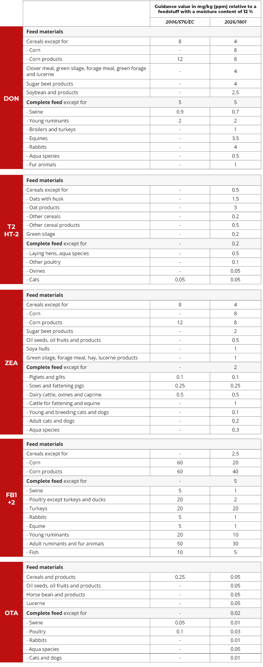
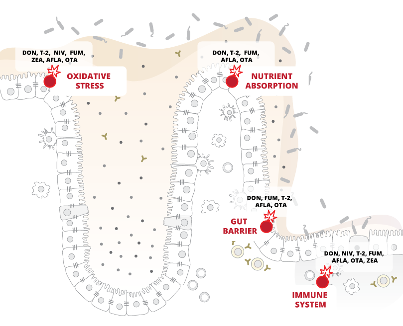

Mycotoxins pose a threat to the horse’s digestive system

Eye Of Arabian Bay Horse

Author: Judith Schmidt, Product Manager On Farm Solutions

Alarm in the gut! Horses have a susceptible digestive system that can quickly become unbalanced. Intestinal disorders in horses are usually associated with colic. Many factors can be responsible for intestinal issues. Have you ever thought about mycotoxins? What can horse owners do to support their horse´s gut health?
The equine stomach is not robust at all. Depending on their age and use, more than half of all horses suffer from stomach pain. Their digestive system is very sensitive and very different from that of other mammals: Horses cannot vomit and often suffer from severe abdominal pain, diarrhea, or cramps if they overeat or ingest spoiled feed.

The horse´s digestive system is complex and sensitive

The horse´s stomach has a relatively small capacity of around twelve to fifteen liters. Depending on the feed’s consistency and composition, it remains in the stomach for around one to five hours before it is pressed through the stomach outlet (pylorus) into the small intestine. The horse´s entire intestine is about ten times its body length.

Figure Digestive TractFigure 1: The horse’s digestive tract

The horse´s gastrointestinal tract is a complex network, reacting extremely sensitively to changes and, therefore, highly susceptible to disorders. It essentially consists of the head intestine (lips, oral cavity, teeth, and esophagus), stomach (blind pouch, fundus, and stomach outlet), small intestine (duodenum, jejunum, and ileum), and large intestine (caecum, colon and rectum). Each section plays a crucial role in the digestive process; any disruption can lead to health issues. Understanding this structure is key to maintaining a horse’s digestive health.

Digestive disorders can have various reasons

Intestinal problems in horses can stem from diverse causes, often a complex interplay of multiple factors. By understanding these causes more deeply, horse owners can be better equipped to prevent and manage these issues. In the following, we delve into several of these causes.

1.   Too long time between the feedings

Usually, a feeding break should be at most four to six hours, as, in nature, a horse is busy eating for at least 18 hours a day. In contrast to humans, who produce stomach acid only after food intake, the horse’s stomach produces gastric acid around the clock. The continuous intake of roughage, intensive chewing, and high saliva production (a horse produces 5 to 10 L of saliva per day) is, therefore, essential to protect the stomach mucosa by neutralizing excess gastric acid.

A too-long time between feedings and, therefore, no saliva production leads to an accumulation of gastric acid in the stomach. Four hours without roughage can already cause inflammation of the mucosa and probably ulcers.

2.   Excessive amounts of concentrated feed

Excessive amounts of concentrates such as wheat or rye, conditioned by less chewing activity, increase gastric acid and histamine production, and the stomach lining can be attacked. Also, in this case, the development of stomach ulcers is possible.

Furthermore, the possibly resulting hyperacidity of the organism can lead to malfunctions of the organs, the skin, and the hooves.

3.   Stress

Stress can also lead to a higher production of gastric acid and, therefore, to gastric ulcers. The horse is a flight animal.  When it is under stress, it prepares for the impending escape, and the muscles are preferably supplied with blood, resulting in a lower blood flow to the mucous membranes. Furthermore, the rising cortisone level reduces the hydrochloric acid-suppressing prostaglandin E. As a result, more stomach acid is produced, irritating the gastric mucosa.

Stress can be triggered, e.g., by transportation, competitions, training, a change of house, a new rider, unsuitable equipment, or poor posture.

4.   Dental diseases

The teeth are essential for digestion. When feed is chewed, it is broken down and mixed with saliva. Chipped teeth cannot chew well, and the feed is not sufficiently salivated or crushed, which has a detrimental effect on digestion.

For this reason, an expert vet should check the horse´s teeth at least once a year.

5.   Administration of painkillers/medication

As with humans, long-term medication administration can promote the formation of stomach ulcers. For this reason, it is essential to ensure that horses are fed a gentle diet on the stomach, especially when using oral pain therapy, and to add stomach protection if necessary.

6. Endotoxins

If pathogens such as E. coli or clostridia proliferate extremely or are killed by an antibiotic, endotoxins can be released. These toxins can cause transformation or inflammation of the gut mucosa. In drastic cases, whole areas of the mucosa can die off. 

7. Mycotoxins – the hidden danger in horse feed

Mycotoxins in plants and horse feed are a common but often unnoticed danger to horses’ health. Mycotoxins are natural, secondary metabolites of molds that have a toxic effect on humans and animals and can trigger mycotoxicosis. Contaminated feed can severely affect the horse’s health and, in the worst case, lead to death.

Over 90 % of the world´s feed production is estimated to be contaminated with at least one mycotoxin (see also Global Mycotoxin Report 2023, EW Nutrition. The intake of mycotoxins via hay, grain, silage, or compound feed can hardly be avoided. Mycotoxins are an increasing problem for all horse owners. Scientific studies show that the mycotoxins DON and ZEA are most frequently found in horse feed and, therefore, are also frequently detected in sports horses’ urine and blood samples.

Due to the highly toxic metabolic products, feed contaminated with molds can lead to severe liver and kidney diseases in horses, affect fertility, trigger colic, and promote digestive issues (diarrhea and watery stools).

Pictures ART
Mycotoxins Horses

Figure 2: Mycotoxins and their impact on horses

How to protect the horse from mycotoxins?

The first measure against the ingestion of mycotoxins is prevention. Correct pasture management and adequate barn and feed hygiene can contribute to preventing the ingestion of toxins.

However, despite the best prophylactic measures, it is impossible to prevent mycotoxin contamination of feed completely. As mycotoxins are not visible, analyzing the feed regarding mycotoxin contamination is recommended.

To protect your horse from mycotoxins, EW Nutrition developed MasterRisk, a tool for evaluating the risk of mycotoxins. Additionally, EW Nutrition has developed a complementary feed specifically for your horse´s needs in the form of granules. The sophisticated formulation of “Toxi-Pearls” is designed to bind mycotoxins and mitigate the adverse effects of mycotoxin contamination.

The pearls contain a mixture of mycotoxin binder, brewer’s yeast, and herbs:

  • The contained mycotoxin binder effectively controls the most important feed myco- and endotoxins. It additionally supports the liver and immune system and strengthens the intestinal barrier.
  • Brewer´s yeast supports the natural strength of the gastrointestinal tract. Due to its high natural content of beta-glucans and mannan-oligosaccharides (MOS), unique surface structure, and the associated high adsorption power, brewer´s yeast has a prebiotic effect on the intestinal microbiome.
  • The additional unique herbal mixture consists of the typical gastrointestinal herbs oregano, rosemary, aniseed, fennel, and cinnamon. The processed beetroot is a true all-rounder. Literature shows that it has an antioxidant effect and strengthens the immune system. It also promotes bile secretion and, therefore, supports fat digestion.

Conclusion

The horse’s digestive tract is highly sensitive and must be supported by all means. Besides failures in management, such as too long breaks between feedings or too high amounts of feed concentrate, mycotoxins present a high risk in horse nutrition. To prevent horses from intestinal issues, feed and stress management, dental care, and medication in the case of disease must be optimized. Particular attention should be paid to possible mycotoxin contamination. Effective toxin risk management, which consists of analysis, risk evaluation, and adequate toxin risk-managing products, should be implemented.