Cryptosporidia in calves – chickens can help

By Lea Poppe, Regional Technical Manager, EW Nutrition

Diarrhea due to infestation with cryptosporidia is one of the most pressing problems in calf rearing. These protozoa, along with rotaviruses, are now considered the most common pathogens in infectious calf diarrhea. Due to their high resistance and thus limited possible control and prevention measures, they have now overtaken other pathogens such as coronaviruses, salmonellae, and E. coli.

Cryptosporidia show complex development

Cryptosporidia are single-celled intestinal parasites. In calves, Cryptosporidium parvum and Cryptosporidium bovis are most commonly found. C. bovis is normally considered nonpathogenic. Accordingly, the disease known as cryptosporidiosis is caused by C. parvum. The rapid tests for determining the diarrheal pathogens, which are increasingly widespread, are usually unsuitable for distinguishing between the individual strains, which can lead to false positive results.

Resistant in the environment, active in the animal

In the environment, cryptosporidia are distributed as oocysts. The oocysts are only about 5 µm in size and have a very resistant shell. They can remain infectious for up to 6 months in high humidity and moderate temperatures. Drought and extreme temperatures (below -18°C and above 65°C) cause the oocysts to die.

After oral ingestion, the oocysts are reactivated by conditions in the gastrointestinal tract (low pH and body temperature): As sporozoites, the parasites attach to the posterior small intestine, causing diarrhea symptomatology. There, they surround themselves with a special protective membrane, and the complex life cycle continues. Only a few days after infection, reproductive forms are detectable in the calf’s intestine, and excretion of infectious oocysts in the feces begins.

Header Calf En
Figure 1 (Olias et al., 2018): Life cycle of cryptosporidia: ingested oocysts release four sporozoites that invade host enterocytes (intestinal epithelial cells). There, they develop into trophozoites before asexual and sexual reproduction ensues, and thin- and thick-walled oocysts are formed. Thick-walled oocysts are excreted through the intestine. Thin-walled oocysts may break apart, and the sporozoites may infect other enterocytes, resulting in relapse or prolonged diarrhea. Infestation of the cells leads to their destruction, resulting in villi atrophy or fusion.

Oocysts bring the disease to the animal

Cryptosporidiosis is transmitted either by direct contact of calves with feces from infected animals or indirectly by ingesting contaminated feed, bedding, or water. Each gram of feces excreted by calves showing symptoms may contain up to 100 million oocysts. According to experimental studies, as few as 17 orally ingested oocysts are sufficient to trigger infection. In addition, some multiplication forms can infect other intestinal cells directly within the intestine and thus further advance the disease by autoinfection.

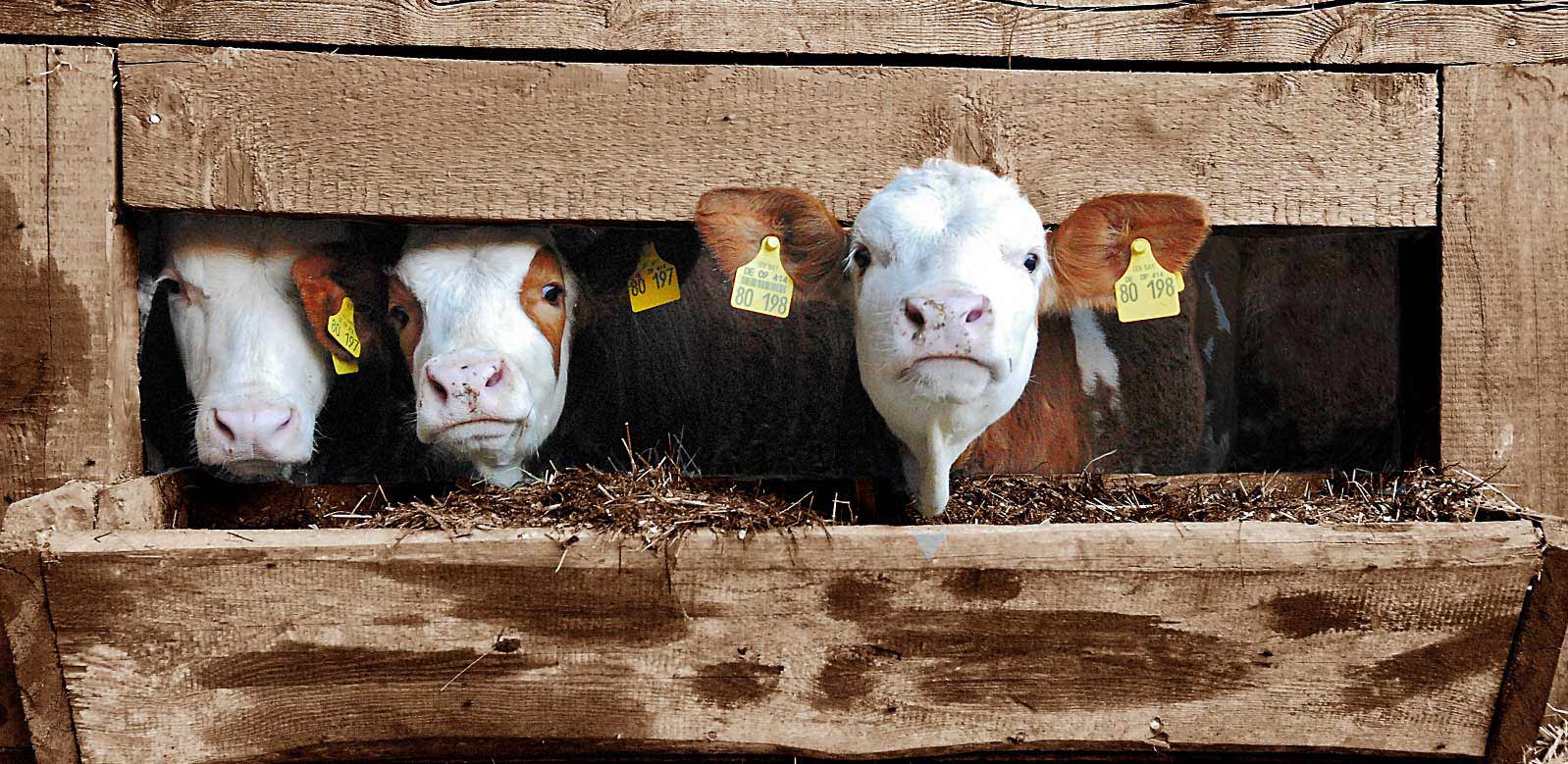
Cryptosporidiosis caused by cryptosporidia often presents with typical diarrhea symptoms and occurs primarily in calves up to 3 weeks of age. Older calves may also be infected with cryptosporidia but usually show no symptoms. Pathogen excretion and, thus, the spread of disease within the herd is nevertheless likely due to the minimal infectious dose.

Damage to the intestinal wall leads to retardation of growth

Attachment of cryptosporidia to the intestinal wall is associated with an inflammatory reaction, regression and fusion of the intestinal villi, and damage to the microvilli. As a result, nutrient absorption in the small intestine is impaired, and more undigested nutrients enter the colon. The microflora starts a fermentation process with lactose and starch, leading to increased lactate levels in the blood and, thus, hyperacidity in the calf. Faintness, unwillingness to drink, recumbency, and growth disorders are the consequences.

Diarrhea often occurs late or not at all and, accordingly, is not considered the main symptom of cryptosporidiosis. When diarrhea occurs, it lasts about 1-2 weeks. The feces are typically watery, greenish-yellow, and are often described as foul-smelling. Due to diarrhea, there is a loss of electrolytes and dehydration.

Studies show: Cryptosporidia are the most prevalent diarrheal pathogens

Several studies in different regions, which examined calf diarrhea and its triggers in more detail, came to a similar conclusion: Cryptosporidia are one of the most common causes of calf diarrhea. In addition, mixed infections often occur.


Country or region Number Age/Health status % Crypto-sporidia % Rota viruses Combined infections with crypto-sporidia Others (%) Source
Switzerland 2 – 21 DL

Ill and healthy

43 46 1 case of E. coli Luginbühl et al., 2012
Switzerland 63 1 – 4 DL

Ill and healthy

————–

7 – 20 DL

————–

26 – 49 DL

34.4

—————

54.0

—————

33.3

3.1

—————-

28.6

—————-

13.3

2 EP – 1.6

4 EP – 3.2

—————-

2 EP – 19

3 EP – 3.2

4 EP – 0

—————-

2 EP – 30

3 EP – 11.7

4 EP – 6.7

Corona 4.7

E. coli 4.7

Giardia 1.6

—————

Corona 0

E. coli 3.2

Giardia 6.3

—————

Corona 0

E. coli 15

Giardia 35


Weber et al., 2016


Weber et al., 2016 EN

Switzerland 147 Up to 3rd WL;

Diarrhea

55 58.7 5.5 % Rota

7.8 % BCV

Lanz Uhde et al., 2014
Sweden 782 1 – 7 DL

Diarrhea

25.3 Detected with Giardia, E. coli, Rota, Eimeria Silverlås et al., 2012
USA (East coast) 503 Pre-weaning 50.3 Santin et al., 2004
USA 30 2 weeks old

1-8 weeks old

3-12 months

12-24 months

96.7

45.8

18.5

2.2

Santin et al., 2008
Germany 521 32 9 Losand et al., 2021
Ethiopia 360 18.6 Ayele et al., 2018
Argentina 1073 n.m. / Ill and healthy 25.5 Lombardelli et al., 2019
UK n.m. Ill ?? 37 25 20 Coccidia 8

E. coli 4

Corona 3

Co infections not including Crypto-sporidia 3

APHA, SRUC, Veterinary investigation diagnosis analysis (VIDA) report (2014)

DL = days of life WL = weeks of life n.m. = not mentioned  EP = enteropathogen

Cryptosporidia reduces profit

Infection with cryptosporidia and sometimes subsequent diarrhea entails treatment of the animals and generates costs (veterinarian, medication, electrolyte drinks). In addition, poorer feed conversion, lower growth, and animal losses result in lower production efficiency.

A Scottish study shows 34 kg less gain in the first six months of life compared to healthy calves in beef calves that experienced severe cryptosporidiosis in the first three weeks of life. Similar results are described in lambs, also a susceptible species to cryptosporidia. These studies suggest a long-term negative effect of cryptosporidia on growth performance and production efficiency.

Here’s how you can support your calves against cryptosporidia

High resistance of the pathogens to environmental influences, a very low necessary infection dose combined with an elevated excretion of infectious oocysts, and the possibility of autoinfection make cryptosporidia tough opponents. This is also reflected in their worldwide distribution.

What is the treatment?

Suitable drugs for the treatment of cryptosporidiosis are currently unavailable on the market. The only medicine that can be used in case of cryptosporidiosis infestation may only be administered to calves that have had diarrhea symptoms for 24 hours or less. Accordingly, this agent is usually used only for prevention. Scientific studies on its effectiveness are contradictory; some suggest that it merely delays the onset of the disease. In addition, it is not always easy to use due to the exact dosage that must be followed. Doubling the dose (sometimes happening already due to incorrectly observed intervals between doses) can lead to a toxic overdose.

Accordingly, only the symptoms of the disease – diarrhea with its accompanying symptoms – can be treated. Electrolyte and water losses must be continuously compensated with the help of a high-quality electrolyte drink. The buffer substances contained also reduce the hyperacidity of the blood caused by faulty fermentation in the intestines. For successful treatment, the electrolyte drink should be given in addition to the milk drink. Under no circumstances should the feeding of milk or milk replacer be discontinued because the sick calf urgently needs energy and nutrients. Opinions to the contrary are outdated.

As always: prevention is better than treatment

To make it more difficult for cryptosporidiosis to spread from the outset, it is worth looking at the risk factors. These include direct contact with other calves and general herd size. Furthermore, organic farms seem to have more problems with cryptosporidia. Weather also influences calves born during warmer and, at the same time, wetter weather periods (temperature-humidity index) often get sick.

Due to the limited possibilities for treatment, prevention is of greater importance. For other diarrheal pathogens such as rotavirus, coronavirus, and E. coli, it has become established practice to vaccinate dams to achieve better passive immunization of the calf. However, commercial vaccination against cryptosporidia is not currently available, making dam vaccination as unavailable as calf vaccination.

Accordingly, optimal colostrum management is the first way to protect the calf from cryptosporidia infection. This also confirms the general discussion on the Failure of Passive Transfer: various studies suggest that calves with poor immunoglobulin supply suffer from diarrhea more frequently than calves with good supply, although a concrete link to cryptosporidia itself cannot always be established with certainty.

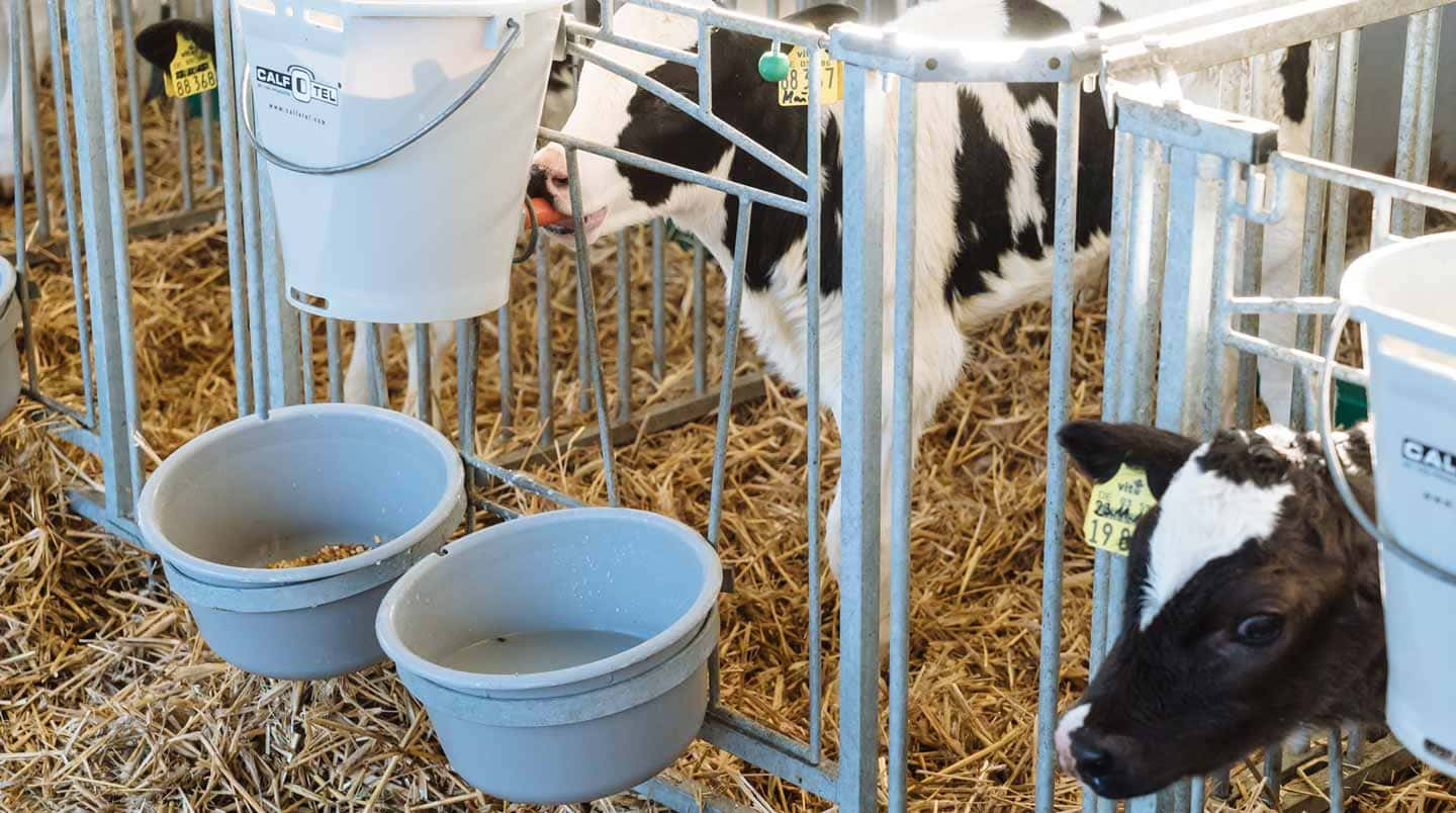
Furthermore, it is essential to break the chain of infection within farms. In addition to the separate housing of the calves, it is necessary to ensure consistent hygiene. One should take advantage of the pathogen’s weakness as well as its sensitivity to high temperatures and ensure that the water temperature is sufficiently high when cleaning the calf pens and calving area. When disinfecting afterward, it is crucial to consider the spectrum of activity of the agent used, as not all are effective against cryptosporidia.

Egg immunoglobulins support animals against cryptosporidia

Egg immunoglobulins were initially designed to help chicks get started. In this process, hens form antibodies against pathogens they are confronted with. As studies have shown, this also works with cryptosporidia. Cama and Sterling (1991) tested their produced antibodies in the neonatal mouse model and achieved a significant (P≤0.001) reduction in parasites there. Kobayashi et al. (2004) registered decreased binding of sporozoites to the intestinal cell model and their decreased viability in addition to oocyst reduction.

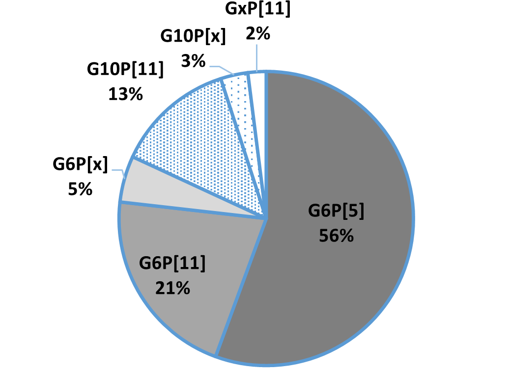
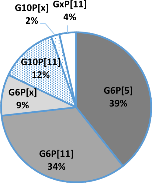
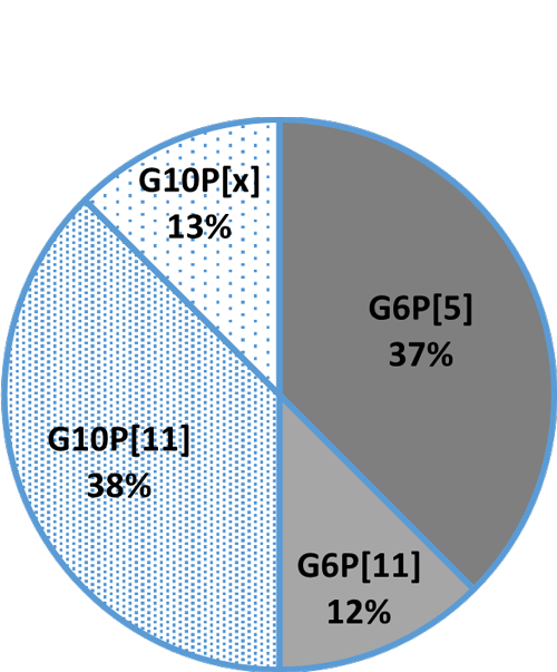
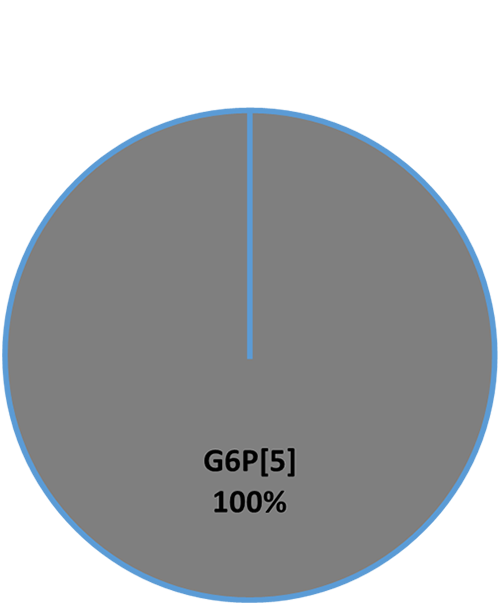
In the IRIG Research Institute (2009, unpublished), feeding egg powder with immunoglobulins against cryptosporidia (10 g/day) to 15 calves reduced oocyst excretion. Before administration, calves excreted an average of 106.42 oocysts/g of feces. After administration of egg powder, only two calves still showed 103.21 oocysts/g feces, and the other 13 of the 15 calves showed no oocyst excretion. All these results are confirmed by positive customer feedback on IgY-based feed supplements.

Egg immunoglobulins and optimal colostrum management as a key solution

Since there are no effective drugs against cryptosporidia, animals must be prophylactically protected against this disease as much as possible. In addition to optimal colostrum management, which means feeding high-quality colostrum (IgG≥50g/L) to the calf as soon as possible after birth, we have products with egg immunoglobulins available to support the calf as a prophylactic against cryptosporidia infestation and thus prevent significant performance losses, especially during rearing.

References

Brainard, J., Hooper, L., McFariane, S., Hammer, C. C., Hunter, P. R., & Tyler, K. (2020). Systemic review of modifiable risk factors shows little evidential support for most current practices in Cryptosporidium management in bovine calves. Parasitology research 119, 3572-3584.

Cama, V. A., and C. R. Sterling. “Hyperimmune Hens as a Novel Source of Anti-Cryptosporidium Antibodies Suitable for Passive Immune Transfer.” University of Arizona. Wiley-Blackwell, January 1, 1991. https://experts.arizona.edu/en/publications/hyperimmune-hens-as-a-novel-source-of-anti-cryptosporidium-antibo.

Kobayashi, C, H Yokoyama, S Nguyen, Y Kodama, T Kimata, and M Izeki. “Effect of Egg Yolk Antibody on Experimental Infection in Mice.” Vaccine 23, no. 2 (2004): 232–35. https://doi.org/10.1016/j.vaccine.2004.05.034.

Lamp, D. O. (25. Januar 2020). Rinder aktuell: Kälberdurchfall durch Kryptosporidien – Hartnäckig und weitverbreitet. BAUERNBLATT, S. 52-53.

Losand, B., Falkenberg, U., Krömker, V., Konow, M., & Flor, J. (2. März 2021). Kälberaufzucht in MV – Alles im grünen Bereich? 30. Milchrindtag Mecklemburg-Vorpommern.

Luginbühl, A., K. Reitt, A. Metzler, M. Kollbrunner, L. Corboz, and P. Deplazes. “Feldstudie Zu Prävalenz Und Diagnostik Von Durchfallerregern Beim Neonaten Kalb Im Einzugsgebiet Einer Schweizerischen Nutztierpraxis.” Schweizer Archiv für Tierheilkunde 147, no. 6 (2005): 245–52. https://doi.org/10.1024/0036-7281.147.6.245.

Olias, P., Dettwiler, I., Hemphill, A., Deplazes, P., Steiner, A., & Meylan, M. (2018). Die Bedeutung der Cryptosporidiose für die Kälbergesundheit in der Schweiz. Schweiz Arch Tierheilkd, Band 160, Heft 6, Juni 2018, 363-374.

Santín, M., Trout, J. M., Xiao, L., Zhou, L., Greiner, E., & Fayer, R. (2004). Prevalence and age-related variation of Cryptosporidium species and genotypes in dairy calves. Veterinary Parasitology 122, 103-117.

Shaw, H. J., Innes, E. A., Marrison, L. J., Katzer, F., & Wells, B. (2020). Long-term production effects of clinical cryptosporidiosis in neonatal calves. International Journal for Parasitology 50, 371-376.

Silverlås, C., H. Bosaeus-Reineck, K. Näslund, and C. Björkman. “Is There a Need for Improved Cryptosporidium Diagnostics in Swedish Calves?” International Journal for Parasitology 43, no. 2 (2013): 155–61. https://doi.org/10.1016/j.ijpara.2012.10.009.

Thomson, Sarah, Carly A. Hamilton, Jayne C. Hope, Frank Katzer, Neil A. Mabbott, Liam J. Morrison, and Elisabeth A. Innes. “Bovine Cryptosporidiosis: Impact, Host-Parasite Interaction, and Control Strategies.” Veterinary Research 48, no. 1 (2017). https://doi.org/10.1186/s13567-017-0447-0.

Uhde, F., Kaufmann, T., Sager, H., Albini, S., Zanoni, R., & Schelling, E. (2008). Prevalence of four enteropathogens in the feces of young diarrhoeic dairy calves in Switzerland. Veterinary Record (163), 362-366.


Coughing calves? How to save costs and prevent respiratory disease

Group of calves peaking through barn opening

By Judith Schmidt, Product Manager On Farm Solutions

There will always be germs in barns. Yet, calves are particularly susceptible to lung viruses and bacteria that attack the respiratory systems. What can we do to prevent calf flu?

Dairy cow and calf on meadow

Coughing in calves is one of the most obvious signs of illness. It should be taken seriously – calves are important for the profitability of farms. Calf flu not only leads to treatment costs but also has long-term consequences, such as weak daily gains, delayed lactation, lower milk yield, reduced fertility, and increased susceptibility to other diseases.

Respiratory disease in calves: recognize the symptoms and protect their lung health

Calves are much more sensitive to respiratory diseases than many other animals. Why? One major cause is that calves are born with immature lungs. The lungs are only fully developed at about one year of age. In addition, calves generally have small lungs relative to their body size. Furthermore, the immunological gaps around the second month of life are decisive. During this phase, the number of maternal antibodies in the calf´s blood decreases, while the calf´s own immune system is still slowly building up.

Symptoms of calf flu

1) Cough

A very easy-to-recognize sign of a developing calf flu is coughing. Coughing can also be caused by changes in weather, stress, or an unsuitable barn climate, but coughing should always be monitored, and animals should be checked for other symptoms.

2) Respiratory distress

Sick calves breathe heavily and show an increased respiratory rate. Even at rest, this can be more than forty breaths per minute, ranging from a slight acceleration of breathing to severe respiratory distress and breathing through the open mouth. Mouth breathing can be the first indication of lung damage.

3) Eye and nose discharge

Calf flu not only shows its symptoms in the internal respiratory tract but also in the eyes and nose through clear, watery discharge. In later stages, bacterial infections can also cause purulent discharge. The animal’s gaze is not clear and rather “sleepy.”

4) Body posture

Calf flu often manifests itself by drooping ears or an overall low head posture, as the calves are dull and weak. They are inactive and separate themselves from the group. They also lie down and standing up is delayed.

5) Reduced water and feed intake

Due to their physical condition, animals suffering from flu tend to take in only little feed and water or do not eat and/or drink at all. The logical consequence is a weakening of the animals. In case of doubt, one should actively water and feed the animals.

Economic significance of respiratory disease in calves

Influenza in cattle and calves is a herd disease and often causes serious financial losses. Losses are caused by pronounced performance decreases, developmental disorders of the animals, and treatment costs. Significantly reduced daily gains have been demonstrated for fattening animals.

Next to diarrheal diseases, calf flu causes the highest treatment and follow-up costs for calves. A study by the Chamber of Agriculture of Lower Saxony (Germany) found that farmers had to spend between 83 and 204 euros per sick calf, depending on the severity of the disease.

4 tips to save costs and tackle calf flu with less antibiotics use

1) Offer a stable climate

Warm, damp barns, as well as overcrowded and poorly ventilated ones, weaken the calf´s defense mechanisms. Temperature fluctuations of more than 10°C between day and night also favor the development of calf flu. It is important to keep the calves’ environment free of dust and draughts. This can be achieved by adjusting the air exchange rate.

In addition, the humidity in barns without a heating system should be between 60 and 80 percent. Data loggers help to keep an eye on the climate in the barn. They make it possible to check how the outdoor climate and ventilation affect the climate conditions in the barn.

2) Hygiene-sensitive calving management

Attention should be paid to calving management. The long-term health of the animal is already predetermined in the calving pen. If several cows calve at the same time or if calving pens are not mucked out regularly, harmful germs will accumulate. In other words: if a calf is born into a dirty box, it will absorb many germs through its mucous membranes.

3) Avoid stress

It is crucial to minimize stress from causes such as transport, re-housing, feed changes, group formation, dehorning, and weaning. These events should be spaced out as far as possible and should never occur simultaneously.

4) Prevention through supplementary feed

In the winter months, when the weather is cold and damp and constantly changing, calf flu incidence skyrockets. Now, it is imperative to strengthen the calf´s respiratory tract from the beginning. EW Nutrition’s Bronchogol Liquid is a herbal concentrate that supports respiration and stabilizes the physiological defense system in the respiratory organs.

Bronchogol liquid supports young calves in stressful situations, such as critical weather transition periods (autumn-winter; winter-spring) and housing changes, and when they suffer from calf flu. The product is based on a proprietary mixture of phytomolecules. By stimulating the cilia in the respiratory tract, the phytomolecules promote the transport of mucus and facilitate expectoration.


IgY supports calves against rotavirus infections

By Kouji Umeda, Production Director, EW Nutrition Japan

Calves are susceptible to infection by pathogens due to their immature congenital immunity. Bovine rotavirus and bovine coronavirus, pathogenic E. coli, Clostridium, Cryptosporidium, and Eimeria spp are the major pathogens of infectious diarrhea in calves less than one month of age. Bovine rotavirus, the most frequently detected in dairy and beef cattle, is responsible for approximately 40% of diarrhea cases. In addition, 60-70% of cases of diarrhea involving bovine rotavirus occur within the first two weeks of life. Symptoms include fever, anorexia, loss of energy, and acute yellow-white watery diarrhea after 12 to 36 hours post infection, which leads to dehydration and metabolic acidosis. In more severe cases, the disease can lead to death and is considered one of the most severe diarrhea-causing pathogens in newborn calves worldwide.

Rotavirus A is a major causative pathogen of diarrhea in calf

Rotaviruses belong to the family of Reoviridae and are classified into species A to J. The rotaviruses in bovines mainly belong to species A, B, and C, which are the leading infectious agents in cattle. Calf diarrhea is primarily caused by rotavirus A (RVA). This virus is transmitted orally through feces, bedding, utensils, or people contaminated with feces. Significant diarrhea caused by the virus is attributed to

  • malabsorption due to the destruction of small intestinal epithelial cells and
  • inhibition of water reabsorption by enterotoxin (NSP4) produced by rotaviruses.

Adult cattle and other host animals have an immune system that protects them from infection and the development of various pathogens. As RVA exists in different genotypes, the antibodies must be specifically against this genotype; otherwise, the virus-neutralizing activity, as well as protection against infection and pathogenesis, is significantly reduced.

The classic method to prevent RVA infection

Besides adequate sanitation in the production facilities, farmers try to “improve” the composition of the maternal colostrum by vaccinating the cow. For this purpose, the cows are inoculated with inactivated, previously isolated bovine RVA. However, the immunization of calves through colostrum may not be effective enough. It also may be difficult to prevent the spread of bovine RVA by barn hygiene alone due to the recent increase in the number of cattle being raised and moved from one farm to another.

Calf diarrhea feces contain G and P genotypes of bovine RVA

In general, the three most common G genotypes of bovine RVA detected in calf diarrhea are G6, G8, and G10, and the three most common P genotypes are P[1], P[5], and P[11]. Based on the results of the genotyping survey in Japan from 1987 to 2000 (Fig. 1) and the one from 2017 to 2020 (figure 2) (Animal Health Research Division of the National Institute of Agrobiological Sciences (NIAH) together with IRIG), the bovine RVA genotypes identified as prevalent and endemic in Japan in recent years were G6P[5], G6P[11], and G10P[11]. However, the percentage of genotypes detected differed among cattle breeds (Fig. 3A, Fig. 3B, Fig. 3C).

Fig.1: Genotyping results from 1987-2000

Fig.2: Genotyping results from 2017-2020


Fig. 3A:Percentage of detection in Holstein

Fig. 3B: Detection rate in crossbreeds
Fig. 3C: Detection rate in beef cattle (Wagyu)

Cow colostrum protects the calf, egg yolk the chick AND the calf

A cow provides the calf with colostrum to ensure immunoglobulin delivery (passive immunity). In poultry, hens transfer immunoglobulins to the egg yolks and pass immunoglobulins to their chicks in this way. This biological mechanism of “immune transfer to the egg yolk” in birds can be used to arbitrarily produce yolk immunoglobulin (IgY) against pathogens of enteric infections in livestock (Ikemori et al., 1992; Ikemori et al., 1997; Yokoyama et al., 1998).


For this purpose, hens must get in contact with the respective pathogens. They produce antibodies against these pathogens – which also works with non-poultry-relevant pathogens such as bovine RVA – and transfer them to the egg (⇒IgY). The eggs with accumulated high levels of useful IgY can be collected almost daily. The immunoglobulins can be fed to livestock animals such as calves to protect them in critical times.

Continuous feeding of milk formulas containing IgY allows the IgY to remain in the intestinal lumen for a long time (Nozaki et al., 2019). There, they bind to the target pathogens and prevent infection by inhibiting their attachment to and cell invasion into intestinal epithelial cells.

IgY and genotype of the virus must match

A study verified that anti-bovine RVA IgY consisting of anti-G6P[1], anti-G6P[5], and anti-G10P[11] shows broad-spectrum virus-neutralizing activity against recent field isolates. Separate trials (see table 1) demonstrated that anti-G6 genotype IgY acted best against the RVA genotypes G6P[1] and G6P[5] and showed less activity against the G10 genotype. Anti-G10P[11] IgY worked optimally against the P[11] genotypes. The trials confirmed that either the G or the P genotype must match to achieve a sufficient virus-neutralizing activity. The IgY mixture is not helpful against bovine RVA strains that match neither the G nor the P genotypes (Odagiri et., 2020).

As the genotyping survey of 2017-2020 showed mainly G6 and G10 genotypes, a mixture of anti- bovine RVA G6P[1] IgY, G6P[5], and G10P[11] has strong virus neutralizing activity against bovine RVA that is currently prevalent and spreading in production sites.

Table 1: Virus-neutralizing activity of field-isolated bovine RVA against various genotypic strains

IgY Virus-neutralizing test strain
SMN 1 HKD 18 SMN 35 HKD 6 HKD 7 HKD 17 KK-3 OKY 31 MYG 1 Dai-10
1978 2018 2018 2017 2017 2017 1983 2017 2017 2007
G6P[1] G6P[5] G6P[5] G6P[11] G6P[11] G6P[11] G10P[11] G10P[11] G8P[14] G24P[33]
anti-G6P[1] 1978 IgY +++ +++ +++ +++ +++ +++ + +
anti-G6P[5] 2018 IgY +++ +++ +++ ++ ++ ++ + + +
Anti-G10P[11] 2017 IgY + + + + ++ ++ +++ +++
Control IgY

+++:Strong virus neutralizing activity; ++:Moderate virus neutralizing activity; +:Weak virus neutralizing activity; -:No virus neutralizing activity

Anti-bovine RVA IgY supports calves against rotavirus infection

To verify the protective effect of oral passive immunization with anti-bovine RVA IgY against bovine RVA infection, a trial with newborn calves was conducted.

Trial design: Eight calves were separated from their mothers immediately after birth without feeding colostrum and moved to a house with infected animals. From the first day, the calves were fed artificial milk supplemented with anti-bovine RVA IgY (n=4) or non-immune IgY (Control IgY; n=4) three times a day.

The parameters observed were fecal score, bovine RVA excretion, and weight gain; data were collected daily. The fecal score was calculated as the cumulative fecal score during the study period: 0 for normal stools, 1 for soft to muddy stools, and 2 for watery stools. Bovine RVA was isolated from daily fecal samples and evaluated by the total number of days of bovine RVA excretion.

Results: The anti-bovine RVA IgY group was found to be effective in reducing the incidence of diarrhea and shortening the duration of virus excretion in the infection test with the bovine RVA G6 genotype strain and the bovine RVA G10 genotype strain (tables 2 and 3).

Table 2: Efficacy of anti-bovine RVA IgY feeding in bovine RVA G6 genotype strain infection

Test Group Diarrhea incidence Cumulative fecal score Bovine RVA excretion days Increase in body weight
(n animals affected/n animals tested) kg %
Anti-bovine RVA IgY     0%   (0/4) 0.0 ± 0.0* 2.3 ± 0.5** 1.3± 0.4** 3.5 ± 0.7**
Control IgY 100%  (4/4) 12.8 ± 4.8 7.8 ± 1.3 – 3.3 ± 1.6 – 7.6 ± 3.6

**: P<0.01; *: P<0.05


Table 3: Efficacy of anti-bovine RVA IgY feeding in bovine RVA G10 genotype strain infection

Test Group Diarrhea incidence Cumulative fecal score Bovine RVA excretion days increase in body weight
(n animals affected/n animals tested) kg %
Anti-bovine RVA IgY 50%   (2/4) 2.3 ± 4.5** 4.3 ± 1.3** 1.1± 0.8** 3.3 ± 3.1**
Control IgY 100%  (4/4) 14.5 ± 3.7 7.3 ± 1.0 – 4.2 ± 0.7 – 11.1 ± 2.1

**: P<0.001

IgY is a valuable tool in rotavirus control

Newborn calves, susceptible to severe diarrhea caused by bovine RVA infection, require passive immunization with antibodies transferred from the colostrum of the mother cow. However, sometimes, calves don’t get enough antibodies which can be the case if

  • the calf does not receive enough colostrum or receives it too late
  • the cow still has not the farm-specific antibodies because of a too short time of being on the farm

To compensate for this lack of immunity, calves have been fed milk formulas containing anti-bovine RVA IgY for some time. Continuous feeding of anti-bovine RVA IgY, which shows strong virus neutralizing activity against each genotype of bovine RVA isolated from recent cases of calf diarrhea, is expected to provide sufficient immunity and be an effective means of bovine RVA control.

In the case of disease outbreaks, it makes sense to utilize IgY with appropriate mechanisms of action in addition to improving the level of quarantine measures, including hygiene control and vaccination.

References:

Ikemori, Yutaka, Masahiko Kuroki, Robert C. Peralta, Hideaki Yokoyama, and Yoshikatsu Kodama. “Protection of Neonatal Calves against Fatal Enteric Colibacillosis by Administration of Egg Yolk Powder from Hens Immunized with K99-Piliated Enterotoxigenic Escherichia Coli.” Amer. J. Vet. Res. 53, no. 11 (1992): 2005–8. PMID: 1466492.

Ikemori, Yutaka, Masashi Ohta, Kouji Umeda, Faustino C. Icatlo, Masahiko Kuroki, Hideaki Yokoyama, and Yoshikatsu Kodama. “Passive Protection of Neonatal Calves against Bovine Coronavirus-Induced Diarrhea by Administration of Egg Yolk or Colostrum Antibody Powder.” Veterinary Microbiology 58, no. 2-4 (1997): 105–11. https://doi.org/10.1016/s0378-1135(97)00144-2.

Nozaki, I., M. Itoh, F. Murakoshi, T. Aoki, K. Shibano, and K. Yamada. “Effect of an Egg Yolk Immunoglobulin(Igy)Product on Oocyst Shedding and Blood and Fecal Igy Concentrations in Cryptosporidium-Infected Calves.” Japanese Journal of Large Animal Clinics 10, no. 2 (2019): 68–72. https://doi.org/10.4190/jjlac.10.68.

Odagiri, Koki, Nobuki Yoshizawa, Hisae Sakihara, Koji Umeda, Shofiqur Rahman, Sa Van Nguyen, and Tohru Suzuki. “Development of Genotype-Specific Anti-Bovine Rotavirus a Immunoglobulin Yolk Based on a Current Molecular Epidemiological Analysis of Bovine Rotaviruses a Collected in Japan during 2017–2020.” Viruses 12, no. 12 (2020): 1386. https://doi.org/10.3390/v12121386.

Yokoyama, Hideaki, Robert C. Peralta, Kouji Umeda, Tomomi Hashi, Faustino C. Icatlo, Masahiko Kuroki, Yutaka Ikemori, and Yoshikatsu Kodama. “Prevention of Fatal Salmonelosis in Neonatal Calves, Using Orally Administered Chicken Egg Yolk Salmonella-Specific Antibodies.” Amer. J. Vet. Res. 59, no. 4 (1998): 416–20. PMID: 9563623.