Nutritional strategies to maximize the health and productivity of sows
Conference Report
During lactation, the focus should be on maximizing milk production to promote litter growth while reducing weight loss of the sow, stated Dr. Jan Fledderus during the recent EW Nutrition Swine Academies in Ho Chi Minh City and Bangkok. A high body weight loss during lactation negatively affects the sow’s performance in the next cycle and impairs her longevity.
Milk production – ‘push’ or ‘pull’?
“Is a sow’s milk production driven by “push” – the sow is primarily responsible for milk production, or “pull” – suckling stimulates the sow to produce milk?” asked Dr. Jan Fledderus at the beginning of his presentation. The answer is that it is primarily a pull mechanism: piglets that suckle effectively and frequently can activate all compartments of the udder, leading to increased milk production. Therefore, the focus should be optimizing piglet suckling behavior (pull) to enhance milk production. This highlights the importance of piglet vitality and access to the udder in maximizing milk yield.”
Modern sows are lean
Modern sows have been genetically selected for increased growth rates and leanness, which alters their body composition. This makes traditional body condition scoring (BCS) methods, which rely on subjective visual assessment and palpation of backfat thickness, less effective. This may not accurately represent a sow’s true condition, especially in leaner breeds where muscle mass is more prominent than fat. Technology, such as ultrasound measurements of backfat and loin muscle depth, provide more accurate assessments of body condition and can help quantify metabolic reserves more effectively than visual scoring.
Due to their increased lean body mass, we must consider adjusted requirements for amino acids, energy, digestible phosphorus, and calcium. Their dietary crude protein and amino acid requirements have increased drastically.
Importance of high feed intake for milk production
Sows typically catabolize body fat and protein to meet the demands of milk production. Adequate feed intake helps reduce this catabolism, allowing sows to maintain body condition while supporting their piglets’ nutritional needs.
Feeding about 2.5kg on the day of farrowing ensures that sows receive adequate energy to support the farrowing process and subsequent milk production. Sows that are well-fed before farrowing tend to have shorter farrowing durations due to better energy availability during labor.
A short interval between the last feed and the onset of farrowing (3 hours) has been shown to significantly reduce the probability of both assisted farrowing and stillbirths without increasing the risk of constipation. To enhance total feed intake, feeding lactating sows at least three times a day is helpful.
Dr. Fledderus recommended a gradual increase in feed intake during lactation, then from day 12 of lactation to weaning, feeding 1% of sow’s bodyweight at farrowing + 0.5 kg/piglet. For example, for a 220kg sow with 12 piglets:
(220 kg x 0.01) + (12 x 0.5 kg) = 2.2 +6 = 8.2 kg total daily feed intake
Energy source – starch versus fat
The choice between starch and fat as an energy source in sow diets has substantial implications for body composition and milk production.
Starch digestion leads to glucose release, stimulating insulin secretion from the pancreas. Insulin is essential for glucose uptake and utilization by tissues. Enhanced insulin response can help manage body weight loss by promoting nutrient storage and reducing the mobilization of the sow’s body reserves.
Sows fed diets with a higher fat supplementation had an increased milk fat, which is crucial for the growth and development of piglets.
Table 1: Effect of energy source (starch vs. fat) on sows’ body composition and milk yield (Schothorst Feed Research)
Diet 1
Diet 2
Diet 3
Energy value (kcal/kg)
2,290
2,290
2,290
Starch (g/kg)
300
340
380
Fat (g/kg)
80
68
55
Feed intake (kg/day)
6.7
6.7
6.8
Weight loss (kg)
15
11
10
Weight loss (kg)
3.1
2.7
2.3
Milk fat (%)
7.5
7.2
7.0
Milk fat (%)
260
280
270
Heat stress impacts performance
The optimum temperature for lactating sows is 18°C. A meta-analysis concluded that each 1°C above the thermal comfort range (from 15° to 25°C) leads to a decrease in sows’ feed intake and milk production and weaning weight of piglets, as shown below.
Effect of heat stress on lactating sows (according to Ribeiro et. al., 2018 Based on 2,222 lactating sows, the duration of lactation was corrected to 21 days)
To mitigate the effects of heat stress, which reduces feed intake, it is beneficial to schedule feeding during cooler times of the day. This strategy helps maintain appetite and ensures that sows consume sufficient nutrients for milk production. Continuous access to cool, clean water can also enhance feed consumption.
Pigs produce much heat, which must be “excreted”. Increased respiratory rate (>50 breaths/minute) has been shown to be an efficient parameter for evaluating the intensity of heat stress in lactating sows.
When sows resort to panting as a mechanism to dissipate heat, this rapid breathing increases the loss of carbon dioxide, resulting in respiratory alkalosis. To prevent a rise in blood pH level, HCO3 is excreted via urine, and positively charged minerals (calcium, phosphorous, magnesium, and potassium) are needed to facilitate this excretion. However, these minerals are crucial for various physiological functions. As their loss can lead to deficiencies that affect overall health and productivity, this mineral loss must be compensated for.
Implications for management
Implementing effective nutritional strategies together with robust management practices is crucial for maximizing the health and productivity of sows. The priority is to stimulate the sow to eat more. This not only enhances milk production and litter growth but also supports the overall well-being of the sow. Regularly assessing sow performance metrics – such as body condition score, feed intake, and litter growth – can help identify areas for improvement in nutritional management.
EW Nutrition’s Swine Academy took place in Ho Chi Minh City and Bangkok in October 2024. Dr. Jan Fledderus, Product Manager and Consultant at the S&C team at Schothorst Feed Research, with a strong focus on continuously improving the price/quality ratio of the diets for a competitive pig sector and one of the founders of the Advanced Feed Package, was a reputable guest speaker in these events.
Ketosis: the most critical metabolic disease in dairy cows
Judith Schmidt, Product Manager On-Farm Solutions
Improvements in genetics, nutrition, and management continue to enhance dairy cows’ performance. However, being high-performance athletes comes at a cost, putting an extremely high burden on the animals’ energy metabolism. Especially around calving and during the first eight weeks of lactation, dairy cows can experience many stress factors: subclinical hypocalcemia, abomasum displacements, herd composition changes, or lameness. The more stress factors put the cows’ organism under pressure, the more likely they will become sick. A common consequence of stress is the occurrence of metabolic diseases, especially ketosis.
Both in terms of animal health and economic aspects, ketosis is probably the most critical dairy cow disease when also considering the correlated diseases. In this article, we explore the causes and consequences of ketosis and highlight prevention strategies that keep this issue under control.
Ketosis: causes and consequences
How ketosis develops
A restricted feed intake capacity and/or reduced energy concentration in the ration lead to a deficit in the animal’s energy balance. This situation occurs, for instance, at calving when the mother animal focuses her resources on the calf and its care. To compensate for the energy deficit, body fat is broken down for energy production. This process creates free fatty acids that accumulate in the liver and are partially converted into ketone bodies. These ketone bodies are a “transport medium” for energy, which various organs can use as an alternative energy source.
The problem arises when the deficiency lasts too long: more and more body fat is broken down, more and more fatty acids reach the liver, which leads to a fatty liver, and too high an amount of ketone bodies is formed and released into the blood. The ketone bodies in the blood inhibit appetite, resulting in less feed consumption and an energy deficit – the vicious cycle of ketosis begins.
Subclinical ketosis
Subclinical ketosis is defined as the stage of the disease at which an increased level of ketone bodies can be detected in the blood, urine, and milk. Furthermore, signs of hypoglycemia, increased levels of non-esterified fatty acid, and decreased hepatic gluconeogenesis can be seen in the blood. These conditions are typically not detected because there are no clinical signs.
Subclinical ketosis is a problem as it does not cause visible symptoms but leads to an increased incidence of subsequent diseases such as lab stomach displacement, clinical ketosis, and uterine inflammation. In addition, there may be loss of milk and fertility problems. Subclinically ill animals cannot be identified by the farmer by observation alone. Therefore, subclinical ketosis must be detected at an early stage to be able to act at the right time: prophylaxis instead of therapy.
There are several test possibilities to find out if an animal suffers from ketosis:
Milk: Milk test for ketosis detection has been available for many years. The results are to be obtained based on a color gamut. In contrast to blood analysis, the milk test does not evaluate exact values but shows a color change of the contained indicator. However, an increased milk cell content of the feeding of poorly fermented silages with a high butyric acid content significantly influences the result. The test often does not adequately reflect the actual conditions.
Urine: Another possibility is the examination of urine samples. Urine can be obtained spontaneously or with the help of a catheter. The results can also be read on a color scale of the urine test stripes. Like the milk test, the urine test only distinguishes different concentration ranges, but these are more finely graded than in the milk tests.
Blood: The most accurate but also most complex and expensive method is a blood test. It has the advantage that not only ketone bodies but also other parameters such as free fatty acids, minerals, and liver enzymes can be analyzed. In addition, the blood analysis results are evaluated in numbers and are more comparable than the color changes of test stripes. A good alternative is a rapid test by using a rapid test device, which is also used for measuring human blood sugar. A result is displayed with a drop of blood on a test strip within a few seconds.
Clinical ketosis
Depending on why there are elevated ketone body levels in the blood, we distinguish between primary and secondary clinical ketosis. For the primary form of clinical ketosis, the energy deficit itself (due to high performance and/or incorrect feeding) causes the condition. This form mainly occurs in susceptible, high-yielding dairy cows between the second and seventh weeks of lactation (Vicente et al., 2014). Secondary ketosis is caused indirectly by other diseases disease. A cow suffering from, for example, a claw disease might no longer consume a performance-based feed ration, leading to an energy deficit.
Typical symptoms
Typical of metabolic diseases, ketosis leads to a broad spectrum of symptoms. The classic symptoms at the beginning of the disease are a loss of appetite and decreased milk performance. As the disease develops, motor skills may be affected, and the excrement’s consistency becomes firmer and darker in color. The respiratory rate of sick animals increases, and they show dyspnea. Dyspnea is the medical description for breathing difficulties. Affected animals suffer from air shortage, which can occur in different situations. Due to the excretion of ketone bodies via the mucous membranes, the animals’ breath smells more or less strongly of acetone (Robinson and Williamson, 1977).
In addition, the animals undergo rapid and severe weight loss, and their general body conditions deteriorate noticeably. Furthermore, cows suffering from ketosis show increased milk fat content or an increased milk fat/protein quotient. Clinical symptoms include reduced general well-being, apathy, blindness, staggering, persistent “absent-minded” licking of the environment or overexcitability, muscle tremors, and aggressiveness (Andersson, 1984).
Effects on animal health and performance
Even in its subclinical form – if untreated – ketosis will engender health risks and reduced performance, negatively impacting milk yield and cows’ fertility. For clinical cases, typical effects include infertility, udder and hoof problems, and a fatty liver. Ketosis during early lactation is usually associated with fatty liver disease. In severe cases, the liver becomes enlarged and more fragile. It then no longer performs its detoxification function, toxic compounds increase, and the central nervous system is damaged. Anorexia or even a total loss of consciousness, the so-called hepatic coma, might ensue, ending in a complete liver function failure.
Direct economic costs range from high veterinary costs to the total loss of the dairy cow, i.e., approximately € 600 to € 1.000 per cow. Moreover, producers face indirect costs from secondary diseases such as fatty liver disease, increased postpartum behavior such as uterine infections, abomasum dislocations, or claw diseases.
Ketosis prevention: feeding and targeted supplementation
Feeding strategy
As part of the preparatory feeding, both dry and pregnant cows should receive rations that lead to an optimal (and not maximum) body condition at the time of calving. Animals with a poorer nutritional status do not have enough body fat reserves to compensate for lack of energy in the first phase of lactation. In more cases, animals have a too high BCS, leading to a risk of difficult births, and the cows have too little appetite at the beginning of lactation. These cows tend to show an excessive mobilization of fat reserves and develop a fatty liver. So prevention of ketosis of the current lactation starts with preventing a too-high BCS in the middle of the previous lactation.
The aim of feeding measures is to keep the lactating cow’s discrepancy between nutrient requirements and nutrient uptake as low as possible when the genetically determined performance potential is exhausted. For this reason, the ration must have a certain minimum energy density (high-quality forage and appropriate concentrate supplements). Also, anything that prevents the cows from ingesting the maximum amount of dry matter should be avoided.
Ket-o-Vital bolus for metabolic support
Another important preventive measure is the specific support of the calving cow’s liver, rumen, and immune system. EW Nutrition’s Ket-o-Vital Bolus was explicitly designed to reduce the risk of ketosis. It contains fast-available glucogenic substances, positively influencing the cow’s energy metabolism. Another advantage the bolus offers is the slow release of the contained cobalt, selenium, niacin, and active yeast:
Cobalt is a trace element important to form cobalamin, the so-called vitamin B12. It is essential for blood formation and the functioning of the nervous system.
Selenium protects cells from oxidative damage and ensures an intact immune defense;
Niacin is a B vitamin that intervenes in energy metabolism and prevents fatty liver syndrome;
And active yeast supports rumen health, preventing rumen acidosis and increasing feed intake.
The application of the Ket-o-Vital Bolus is profitable and straightforward. Only one bolus per application is required.
Ketosis control: be one step ahead
High-performance dairy cows are at risk of ketosis, which results in involuntary culling, poor health, and performance losses. Advanced feed management practices combined with the targeted use of the Ket-o-Vital bolus offer a solution for preventing this debilitating disease. The bolus protects the cows from clinical and subclinical ketosis, reduces metabolic disorders, increases appetite, and improves health – leading to a quick recovery and ensuring profitable production.
References
Vicente, Fernando, María Luisa Rodríguez, Adela Martínez-Fernández, Ana Soldado, Alejandro Argamentería, Mario Peláez, and Begoña de la Roza-Delgado. “Subclinical ketosis on dairy cows in transition period in farms with contrasting butyric acid contents in silages.” The Scientific World Journal 2014 (November 25, 2014): 1–4. https://doi.org/10.1155/2014/279614.
Andersson, L. “Concentrations of blood and milk ketone bodies, blood isopropanol and plasma glucose in dairy cows in relation to the degree of hyperketonaemia and clinical signs*.” Zentralblatt für Veterinärmedizin Reihe A 31, no. 1-10 (1984): 683–93. https://doi.org/10.1111/j.1439-0442.1984.tb01327.x.
Robinson, A. M., and D. H. Williamson. “Effects of acetoacetate administration on glucose metabolism in mammary gland of fed lactating rats.” Biochemical Journal 164, no. 3 (1977): 749–52. https://doi.org/10.1042/bj1640749.
From sub-acute ruminal acidosis to endotoxins: Prevention for lactating cows
by Technical Team, EW Nutrition
Sub-acute acidosis (SARA) is linked to high levels of ruminal LPS. The LPS cause inflammation and contribute to different metabolic conditions and diseases. Various strategies and solutions can be applied to modulate the rumen microbiota and prevent this risk.
In sub-acute rumen acidosis (SARA), the quantity of free lipopolysaccharides (LPS) coming from Gram- bacteria increases considerably. These LPS cross the ruminal wall and intestine, passing into the bloodstream. The negative consequences on the health of the animal are then reflected in decreased productive and reproductive performance.
The LPS are released during the lysis of GRAM- bacteria which die due to the low pH, and these bacteria are mainly responsible for the production of propionic acid for the energy yield that is obtained. It is essential to preserve ruminal balance between Gram+ and Gram- such that there is no excess of LPS.
Nutritional needs of lactating cows with SARA
In the first phase of lactation (from 1 week after calving to 80 – 100 days of lactation), the cow needs a high energy level to meet the large demand for milk production. This energy demand is often not fully satisfied and feed intake falls short. This deficit leads to the need to provide as much energy as possible per feed ration.
Imagine a 650 kg live weight cow, producing about 35 kg of milk per day with a fat percentage of 3.7 and a protein percentage of 3.2. To achieve this production level and fulfill its maintenance requirements, this animal needs a feed intake of 22 kg of dry matter (DM) per day, with an energy level of 21 UFL equal to 36,000 Kcal/day of NE l (Net Energy Lactation)).
To obtain an energy supply of this type, it is necessary to provide rations with a high content of cereals rich in nonstructured carbohydrates (NSC). This will allow the animals to obtain the maximum efficacy in getting the NE I from the metabolizable energy (ME) expressed as kl*.
*kl expresses the effectiveness in passing from EM to EN l net of the heat dissipated by the animal, therefore kl = ENl/EM (Van Es 1978).
Compared to a diet rich in NDF (Neutral Detergent Fiber), this type of diet promotes and stimulates certain strains of bacteria to the detriment of others, shifting the balance towards a greater population of bacteria that produce propionic acid instead those which produce acetic acid. This change also determines a greater share of Gram- compared to Gram+.
What is rumen acidosis?
Rumen acidosis is that “pathology” whereby the volume of SCFA (Short Chain Fatty Acids) produced by the rumen bacteria is greater than the ability of the rumen itself to absorb and neutralize them. Rumen acidosis is mainly caused by the amylolytic and saccharolytic bacteria (Streptococcus bovis; Selenomonas ruminantium, Bacteroides amylophilus, Bacteroides ruminicola and others) responsible for the production of lactic acid. Unlike the other most representative volatile fatty acids (acetic, butyric and propionic), lactic acid has a lower pKa: 7 (3.9 versus 4.7). This means that for the same amount of molecules produced, lactic acid releases a number of ions H+ in the fluid ten times greater than other VFAs, with evident effects on the pH.
Ruminal acidosis can be characterized as acute or subacute. During acute ruminal acidosis, the pH in the rumen drops below 4.8 and remains low for an extended period of time. Acute acidosis leads to complete anorexia, abdominal pain, diarrhea, lethargy, and eventually death. However, the prevalence of acute acidosis in dairy is very low.
Consequences of rumen acidosis
In such situations, a series of negative consequences can be triggered in the lactating cow. Investigations (for instance, using fistulated cows) can reveal, among others, the following alteration in the rumen:
Shift in total microbiome rumen profile (density; diversity; community structure)
Shift in protozoa population (increase in ciliates protozoa after 3 weeks of SARA; increase in the GNB population)
Shift in fungi population (decreasing the fungi population with high fibrolytic enzymes, which are sensitive to low pH)
Rise in LPS rumen concentration (increasing the GNB strain and their lysis)
Influence on the third layer of Stratified Squamous Epithelium (SSE) (desmosomes and tight junctions)
Lower ruminal fiber degradation (reduction in the number of cellulolytic bacteria which are less resistant to acid pH)
Reduction of the total production of fatty acids (propionic, acetic, butyric), therefore less available energy
Lower rumen motility (also as a consequence of the smaller number of protozoa)
The increased acid load damages the ruminal epithelium
Acid accumulation increases the osmotic pressure of the rumen inducing an higher flux of water from the blood circulation into the rumen, causing swelling and rupture of rumen papilla as well as a greater hemoconcentration
The last points are extremely important, as it enables an easier passage of fluids from the blood to the pre-stomachs, greatly influencing the fermentation processes.
Furthermore, with diets low in NDF, the level of chewing and salivation is certainly lower, with a consequent lower level of salivary buffers that enter the rumen and which would maintain an appropriate pH under normal conditions.
Rumen sub-acute and acute acidosis: a fertile ground for LPS
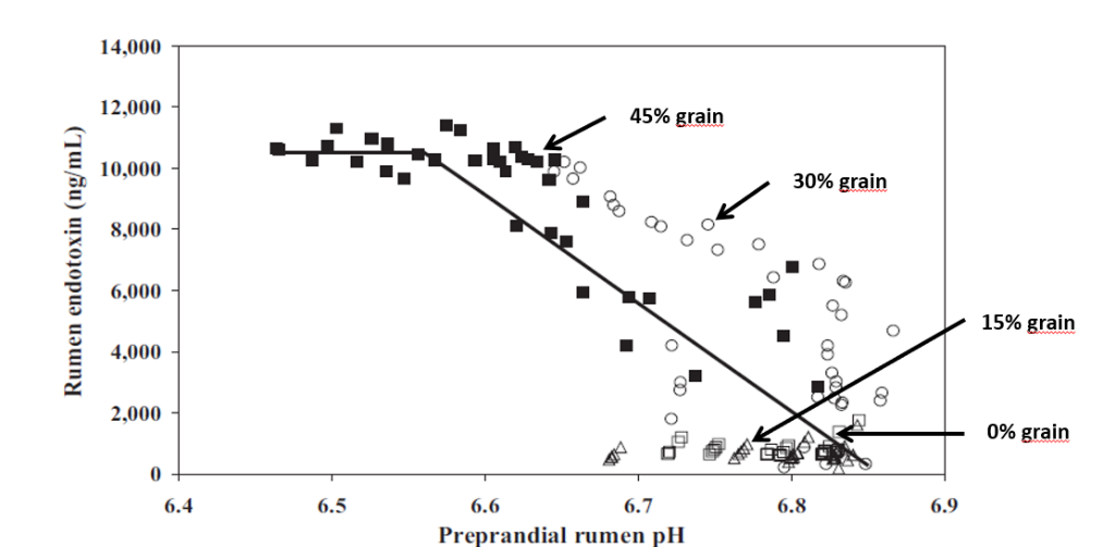
Studies inducing SARA in dairy cows have shown that feeding high levels of grain causes the death and cell lysis of Gram- bacteria, resulting in higher concentration of free LPS in the rumen. In a trial conducted by Ametaj et al., in 2010 (Figure 1), a lower ruminal pH and an increase in the concentration of LPS in the rumen fluid -measured as ng / ml (nanograms / milliliter)-, was the result of increasing of NSC present in the diet (% of grains).
Figure 1. The increase in the level of endotoxins in the rumen is directly correlated with an increase in ration concentrates
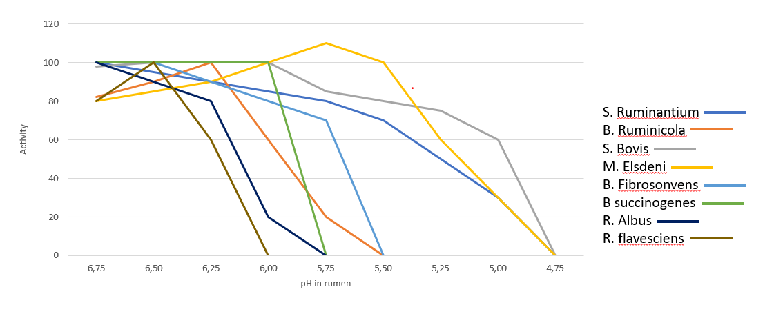
In the rumen, the presence of Gram- is very significant, however the dietary changes towards high energy concentrates, reduce the substates necessary for them to thrive, leading to their lysis and favoring gram-positive bacteria (Gram+). Gram+ also produce bacteriocins against a wide variety of bacteria, including many Gram-. Figure 2 shows the influence of ruminal pH in the population of different bacteria, many of which are are crucial for the production of SCFA and therefore of energy.
Figure 2. Activity of main bacteria in the rumen in function of pH (Daniele Cevolani Edizioni Agricole di New Business Media srl 2020)
It is therefore necessary to pay close attention to the energy level of the ration as an energy input (generally around 1500 – 1700 Kcal/kg of DM intake). At the same time, we need to ensure that the animal does receive and ingest that daily amount of DM. If ingestion is negatively influenced by acidosis (clinical or sub-clinical), this can lead to endotoxemia, with harmful consequences for the animal’s health and production performance.
We can therefore note that the level of LPS (endotoxins) present in the rumen is directly correlated with the pH of the rumen itself and with a symptomatologic picture dating back to SARA. This occurs when the mortality and lysis of Gram- bacteria (GNB) is high and through the consequent imbalance created with diets containing excess fermentable starches, compared to diets with higher fiber content.
In fact, it was shown that the transition from a concentrated fodder ratio of 60:40 to a more stringent ratio of 40:60 caused the level of free LPS in the rumen to go from 410 to 4.310 EU / ml.
Endotoxemia: Pathological consequences in dairy cows
Once the LPS enter the bloodstream, they are transported to the liver (or other organs) for the detoxification. However, sometimes this is not enough to neutralize all the endotoxins present in blood. The remaining excess can cause issues such as the modification of the body’s homeostasis or cause that cascade of inflammatory cytokines responsible for the most common pathologies typical in cows in the first phase of lactation. The most common symptoms are the increase of somatic cells in milk or claws inflammation.
Pro-inflammatory cytokines as TNF, IL6 and IL8 induced by LPS-related inflammation are able to stimulate the production of ACTH (adrenocorticotropic hormone).
ACTH, together with cortisol and the interleukins, inhibit the production of GnRH and LH, with serious effects on milk production. The productivity and the fertility of the animal are thus compromised.
Moreover, prostaglandins are as well stimulated by LPS, and are linked with fever, anorexia and ruminal stasis. This not only limits the amount of energy available for production and maintenance functions, but also induces a higher susceptibility to disease and adds-up to the emergence of other metabolic conditions, such as laminitis and mastitis.
Preventing rumen acidosis
The solution to these massive risks is a prudent and proactive approach by the nutritionist towards all situations that can cause a rapid increase of Gram- in the rumen.It is therefore necessary to avoid cases of clinical and sub-clinical acidosis (SARA) in order to avoid the issues listed above. This would also help avoid stressful conditions for the animal that would lead to decreased performance and health.
To maintain balance and a healthy status of the animal, the use of additives such as phytomolecules and binders is suggested in the first phase of lactation, starting from 15 days before giving birth.
Activo Premium (a mix of phytogenic substances) has given excellent results in decreasing the acetic/propionic acid ratio, while safeguarding the population of Gram+ bacteria. This is in contrast to treatments with ionophores, which, as is well known, interfere with the Gram+ population.
Case study. Acetic acid:propionic acid ratio with Activo Premium
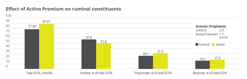
In a study conducted at the the University of Lavras and the Agr. Res. Comp. of Minas Gerais (both Brazil), 30 Holstein cows were allocated to two groups considering parity and milk production. One group was fed the standard feed (control), the other group received standard feed containing 150mg of Activo Premium/kg of dietary dry mass (DM). The following parameters were measured or calculated: intake of DM and milk production, milk ingredients such as fat, protein, lactose every week, body weight and body condition score every two weeks, and ruminal constituents (ph and SCFAs) through oesophaeal samples at day 56.
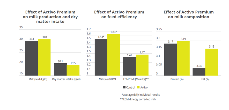
Activo Premium was able to decrease the ratio between acetic acid and propionic acid, and at the same time maintain the level of Gram+ bacteria in the rumen, thus reducing the risk of endotoxins.The same trial carried out at the University of Lavras demonstrated how the performance of the animals was superior in the group fed with Activo Premium compared to the control group (see below).
Figure 3. Effect of Activo Premium on ruminal constituents
Figure 4. Effect of Activo Premium on animal performance
Solution: Preserve Gram+ bacteria levels while decreasing free LPS
We have therefore seen how important it is to decrease the acetic:propionic ratio in the rumen to obtain a greater share of available energy. However, the level of endotoxins in the rumen must remain low in order to avoid those problems of endotoxemia linked to very specific pathologies typical of “super productive cows”. These pathologies (always linked to inflammatory manifestations) can be prevented by decreasing the level of free LPS in the rumen with a product that can irreversibly bind the LPS and thus make them inactive.
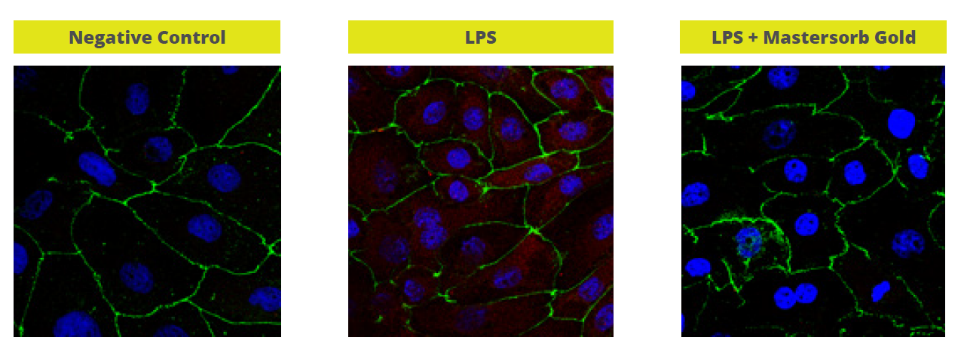
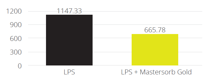
In a trial with porcine intestinal cells (IPEC-J2) challenged by E. coli LPS, a decrease in the intensity of inflammation was observed when Mastersorb Gold was added. This decrease could be shown through a lower amount of phosphorylated NF-kB in an immunofluorescence trial, as well as through the reduced production of Interleukin (IL)-8 in the cells measured by ELISA.
The fact that pig intestine tissue was used does not affect the adsorption concept. In this case, these intestinal cells are only a vehicle to demonstrate that in an aqueous solution containing 50 ŋg of LPS / ml and in the same solution with the addition of Mastersorb Gold, the level of LPS actually active is decreased, as a part of the LPS was tied up by Mastersorb. The solution with a lower level of LPS gave minor “inflammatory” reactions to intestinal cells, and this can be statistically reported in dairy cows.
Figure 5. Immunofluorescence in PEG-J2: Challenge with LPS without (in the middle) and with Mastersorb Gold (right)
Figure 6. IL-8 AP secretion after incubation with LPS 0111:B4 for 24h without and with Mastersorb Gold
Conclusions
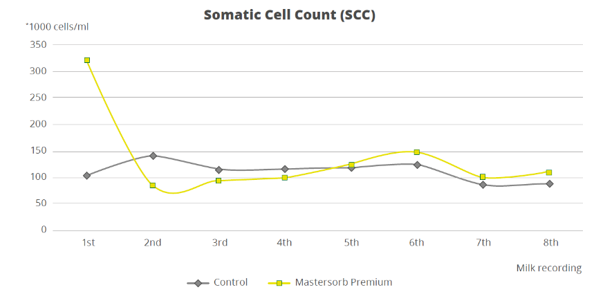
To demonstrate how the decrease in the level of LPS in the rumen is directly correlated with inflammatory states in general, a trial with a total of 60 dairy cows shows that the inclusion of 25g of Mastersorb Premium/animal/day increases milk yield and improves milk quality by decreasing somatic cell count. Adsorbing substances contained in Mastersorb Premium tie up the LPS produced in the rumen in different cow lactation phases.
Normally, the rise in the level of somatic cells in milk depends on etiological agents such as Streptococcus spp, Staphylococcus spp, mycoplasma and more. LPS stress is not the sole agent responsible for raising somatic cell counts, but also other factors among which:
Lactation stage and age of the animal
Season of the year (in summer the problem is increased)
Milking plant (proper maintenance)
General management and nutrition
However, by reducing the level of LPS, Mastersorb provides an important aid to decrease somatic cell count.
Figure 7. Effect of Mastersorb Premium on somatic cell count
Prevent escalation with rumen balance
In the end, ruminant producers are, like all livestock operations, interested in producing healthy animals that can easily cope with various stressors. Ensuring a proper diet, adjusted to the energy requirements of various production stages, is a first step. Providing the animal with the ingredients that modulate the microbiota and reduce the negative impact of stress in the rumen is the next essential step in efficient production.
Nowadays, dairy cows are real top athletes. This comes with additional challenges for their health and for on-farm management. Many of these problems can be traced back to supply deficits and can be easily managed with appropriate feed supplements.
Milk fever is a disease that occurs mainly in cows around calving. It is caused by an insufficient amount of calcium in the blood and particularly affects cows with a very high milk yield.
The link between calcium and milk fever
Calcium performs essential functions in the body. It is particularly important for the nervous system and muscle cells, and plays a central role in muscle contraction. If the calcium content in the blood is too low, the muscles can no longer contract. When this happens, the cows cannot move or stand up.
While mild cases may not be easily detectable, they still trigger productivity loss. If undetected, long-term calcium deficiency can even lead to cardiac arrest and thus to the death of the animal.
The development of milk fever
The cause of milk fever is a lack of sufficient calcium in the blood serum (hypocalcemia). The dairy cow has to abruptly change its metabolism at the end of the dry period, going from the resting phase to a high performance phase. During the dry period, cows have a relatively low need for calcium.
When lactation starts, the need for calcium suddenly almost doubles, as large amounts of calcium are required for the production of colostrum (2.3 g/l). The calcium is generally drawn from feed or from the bones. In older cows, the mobilization mechanism often does not start quickly enough. The supply from the bones and feed is insufficient and the body draws the missing calcium from the muscles. This ultimately leads to symptoms of paralysis and overstimulation of the nervous system.
Phases of milk fever
Stage One
In the initial phase of milk fever, the initial signs are
muscle tremors
restlessness
stiff gait
slightly elevated temperature
Stage Two
At this point, the cows lie on the stomach with an extended neck or the head is lying on the flank. Early symptoms of paralysis appear:
fast, flat pulse
cold body surface
dilated pupils
flatulence
Stage Three
In the last phase of milk fever, the cow lies on its side, loses consciousness and falls into a coma. The third phase often leads to death (the mortality rate averages 2 – 5%).
While the second phase of milk fever is easy to recognize due to the clear symptoms, the consequences of a “slight” calcium deficiency (Stage One) are often underestimated. Feed intake diminishes, the negative energy and protein balance is increased, and the cows barely move. The impairment of the muscles can cause problems in the udder (mastitis) or in the gastrointestinal tract.
Prevention and solutions
As cases of hypocalcemia immediately after calving may be as high as 50% among second- or third-lactation cows, it is important to act preventively to keep potential milk fever from developing. The dairy farmer´s aim is to support the dairy cows that are at higher risk of milk fever, especially around the critical time of calving. The cows must be enabled to quickly release calcium from the bones after calving, or they must be supplied with calcium that can be easily metabolized.
Upfront prophylaxis
An energy and protein oversupply during the dry period should be avoided. In addition, an application of Vitamin D3 at the end of the pregnancy makes sense.
To stimulate the active regulatory mechanisms of calcium metabolism, the calcium content in the feed should be reduced three to four weeks before calving. In practice, however, this often is not properly observed and feed with a relatively high calcium content is still given out during this period.
There are, no doubt, farms where these above-mentioned preventive measures cannot be carried out due to operational reasons, just as there are animals that are particularly susceptible due to factors such as age, breed or healthy history.
To protect the cow from milk fever around calving, oral administration of calcium salts is widespread in practice. Vitamin D also plays a central role in calcium metabolism. It ensures that the absorption of calcium from the intestines and bones is increased.
When administering oral calcium supplements, there are three important points:
– The cow must have sufficient calcium available per dosage
– The calcium must be available immediately
– Administration must be appropriate for the animals and farmers
Methods of calcium supplementation
To support the cow, oral supplements such as pastes and gels are widely used. They are useful, however they are also relatively difficult to administer, as they require handling the animal in relatively difficult ways.
Liquids are another way of administering calcium supplements. When administering liquids, it is important to make sure the animal does not choke so that the liquids do not get into the lungs.
Boluses are probably the easiest and safest method of supplementation to prevent milk fever. The bolus must naturally be carefully inserted, however the process is easy and requires minimal handling of the animal.
EW Nutrition´s Calzogol Bolus is a dietetic mineral feed with a high level of calcium from of highly available calcium salts and vitamin D3. The Calzogol Bolus contains several calcium sources with different release rates. One major advantage is the very high mucous membrane compatibility, which helps avoid irritation of the mouth, esophagus and rumen. Furthermore, the Calzogol Bolus does not contain caustic calcium chloride. The application is simple and economical, as only one bolus per dose must be administered at the time of calving.
Conclusion
Milk fever is very common in dairy herds. When a cow has milk fever, the farm can incur costs of approx. €350. This is reflected in the loss of milk yield up to 600 kg, losses due to unusable milk, and veterinary and medication costs.
Time resources are also to be taken into account: The economic repercussions represent a significant factor, however they come on top of the extra workload due to the increased need for care of animals.
Cows that suffer from calcium deficiency are also much more susceptible to other diseases. For the farmer, the best strategy is to avoid losses through prophylaxis. Feeding plays a central role; to ensure the best possible production conditions, oral calcium administrations, such as Calzogol Bolus, have proven themselves in practice.
by Judith Schmidt, Product Manager, On Farm Solutions
References:
Rérat, M. (2005): Milchfieber bei der Milchkuh. ALP aktuell. Nr. 20.
Spiekers, H., Potthast, V. (2004): Erfolgreiche Milchviehfütterung. DLG-Verlag, Frankfurt a. M.
Kirchgeßner, M., Roth, F. X., Schwarz, F. J., Stangl, G. I. (2008): Tierernährung. 12. Auflage. DLG-Verlag, Frankfurt a. M.