Feed hygiene protects animals and humans
By Vaibhav Gawande, Assistant Manager Technical Services, Dr. Inge Heinzl, Editor, and Marisabel Caballero, Global Technical Manager Poultry, EW Nutrition

The utility value of feed consists of the nutritional value and the quality. The first covers all characteristics concerning the essential nutrients and is important for feed formulation and the adequate supply of the animals.

Feed quality comprises all characteristics of a feed influenced by treatment, storage, conservation, hygiene, and its content of specific substances. For many factors, guidance and threshold values are available which should be met to guarantee animal health and welfare, as well as to protect public health, since some undesirable substances can be transferred to animal products such as meat, eggs, and milk.

In this article, we will focus on feed hygiene. We will talk about the consequences of low feed quality, how to understand it, its causes, and possible solutions.

What are the effects of deficient feed hygiene?

The consequences of deficient feed hygiene can be divided into two parts, impurities and spoilage.

Impurities comprise:

  • the presence of soil, sand, or dust
  • contamination with or residues of heavy metals, PCB, dioxins, pesticides, fertilizers, disinfectants, toxic plants, or banned feed ingredients

In the case of spoilage, we see:

  • degradation of organic components by the action of molds and bacteria
  • growth of pathogens such as E. coli, salmonella, etc.
  • accumulation of toxins such as mycotoxins or bacterial toxins (Hoffmann, 2021)

Bad feed hygiene can also negatively impact the feed’s nutritional value by leading to a loss of energy as well as decreasing the bioavailability of vitamins A, D3, E, K, and B1.

But, how can all signs of deficient feed hygiene be recognized? Soil, sand, and probably dust can be seen in well-taken samples and impurities can be analyzed. But is it possible to spot spoilage? In this case, agglutinated particles, rancid odor, moisture, and discoloration are indicators. Sometimes, also the temperature of the feed or ingredient increases. However, spoilage is not always obvious and an analysis of the feed can give more information about the spoilage-related organisms present. It also helps to decide if the feed is safe for the animals or not. In the case of obvious alterations, the feed should not be consumed by any animal.

Different organisms decrease feed quality and impact health

Several organisms can be responsible for a decrease in feed quality. Besides the visible pests such as rats, mice, or beetles, which can easily be noticed and combatted, there are organisms whose mastering is much more difficult. In the following part, the different harmful organisms and substances are described and solutions are presented.

Enteropathogens can cause diarrhea and production losses

In poultry, different bacteria responsible for high production losses can be transferred via the feed. The most relevant of them are Clostridium perfringens, Escherichia coli, and some strains of Salmonella.

Clostridium perfringens, the cause of necrotic enteritis

Clostridium perfringens is a Gram-positive, anaerobic bacterium that is extremely resistant to environmental influences and can survive in soil, feed, and litter for several years and even reproduce. Clostridium perfringens causes necrotic enteritis mainly in 2-16 weeks old chickens and turkeys, being more critical in 3-6 weeks old chicks.

There is a clinical and a subclinical form of necrotic enteritis. The clinical form can be detected very well due to clear symptoms and mortality rates up to 50%. The subclinical form, while harder to detect, also raises production costs due to a significant decrease in performance. The best prophylaxis against clostridia is the maintenance of gut health, including feed hygiene.

Clostridia can be found in animal by-products, as can be seen in table 1.

Sr. No. Sample details Clostridium perfringens contamination Total number of samples Positivity %
Positive Negative
1 Meat and bone meal 39 52 91 42.86
2 Soya meal 0 3 3 0
3 Rape seed meal 0 1 1 0
4 Fish meal 21 17 38 55.26
5 Layer Feed 21 71 93 22.58
6 Dry fish 5 8 13 38.46
7 De-oiled rice bran 0 2 2 0
8 Maize 0 2 2 0
9 Bone meal 13 16 29 44.83

Table 1: Isolation of Clostridium perfringens from various poultry feed ingredients in Tamil Nadu, India (Udhayavel et al., 2017)

Salmonella is harmful to animals and humans

Salmonella is a gram-negative enterobacterium and can occur in feed. There are only two species – S. enterica and S. bongori (Lin-Hui and Cheng-Hsun, 2007), but almost 2700 serotypes. The most known poultry-specific Salmonella serotypes are S. pullorum affecting chicks and S. gallinarum affecting adult birds. The other two well-known serotypes, S. enteritidis and S. typhimurium are the most economically important ones because they can also infect humans.

Salmonella enteritidis, in particular, can be transferred via table eggs to humans. The egg content can be infected vertically as a result of a colonization of the reproductive tract of the hen (De Reu, 2015). The other possibility is a horizontal infection, as some can penetrate through the eggshell from a contaminated environment or poor egg handling.

Salmonella can also be transferred through meat. However, as there are more production steps where contamination can happen (breeder and broiler farm, slaughterhouse, processing plants, food storage…), traceability is more complicated. As feed can be vector, feed hygiene is crucial.

Moreover, different studies have found that the same Salmonella types found in feed are also detected – weeks later – in poultry farms and even further in the food chain, as reviewed by Ricke and collaborators (2019). Other researches even imply that Salmonella contamination of carcasses and eggs could be significantly reduced by minimizing the incidence of Salmonella in the feed (Shirota et al., 2000).

E. coli – some are pathogenic

E. coli is a gram-negative, not acid-resistant bacterium and most strains are inhabitants of the gut flora of birds, warm-blooded animals, and humans. Only some strains cause disease. To be infectious, the bacteria must have fimbriae to attach to the gut wall or the host must have an immune deficiency, perhaps due to stress. E. coli can be transmitted via contaminated feed or water as well as by fecal-contaminated dust.

Escherichia coli infections can be found in poultry of all ages and categories and nearly everywhere in the bird. E. coli affects the navel of chicks, the reproductive organs of hens, several parts of the gut, the respiratory tract, the bones and joints, and the skin and are part of the standard control.

The feed microbiome can contribute to a balanced gut microbial community. The origins of pathogenic E. coli in a flock can also be traced to feed contamination (Stanley & Bajagai, 2022). Especially in pre-starter/starter feeds, E. coli contamination can be critical as the day-old chick’s gut is starting to be colonized. Especially in this phase, maintaining a low microbial count in feed is crucial.

Molds cause feed spoilage and reduce nutritional value

Molds contaminate grains, both in the field and during storage, and can also grow in stored feed and even in feed stored or accumulated in storage facilities in animal production farms.

The contamination of feed by molds and their rapid growth can cause heating of the feed. As molds also need nutrients, their growth results in a reduction of energy and the availability of vitamins A, D3, E, K, and B1, thus decreasing the feed’s nutritional value. This heating occurs in most feeds with a moisture content higher than 15 /16%. Additionally, mold-contaminated feed tends to be dusty and has a bad taste impacting palatability and, as a consequence, feed intake and performance.

Molds produce spores that can, when inhaled, cause chronic respiratory disease or even death if the animals are exposed to contaminated feed for a longer time. Another consequence of mold contamination is the production of mycotoxins by several mold species. These mycotoxins can affect the animal in several ways, from decreasing performance to severe disease (Esmail, 2021; Government of Manitoba, 2023).

With effective feed hygiene management, we want to stop and prevent mold growth, as well as all its negative consequences.

Prevention is better than treatment

It is clear that when the feed is spoiled, it must be removed, and animal health supporting measures should take place. However, it is better to prevent the consequences of low feed hygiene on animals. Proper harvest and adequate storage of the feed are basic measures to stop mold growth. Additionally, different tools are available to protect the animals from feed bacterial load and other risk factors.

Solutions are available to support feed hygiene

There are several solutions to fight the organisms which decrease feed quality. Some directly act against the harmful substances / pathogens, and others act indirectly, meaning that they change the environment to a non-comfortable one for the organism.

Formaldehyde and propionic acid – an unbeatable team against bacteria

A combination of formaldehyde and propionic acid is perfect to sanitize feed. Formaldehyde results in bacterial DNA and protein damage, and propionic acid is active against bacteria and molds. Together, they improve the microbiological quality of the feed and reduce the risk of secondary diseases such as necrotic enteritis or dysbiosis on the farm. In addition to the pure hygienic aspect, organic acids support digestion.

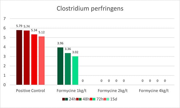
An in-vitro trial was conducted to evaluate the effect of such a combination (Formycine Gold Px) against common poultry pathogens. Poultry feed was spiked with three different bacteria, achieving very high initial contamination of 1,000,000 CFU/g per pathogen. One batch of the contaminated feed served as a control (no additive). To the other contaminated batches, 1, 2, or 4 kg of Formycine per ton of feed were added. The results (means of triplicates) are shown in figures 1 a-c.
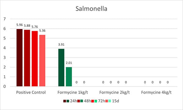
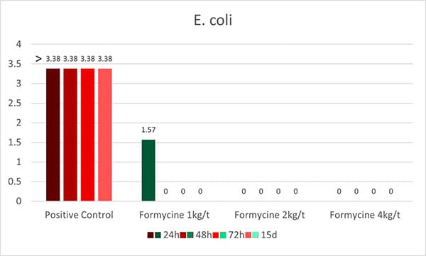
Figure A Salmonella

Figure B E

Figure C Clostridium PerfringensFigures 1 a-c: Reduction of bacterial count due to the addition of Formycine

Formycine Gold Px significantly reduced the bacterial counts in all three cases. A clear dose-response-effect can be seen and by using 2 kg of Formycine / t of feed, pathogens could not be detected anymore in the feed.

A further trial showed the positive effects of feeding Formycine Gold Px treated feed to the animals. Also here, the feed for both groups was contaminated with 1,000,000 CFU of Clostridium/g. The feed of the control group was not treated and to the treatment group, 2 kg of Formycine per t was added.
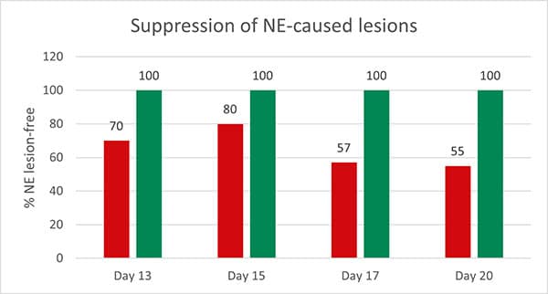
Figure Preventive EffectFigure 2: Preventive effect of Formycine Gold Px concerning necrotic enteritis gut lesions
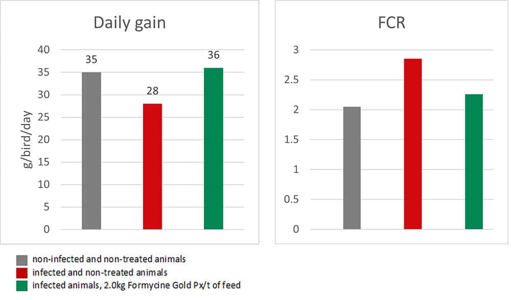
Figure A Daily GainFigure 3a and 3b: Performance-maintaining effect of Formycine Gold Px

The trial showed that Formycine Gold Px reduced the ingestion of the pathogen, and thus could prevent the lesions caused by necrotic enteritis (Fig. 2). The consequence of this improved gut health is a better feed conversion and higher average daily gain (Fig.3a and 3b).

Products containing formaldehyde may represent a risk for humans, however, the adequate protection equipment helps to reduce/avoid exposure.

A combination of free acids and acid salts provides optimal hygienic effects

Additionally, another blend of organic acids (Acidomix AFG) shows the best effects against representatives of relevant feed-borne pathogens in poultry. In a test, 50 µl solution containing different microorganisms (reference strains of S. enterica, E. coli, C. perfringens, C. albicans, and A. niger; concentration 105 CFU/ml, respectively) were pipetted into microdilution plates together with 50 µl of increasing concentrations of a mixture of organic acids (Acidomix) After incubation, the MIC and MBC of each pathogen were calculated.

The test results show (figure 4, Minimal Bactericidal Concentration) that 0.5% of Acidomix AFG in the medium (≙ 5kg/t of feed) is sufficient to kill S. enterica, C. albicans, and A. niger and even only 2.5kg/t in the case of E. coli. If the pathogens should only be prevented to proliferate, even a lower amount of product is requested (figure 5, Minimal Inhibitory Concentration – MIC)
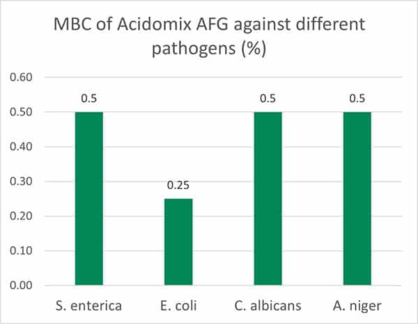
Figure MbcFigure 4: MBC of Acidomix AFG against different pathogens (%)
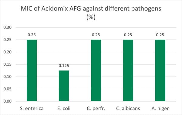
Figure MicFigure 5: MIC of Acidomix AFG against different pathogens (%)

In addition to the direct antimicrobial effect, this product decreases the pH of the feed and reduces its buffering capacity. The combination of free acids and acid salts provides prompt and long-lasting effects.

Feed hygiene: a critical path to animal performance

Feed accounts for 65-70% of broiler and 75-80% of layer production costs. Therefore, it is essential to use the available feed to the utmost. The quality of the feed is one decisive factor for the health and performance of the animals. Proper harvesting and storage are in the hands of the farmers and the feed millers. The industry offers products to control the pathogens causing diseases and the molds producing toxins and, therefore, helps farmers save feed AND protect the health and performance of their animals.

References:

Dinev, Ivan. Diseases of Poultry: A Colour Atlas. Stara Zagora: Ceva Sante Animal, 2007.

Esmail, Salah Hamed. “Moulds and Their Effect on Animal Health and Performance.” All About Feed, June 17, 2021. https://www.allaboutfeed.net/all-about/mycotoxins/moulds-and-their-effect-on-animal-health-and-performance/.

Government of Manitoba. “Spoiled Feeds, Molds, Mycotoxins and Animal Health.” Province of Manitoba – Agriculture. Accessed March 16, 2023. https://www.gov.mb.ca/agriculture/livestock/production/beef/spoiled-feeds-molds-mycotoxins-and-animal-health.html.

Hoffmann, M. “Tierwohl Und Fütterung.” LKV Sachsen: Tierwohl und Fütterung. Sächsischer Landeskontrollverband e.V., January 25, 2021. https://www.lkvsachsen.de/fuetterungsberater/blogbeitrag/artikel/tierwohl-und-fuetterung/.

Ricke, Steven C., Kurt Richardson, and Dana K. Dittoe. “Formaldehydes in Feed and Their Potential Interaction with the Poultry Gastrointestinal Tract Microbial Community–A Review.” Frontiers in Veterinary Science 6 (2019). https://doi.org/10.3389/fvets.2019.00188.

Shirota, Kazutoshi, Hiromitsu Katoh, Toshihiro Ito, and Koichi Otsuki. “Salmonella Contamination in Commercial Layer Feed in Japan.” Journal of Veterinary Medical Science 62, no. 7 (2000): 789–91. https://doi.org/10.1292/jvms.62.789.

Stanley, Dragana, and Yadav Sharma Bajagai. “Feed Safety and the Development of Poultry Intestinal Microbiota.” Animals 12, no. 20 (2022): 2890. https://doi.org/10.3390/ani12202890.

Su, Lin-Hui, and Cheng-Hsun Chiu. “Salmonella: Clinical Importance and Evolution of Nomenclature.” Chang Gung Med J 30, no. 3 (2007): 210–19.

Udhayavel, Shanmugasundaram, Gopalakrishnamurthy Thippichettypalayam Ramasamy, Vasudevan Gowthaman, Shanmugasamy Malmarugan, and Kandasamy Senthilvel. “Occurrence of Clostridium Perfringens Contamination in Poultry Feed Ingredients: Isolation, Identification and Its Antibiotic Sensitivity Pattern.” Animal Nutrition 3, no. 3 (2017): 309–12. https://doi.org/10.1016/j.aninu.2017.05.006.

Mind the immunity gap: egg proteins bolster the piglets’ immune system

EW Nutrition article piglets 6

egg immunoglobulins bolster piglets’ immune system

In contrast to humans, piglets do not receive any maternal immunoglobulins via the placenta. It is therefore of vital importance for these young animals to receive maternal antibodies via the colostrum as soon as possible after birth. Otherwise, they are more vulnerable to illnesses in their early stages of life.

In this article, we look in-depth at how the immune system works and which role antibodies play in it.  We then consider why egg proteins might potentially be a powerful tool for supporting young animals immunologically, allowing producers to maintain young animals’ health and to promote their performance.

How the immune system defends the body: three barriers

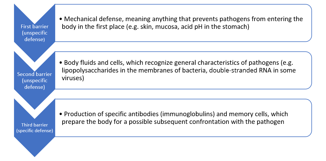
The immune system aims to prevent pathogens such as viruses, bacteria, and fungi from entering the body or to eliminate them when they have already entered. Furthermore, it seeks to prepare the body for quicker reactions, in case of subsequent infections, by building an immunological memory. Generally, in case of an attack by pathogens, there are three barriers against the “enemy” (Figure 1).

Figure 1: The three barriers of the immune response

First barrier: the immediate, physical defense upon contact with pathogens

The animal body has several anatomical features that prevent pathogens from entering in the first place, such as cilia and mucus. Skin, intestines and nose lining are colonized by a community of beneficial micro-organisms that form a physical barrier against pathogens. Other barriers include the urinary system, the acid pH of the stomach, as well as tears and saliva, which contain antibacterial lysozymes.

Second barrier: the unspecific, native defense that does most of the work

If the mechanical mechanisms of defense were not successful, the unspecific, innate immune defense enters into play (Murphy and Weaver, 2018, 47ff.). At this stage, the body needs to differentiate between “known” and “alien” agents, and between “potentially harmful” and “harmless” ones.

To identify alien, potentially harmful agents, the unspecific defense looks for so-called PAMPs (pathogen associated molecular patterns). These are general characteristics often displayed by pathogens, such as lipopolysaccharides in the bacterial membrane or double-stranded RNA in viruses. Everything that shows PAMPs is heavily targeted.

The unspecific defense can be further divided into the humoral and the cellular defense. The humoral defense consists of substances dissolved in the body fluids, such as enzymes, reactive oxygen compounds, signal molecules and a whole cascade of proteins. Some of these substances kill pathogens directly; others “mark” the pathogens and “call for” the help of leucocytes.

The cellular defense consists of different leucocytes, also known as white blood cells (because they do not contain any red hemoglobin). The main task of leucocytes is the defense of the body against pathogens, hence many leucocytes are capable of phagocytosis (the ingestion of other cells). To prevent phagocytes from accidentally ingesting the body’s own cells, these own cells are marked with the so-called major histocompatibility complex (MHC). This acts as a red flag, saying “I belong to the body!”.

The cellular defense consists of:

  • Neutrophil granulocytes (60-70% of the leucocytes), which mainly act against bacteria
  • Eosinophil and basophil granulocytes (1.5% of the leucocytes), which mainly act against parasites
  • Natural killer cells, which mainly act against viruses
  • Monocytes (3-8% of the leucocytes; they differentiate into macrophages and dendritic cells)

Figure 1: The three barriers of the immune response


The monocytes, as well as their macrophage and dendritic cell “offspring”, are the bridge to the next step, the specific defense. When these phagocytes digest pathogens, minuscule protein structures (antigens) of the pathogens remain. These antigens are unique to each pathogen. During a process called antigen presentation, the antigens are tied to the cell’s MHC and transported to the cell surface. This triggers the production of specific antibodies, the immune system’s third barrier.

Third barrier: the specific immune defense that creates antibodies and immunological memory

The specific (also called adaptive or acquired) immune response kicks in a few days after contact with specific pathogens and is mostly carried out by lymphocytes called T and B cells (Murphy and Weaver, 2018, 177ff.). They are active at the cellular and the humoral level, respectively.

T cells possess receptors on their surface through which they can recognize the antigens presented to them by phagocytes. What they do subsequently depends on the subtype of the T cell:

  • Cytotoxic T cells (CD8+) directly destroy the antigen-phagocyte-combination
  • T helper cells (CD4+) attract other cells that can destroy the pathogens (e.g. macrophages) and stimulate B cells to produce antibodies against them

B cells also possess receptors through which they can recognize antigens. Once they spot an antigen (and T helper cells “confirm” that an immune response is required), they divide and mature into so-called plasma cells. Plasma cells, in turn, secrete plenty of antibodies (or immunoglobulins) into the bloodstream and the lymphatic system. Antibodies are protein structures that lock onto and neutralize antigens through different mechanisms.

The chemical reaction between antibodies and antigens is the body’s most powerful immune response through which it can protect itself from pathogens and their toxins. Antibody production continues for several days to remove the antigens, and antibodies usually remain in circulation for a few months.

Moreover, certain T and B cells memorize the first attack of a pathogen and turn into memory cells. The T memory cells CD4+CD8+, for instance, match the antigens from certain past, latent, and particularly persistent viral infections. This immunological memory, created by acquired immunity, can be thought of as a library of antibodies that the body adds to whenever it deals with a new pathogen or receives a vaccine. In case of a subsequent contact with the pathogen, the right antibody “model” already exists and mass production can start up very quickly.

Why young animals’ immune defense is so vulnerable – and what IgY can do about that

Building one’s immunological memory takes time. A lot of new-born animals are in a vulnerable position: they have not had time yet to acquire immunity of their own, but they are also particularly fragile and susceptible to being attacked by commonly occurring pathogens such as corona and rotaviruses, E. coli and clostridia. The toxins that E. coli and clostridia, for instance, release, may cause diarrhea, edema, endotoxic shock, and even death.

To be protected during the first critical days of their lives, new-born animals thus need to receive a foundational stock of antibodies (passive immunity) from their mother. Humans receive maternal immunoglobulins via the placenta. Piglets, because a sow’s placenta is constructed differently, are dependent on receiving them through the colostrum after birth. If this is not the case – due to inadequate quantity or quality of the colostrum – they need to receive immune support in a different way.

Egg-yolk antibodies have been proposed as a powerful tool for supporting young animals during the critical period after birth. These special proteins support the colostrum supply and guarantee that every animal in the herd has some degree of protection. This protection mostly takes place in the gut. The IgY recognize and tie up pathogens and render them ineffective.

Trial: can egg proteins support piglet immunity?

In 2009, research at the National Veterinary Research Institute in Pulawy, Poland, was conducted to probe this hypothesis. The objective of the trial was to evaluate whether an oral application of egg proteins would have a quantifiable, positive influence on the immune system of the piglets. Different immunological parameters were measured, including different types of leucocytes.

Trial design

The test consists of 6 litters with 67 piglets in total divided into two groups. The control group (n=32) received the prophylaxis customary on the farm; the trial group (n=35) additionally received a product based on egg powder (EP)[1], applied at the inclusion rate recommended by the producer. Blood samples were taken on days 0 (before application of the product), 7, 14, and 28. They were analyzed with respect to the percentages of different types of lymphocytes.

Trial results

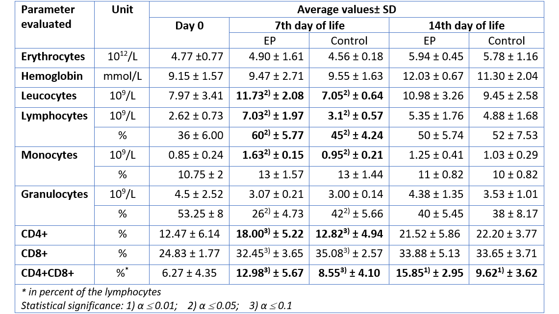
For the group receiving egg powder, the number of leucocytes in peripheral blood was significantly elevated compared to the control group on the 7th day of life (table 1). The amounts of lymphocytes and monocytes – indicators for the specific immunological defense – were also significantly increased on day 7, whereas the total amount of granulocytes – indicator for the innate, unspecific immune defense – remained constant. Hence, already during the first days, the piglets supplied with EP disposed of a higher level of adaptive (specific) immune defense, compared to the animals in the control group. In addition, there was a significant increase in the number of CD4-positive (CD4+) and CD4-CD8-double positive (CD4+CD8+) T cells in the EP group, compared to the control animals, indicating an active stimulation of the immune system.

Except for CD4+CD8+ T cells (which remained elevated in the EP group), on day 14, the differences in cellular immune response were no longer significant. This is most probably the case because by that time the immune system of the control group had activated its own protective response. The EP product therefore supported the young animals precisely when it was necessary, during the critical first days of life.

Table 1: Hematological parameters measured in piglets after prophylactic application of an egg powder based product (EP1)

1Ig-PRO P (EW Nutrition)

The improvement of immune status, as indicated by the presence of the specific immune cells, was confirmed by the results for the incidence of diarrhea and mortality (table 2). The animals of the control group showed a nearly 1.5 times higher incidence of diarrhea and a 1.6 times higher rate of mortality. Another explanation of these results could be the mode of action of egg immunoglobulins: by neutralizing the pathogens directly in the gut, they prevent them from causing diarrhea in the first place.

Table 2: Incidence of diarrhea and mortality

Frequency of diarrhoea and mortality of piglets

In conclusion, this trial demonstrates that natural egg proteins effectively support the immune system of piglets during the critical period of the first days of life.
Thanks to the stimulation of the young animals’ specific immune defense and the direct neutralization of pathogens in the gut, the incidence of diarrhea – one of the main causes of losses during the first weeks of life – decreases. Hence, mind the immunity gap: providing piglets with a suitable egg powder based product sets them up for long-term health, growth, and performance.

By I. Heinzl and S. Regragui Mazili


References:

Foley, J. A., and D. E. Otterby. “Availability, Storage, Treatment, Composition, and Feeding Value of Surplus Colostrum: A Review 1, 2.” Journal of Dairy Science 61, no. 8, 1033-1060. doi.org/10.3168/jds.S0022-0302(78)83686-8.
Heinzl, Inge, and Fellipe Barbosa. “Egg Antibody Technology for Nursery Pig Application.” Swineweb.com. June 24, 2019. Accessed July 17, 2019. http://www.swineweb.com/egg-antibody-technology-for-nursery-pig-application/.
Marquardt, Ronald R., L. Z. Jin, Jung-Woo Kim, Lin Fang, Andrew A. Frohlich, and Samuel K. Baidoo. “Passive Protective Effect of Egg-yolk Antibodies against Enterotoxigenic Escherichia Coli K88 Infection in Neonatal and Early-weaned Piglets.” FEMS Immunology and Medical Microbiology 23, no. 4 (1999): 283-288. https://doi.org/10.1111/j.1574-695X.1999.tb01249.x.
Murphy, Kenneth M., and Casey Weaver. 2018. Janeway Immunologie. 9th ed. Translated by Lothar Seidler. Berlin: Springer.
Nascimbeni, Michelina, Eui-Cheol Shin, Luis Chiriboga, David E. Kleiner, and Barbara Rehermann. “Peripheral CD4 CD8 T Cells Are Differentiated Effector Memory Cells with Antiviral Functions.” Blood 104, no. 2 (2004): 478-486. doi:10.1182/blood-2003-12-4395.
Yokoyama, Hideaki, Robert C. Peralta, Roger Diaz, Sadako Sendo, Yutaka Ikemori, and Yoshikatsu Kodama. “Passive Protective Effect of Chicken Egg Yolk Immunoglobulins.” Infection and Immunity 60, no. 3 (March 1992): 998-1007. https://iai.asm.org/content/iai/60/3/998.full.pdf.


Diarrhoea? Egg powder to the rescue

Calf lying Stable Fotolia 84943524 XL

Another tool to reduce the use of antibiotics is the use of immunoglobulins from eggs.
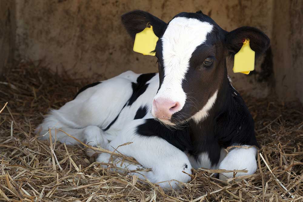
Trials showed that this product is effective to support a calf’s start in life and also to offer support when challenged by various forms of diarrhoea.

The main cause for calf losses during the first two weeks of life is diarrhoea. In general diarrhoea is characterised by more liquid being secreted than that being resorbed. However, diarrhoea is not a disease, but actually only a symptom. Having diarrhoea has a protective function for the animal, because the higher liquid volume in the gut increases motility and pathogens and toxins are excreted faster. Diarrhoea can occur for several reasons. It can be caused by incorrect nutrition, but also by pathogens such as bacteria, viruses and protozoa.

Bacteria in the gut
E. coli belong to the normal gut flora of humans and animals and can be mainly found in the colon. Only a fraction of the serotypes causes diseases. The pathogenicity of E.coli is linked to virulence factors. Decisive virulence factors are for example the fimbria used for the attachment to the gut wall and the bacteria’s ability to produce toxins.

Salmonella in general plays a secondary role in calf diarrhoea, however, salmonellosis in cattle is a notifiable disease. Disease due to Clostridia is amongst the most expensive one in cattle farming globally. In herbivores, clostridia are part of the normal gastro-intestinal flora, only a few types can cause serious disease. In calves, Clostridium perfringens occurs with the different types A, C, and D. Rotaviruses are the most common viral pathogens causing diarrhoea in calves and lambs. They are mainly found at the age of 5 to 14 days. Coronaviruses normally attack calves at the age of 5 to 21 days. Cryptosporidium parvum is a protozoa and presumed to be the most common pathogen causing diarrhoea (prevalence up to more than 60 %) in calves.

Undigested feed and incorrect use of antibiotics
Plant raw materials (mainly soy products) are partly used in milk replacers as protein sources. These products contain carbohydrates, that cannot be digested by calves which can lead to diarrhoea. The transition from milk to milk replacer can also be a reason.

An early application of tetracyclines and neomycin to young calves can lead to a change in the villi, malabsorption and therefore to slight diarrhoea. Longer therapies using high dosages of antibiotics can also lead to a bacterial superinfection of the gut. The problem is that in a disease situation, antibiotics are often used incorrectly. The use of antibiotics only makes sense when there is a bacterial diarrhoea and not due to viruses, protozoa or poor feed management. To keep the use of antibiotics as low as possible, alternatives need to be considered.

Egg powder to add immunoglobulins
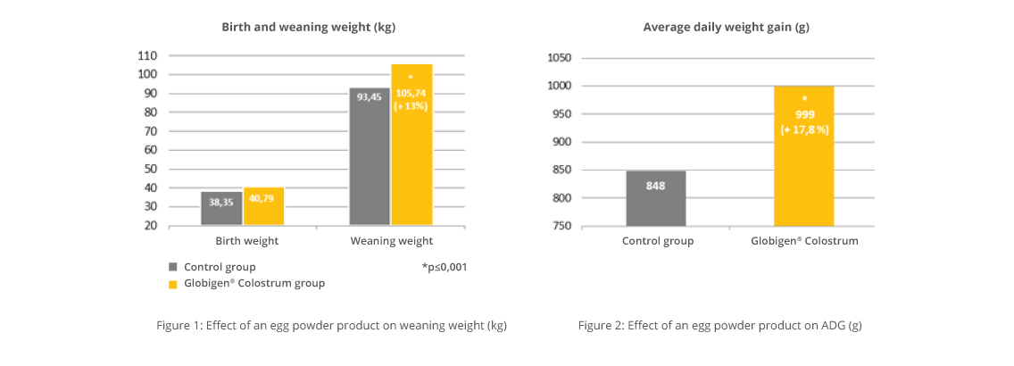
In order to achieve optimal results in calf rearing two approaches are possible. Firstly, the prophylaxis approach. This is the method of choice as diarrhoea can mostly be prevented. Therefore, it is necessary to supply the calf with the best possible equipment. As antibodies are one crucial but limiting factor in the colostrum of the “modern” cow, this gap needs to be minimised.  A study conducted in Germany in 2015 demonstrated that more than 50% of the new-born calves had a deficiency of immunoglobulins in the blood. Only 41% of the calves showed an adequate concentration of antibodies in the blood (>10 mg IgG/ml blood serum). Immunoglobulins contained in hen eggs (IgY) can partly compensate for poor colostrum quality and serve as a care package for young animals. A trial was conducted with an egg powder product* on a dairy farm (800 cows) in Brandenburg, Germany. In total 39 new-born calves were observed until weaning (65th day of life). Before birth, the calves were already divided into control and trial group according to the lactation number of their mother cow. All calves were fed the same and received four litres of colostrum with ≥ 50 mg IgG /ml on the first day of life.

Control (n=20):            no additional supplementation
Trial group (n=19):      day 1 – 5: 100 g of the egg powder product per animal per day mixed into the colostrum or milk.

It was shown that the calves in the trial group showed a significantly higher (13%) weaning weight (105.74 kg compared to 93.45 kg in the control group) and 18%  higher average daily gain (999 g compared to 848 g in the control group) (Figure 1 and Figure 2).

Support during acute diarrhoea
When diarrhoea occurs, the calf has to be treated. So the second approach is to find the best and quickest solution. It is not always necessary to use antibiotics, as they do not work against virus or protozoa. Egg antibodies can be an answer when combined with electrolytes as the following trial shows. On a dairy farm (550 cows) in Germany a feeding trial with a product based on egg powder and electrolytes** was conducted from December 2017 to May 2018. Two groups of calves were used. Before birth the animals were allocated into the two groups according to the calving plan and were examined from day one until weaning (77th day of life). All calves suffering from diarrhoea (38 in total, 17 in the control and 21 in the trial group) were treated as follows:

Control (n=17):            Application of electrolytes
Trial group (n=21):      50 g of the egg powder and electrolytes product twice daily, stirred into the milk replacer until diarrhoea stopped.

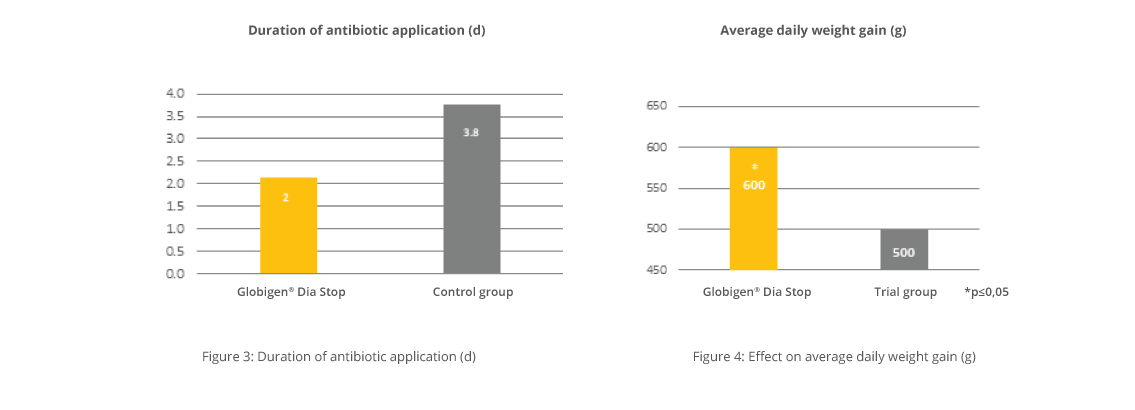
If the diarrhoea did not stop or even got worse, the animals were treated with antibiotics. It was shown that in the control group the antibiotic treatment necessary was nearly twice as long as needed in the trial group (Figure 3). This means also that nearly twice the amount of antibiotics were used. This leads to the conclusion that calves in the trial group had an improved health status compared to calves in the control group. A further result from the improved health status was an increase in performance in the trial group (Figure 4).

The average daily weight gain of the trial group was 20% higher than in the control (600 vs. 500 g per day) leading to a significantly higher weaning weight (87.8 kg) than in the control (80.7 kg).

By Franziska Stemmer and Dr. Inge Heinzl, EW Nutrition, Germany
Published in Dairy Global (Online and Printed), 10/2018


Fewer pathogens with egg immunoglobulins

1487476672 c92daf8f6d o TRATADA 1 1024x474

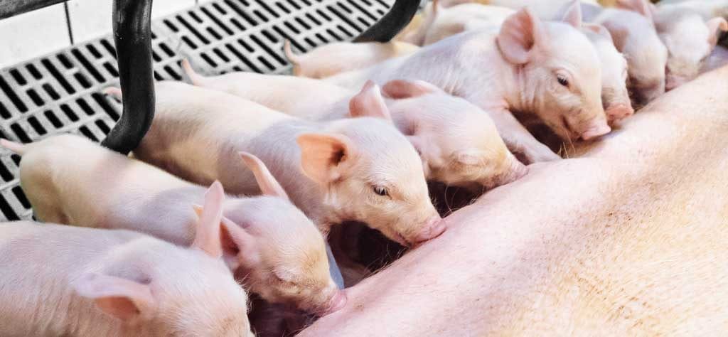
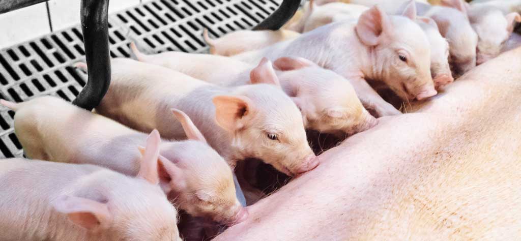
Piglets nursing

For newborn pigs there are often a host of different challenges – think of crushing or contamination of the farrowing pen.
For the last problem, solutions exist. A dietary approach can help to relieve pathogenic pressure through sow manure.

The main objective of a piglet producer is to maximise the number of healthy weaned piglets per animal per year. Nowadays, it is not difficult to find production systems delivering more than 30 piglets weaned/sow/year. Combining strategies on management, feeding, and health of both piglets and sows, is crucial for increasing sow’s productivity. A unique environment that can determine the success of a piglet farm is the farrowing unit. It is important to reduce as much as possible losses during this period. Pre-weaning mortality must always be monitored and targets must be set. In European conditions, it ranges between 8-10%.

One important driver in reducing pre-weaning mortality is understanding the fragility of newborn piglets. At birth, the resources of a piglet are very scarce: low energy reserves and practically no immune defence against existing pathogens in their new environment. Problems are prone to happen and will be mostly caused by pathogens present in the environment, in the feed, in the water and most important, in the faeces of the sow. The main contamination source for newborn piglets is their mother’s manure. And this first contamination can be quite severe causing diarrhoea and increasing piglet mortality.

Together with crushing, diarrhoea definitely causes a high percentage of total losses during the first days of life. In most of the cases, the disease is caused not only by one agent but by a combination of enteric infections from different pathogens or at least different strains of a pathogenic species. E. coli and clostridia are two of the most important diarrhoea causing pathogens during the first weeks after birth.

Pathogens during the first days
E. coli is well known as one of the main responsible pathogens for pre-weaning diarrhoea. And although it belongs to the normal intestinal flora of pigs, part of the different E. coli strains are pathogenic. E. coli cause about 80% of diarrhoeas in piglets and 50% of losses in piglet production. The factors making E. coli pathogenic, the so-called virulence factors include e.g. fimbria to attach to the intestinal wall and the capacity to produce toxins.

The Clostridium species are another important pathogen class. During the suckling phase, piglets are quite susceptible to Clostridium perfringens type C. This bacteria causes necrotic enteritis in piglets and the clinical symptoms appear during the first days of life. This disease provokes serious disturbances in the organism with a mortality up to 100%. It causes significant decrease in daily gain and in weaning weight.

Strategy to protect the piglets
In order to maximise the sow’s performance – measured in piglets weaned per year – it is crucial to provide the best possible conditions to the piglets. Therefore the reduction of the pathogenic pressure in the farrowing unit ranks first. Cleaning of the pen is a way to get rid of germs like E. coli and Clostridium species, the most important pathogens during the first days. This should be completed by an effective gut health management in sow and piglets. For this purpose natural ingredients can be used. Supplying natural and active immune cells, the so called antibodies, has been proven to be quite efficient in supporting gut health. Applied to piglets, immunoglobulins from the egg bind to pathogens within the intestinal tract. They show efficiency in supporting piglets’ performance, decreasing the incidence of diarrhoea, mortality and increasing daily gain.

The idea was to check if these immunoglobulins from the egg could also bind pathogens in the sow’s gut and generate harmless complexes. That way pathogenic pressure for the piglets could be reduced. Thus a trial was conducted in Japan to check this thesis.


*Globigen Sow

Trial
In the trial two groups contained eight sows each. The sows of the control group received standard lactation feed, the trial group was also fed standard feed with a supplement containing egg immunoglobulins (Globigen Sow, EW Nutrition, at a dosage of 5 g/sow twice daily) on top during the last ten days before and the first seven days after delivery. The faeces of the sows were obtained by rectal stimulation (in order to get no contamination from the environment) on day 10 before and day 7 after delivery. The amount of colony forming units (CFU) of total E. coli, E. coli O141 and Clostridium perfringens were determined.

Results are shown in Figure 1. At the beginning of the trial, before the application of the immunoglobulin supplement, both groups showed nearly the same level of the evaluated pathogens with a slight disadvantage for the supplement group. After 17 days of applying the product based on egg immunoglobulins, a reduction of the colony forming units of total E. coli, E. coli O141 and of Clostridium perfringens could be seen. The sows of the supplement-fed group showed a lower level of pathogens in their excrements than the sows of the control group.

Conclusion
It is important for swine producers to understand what adversely influences the results on the farm. One consideration is to improve farrowing unit conditions of the piglets, aiming to reduce pre-weaning mortality. The results of the trial showed that a supplement based on egg immunoglobulins supplied on top of standard sow diets substantially reduced the amount of pathogenic colonies in sow manure. The reduction on pathogenic pressure and therefore the incidence of diarrhoea may be an alternative for increasing the profitability of piglet producers by increasing the number of healthier piglets weaned/sow/year.

*References are available on request.

By Dr Inge Heinzl.
Published on PigProgress | 20th July, 2018.


Secondary Plant Compounds (SPC’s) to reduce the use of antibiotics?

shutterstock 794137729 lab ecoli website

Initial in vitro trials give reason for hope

Antibiotic Resistance

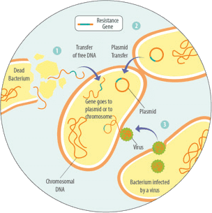
Some bacteria, due to mutations, are less sensitive to certain antibiotics than others. This means that if certain antibiotics are used, the insensitive ones survive. Because their competitors have been eliminated, they are able to reproduce better. This resistance can be transferred to daughter cells by means of „resistance genes“. Other possibilities are the intake of free DNA and therefore these resistance genes from dead bacteria 1, through a transfer of these resistance genes by viruses 2 or from other bacteria by means of horizontal gene transfer 3 (see figure 1). Every application of antibiotics causes a selection of resistant bacteria.  A short-term use or an application at a low dosage will give the bacteria a better chance to adapt, promoting the generation of resistance (Levy, 1998).

Antibiotics are promoting the development of resistance:

  • Pathogenic bacteria possessing resistance genes are conserved and competitors that do not possess these genes are killed
  • Useful bacteria possessing the resistance genes are conserved and serve as a gene pool of antibiotic resistance for others
  • Useful bacteria without resistance, which probably could keep the pathogens under control, are killed

Reducing the use of antibiotics
Ingredients from herbs and spices have been used for centuries in human medicine and are now also used in modern animal husbandry. Many SPC’s have antimicrobial characteristics, e.g. Carvacrol and Cinnamon aldehyde. They effectively act against Salmonella, E. coli, Pseudomonas aeruginosa, Klebsiella pneumoniae, Entero and Staphylococcus, and Candida albicans. Some compounds influence digestion, others act as antioxidants. Comprehensive knowledge about the single ingredients, their possible negative but also positive interaction (synergies) is essential for developing solutions. Granulated or microencapsulated products are suitable for addition to feed, liquid products would be more appropriate for an immediate application in the waterline in acute situations.

SPC’s (Activo® Liquid) against livestock pathogens in vitro
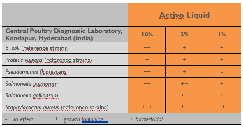
In “agar diffusion tests”, the sensitivity of different strains of farm-specific pathogens was evaluated with different concentrations of Activo® Liquid. The effectiveness was determined by the extent to which they prevented the development of bacterial overgrowth. The larger the bacteria-free zone, the higher the antimicrobial effect.

In this trial, Activo® Liquid showed an antimicrobial effect on all bacteria tested. The degree of growth inhibition positively correlated with its concentration.

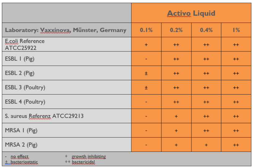
Table 1: Inhibition of field isolated standard pathogens by different concentrations of Activo® Liquid


Activo® Liquid against antibiotic resistant field pathogens in vitro
It cannot be excluded that resistant pathogens not only acquired effective weapons to render antibiotics harmless to them but also developed general mechanisms to rid themselves of otherwise harmful substances. In a follow-up laboratory trial, we evaluated whether the Activo® Liquid composition is as effective against ESBL producing E. coli and Methicillin resistant S. aureus (MRSA) as to non-resistant members of the same species.

Trial Design: Farm isolates of four ESBL producing E. coli and two MRSA strains were compared to nonresistant reference strains of the same species with respect to their sensitivity against Activo® Liquid. In a Minimal Inhibitory Concentration Assay (MIC) under approved experimental conditions (Vaxxinova Diagnostic, Muenster, Germany) the antimicrobial efficacy of Activo® Liquid in different concentrations was evaluated.

The efficacy of SPC’s (Activo® Liquid) against the tested strains could be demonstrated in a concentration-dependent manner with antimicrobial impact at higher concentrations and bacteriostatic efficacy in dilutions up to 0,1% (ESBL) and 0,2% (MRSA)(table 2).

Conclusion:
To contain the emergence and spread of newly formed resistance mechanisms it is of vital importance to reduce the use of antibiotics. SPC’s are a possibility to decrease antibiotic use especially in pro- and metaphylaxis, as they show good efficacy against the common pathogens found in poultry, even against resistant ones.


I. Heinzl 


Secondary plant compounds against antibiotic-resistant E. coli

Poultry drinking line

Due to incorrect therapeutic or preventive use of antibiotics in animal production as well as in human medicine, occurrence of antibiotic resistant pathogens has become a widespread problem. Enterobacteria in particular (e.g. Salmonella, Klebsiella, E. coli) possess a special mechanism of resistance. By producing special enzymes (ß-lactamases), they are able to withstand the attack of so-called ß-lactam antibiotics. The genes for this ability (resistance genes) can also be transferred to other bacteria resulting in a continuously increasing problem. Divers point mutations within the ß-lactamase genes lead to the occurrence of „Extended-Spectrum-Beta-Lactamases“ (ESBL), which are able to hydrolyse most of the ß-Lactam-antibiotics. AmpC Beta-Lactamases (AmpC) are enzymes, which express a resistance against penicillins, cephalosporins of the second and third generation as well as cephamycins.

What are ß-lactam antibiotics?
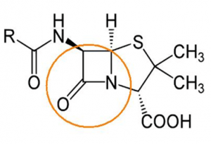
The group of ß-lactam antibiotics consists of penicillins, cephalosporins, monobactams and carbapenems. A characteristic of these antibiotics is the lactam ring (marked in orange):

Mode of action of ß-lactam antibiotic
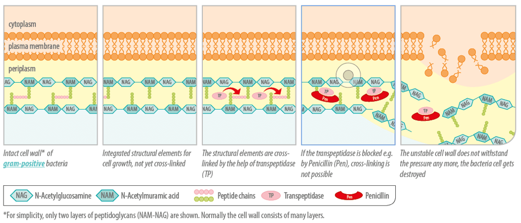
If a bacterial cell is growing, the cell wall also has to grow. For this purpose, existing conjunctions are cracked and new components are inserted. ß-lactam-antibiotics disturb the process of cell wall construction by blocking an enzyme needed, the transpeptidase. If crosslinks necessary for the stability of the cell wall cannot be created, the bacteria cannot survive. Resistant bacteria, which are able to produce ß-lactamases, destroy the ß-lactam antibiotics and prevent their own destruction.

Secondary plant compounds
Secondary plant compounds and their components are able to prevent or slow down the growth of moulds, yeasts, viruses and bacteria. They attack at various sites, particularly the membrane and the cytoplasm. Sometimes they change the whole morphology of the cell. In the case of gram-negative bacteria, secondary plant compounds (hydrophobic) have to be mixed with an emulsifier so that they can pass the cell wall which is open only for small hydrophilic solutes. The modes of action of secondary plant compounds depend on their chemical composition. It also depends on whether single substances or blends (with possible positive or negative synergies) are used. It has been observed that extracts of spices have a lower antimicrobial efficacy than the entire spice.

The best explained mode of action is the one of thymol and carvacrol, the major components of the oils of thyme and oregano. They are able to incorporate into the bacterial membrane and to disrupt its integrity. This increases the permeability of the cell membrane for ions and other small molecules such as ATP leading to the decrease of the electrochemical gradient above the cell membrane and to the loss of energy equivalents of the cell.

Trial (Scotland)

Design
Two strains of ESBL-producing and AmpC respectively, isolated from the field, a non-resistant strain of E. coli as control. Suspensions of the strains with 1×10⁴ KBE/ml were incubated for 6-7 h at 37°C together with different concentrations of Activo® Liquid or with cefotaxime, a cephalosporin. The suspensions were then put on LB-Agar plates and bacteria colonies were counted after a further 18-22h incubation at 37°C. Evaluation of the effects of Activo® Liquid on ESBL-producing as well as on E. coli resistant for aminopenicillin and cephalosporin (AmpC)

Results
The antimicrobial efficacy of the blend of secondary plant compounds depended on concentration with bactericidal effect at higher concentrations and bacteriostatic at dilutions up to 0,1%. It is also possible that bacteria could develop a resistance to secondary plant compounds; the probability is however relatively low, due to the fact that essential oils contain hundreds of chemical components (more than antibiotics) making it difficult for bacteria to adapt.


Phytogenics can positively influence the efficacy of antibiotics

phytgenics spice powder

Many veterinary antibiotics are applied via the waterline, where they are dosed in combination with other feed additives. Amongst those are mixtures of secondary plant compounds with a proven antimicrobial efficacy against veterinary pathogenic bacteria. However, little research has been done to evaluate any effect that antibiotics and phytogenics may have on each other. A possible influence of phytogenics on the efficacy of antibiotics through the combined administration would require a change in application recommendations of antibiotics and phytogenic feed additives. In the case of no interaction, no changes would be necessary. If they were to interact in a positive way, the dosages could be lowered and if they interact in a negative way, a combined application would be avoided.

Antibiotics and SPC’s in co-incubation
There are different groups of antibiotics depending on the chemical structure and on the pathogen they target. Some impair the cell wall or the cytoplasmic membrane (polymyxins, ß-lactam antibiotics) and some affect protein synthesis (macrolides, Chloramphenicol, Lincospectin, tetracyclines, aminoglycosides). Others compromise DNA and RNA synthesis (fluorchinolones, ansamycines) and some disturb the metabolism of e.g. folic acid (Trimethoprim).
The intention of a trial with these different groups of antibiotics was to evaluate possible interactions they may have with a combination of secondary plant compounds. Four ESBL producing E. coli field isolates from poultry flocks were experimentally assessed  as well as a ß-lactamase positive and a ß-lactamase negative reference strain as quality control strains for antimicrobial susceptibility testing.
Two-fold serial dilutions of antibiotics and the liquid product based on secondary plant compounds were co-incubated in a checkerboard assay. The highest concentration of the antibiotic was chosen according to CLSI standard recommendations. The control of the serial dilution of SPC’s was made without antibiotics and vice versa.

Lowering the antibiotic dosage by the use of SPC’s
In the experiment all field isolates proved resistant against the ß-lactam antibiotics, two field isolates and one reference strain were resistant against tetracyclines and macrolides and one field isolate and one reference strain against aminoglycocides.
The results showed that there was no negative influence of the antibiotics on the SPC’s and vice versa. Moreover, for several classes of antibiotics an additive to synergistic effect was observed to such an extent that an antibiotic effect could be achieved with half or even one quarter of the former effective dosage. The dosage of the SPC-mixture could also be reduced. Based on the results of this in vitro experiment it can be stated that in the case of antibiotic resistance, the option exists to apply a phytogenic product with broad antimicrobial efficacy. Even more, for most combinations between antibiotics and Activo® Liquid, a defined mixture of secondary plant compounds, their combined use potentiates the individual efficacy of either compound class against E.coli strains in vitro. This adds further benefits to the improvements in animal performance and health, for which a number of phytogenic feed additives have already proven effective.


Using egg immunoglobulins to enhance piglet survival

sow management

The number of healthy piglets weaned is the most important factor for the calculation of profit in piglet production.

Losses in the farrowing unit normally occur during the first seven days of life as piglets are born with very little protection in the form of immunity. The intake of immunoglobulins from colostrum is therefore of vital importance. Besides cleanliness and special feeding, piglets can be additionally supported by two strategies that mimick the effect of colostrum:
– a direct one, meaning the feeding of immunoglobulins (IgY from eggs) to piglets that would support the immune system in the gut or
– an indirect one, meaning a supply of IgY to the sow to keep the pathogenic pressure in the farrowing unit as low as possible.

Piglets are born with no immune protection and very low energy reserves
It is well known that piglets are physiologically immature at birth. Their energy reserves are very low with only 1 – 2% body fat comprising mainly of structural and subcutaneous fat. Therefore, in the first hours of life they rely on the glucose supply from glycogen from the liver as their main energy source. However, this will only cover their needs for a few hours.
Due to the construction of the sow’s placenta, a transfer of immunoglobulins (antibodies) within the uterus is not possible. This means that piglets are born with practically no immune protection and depend on the immediate intake of immunoglobulins from colostrum. The immunoglobulins can be absorbed in the gastrointestinal tract and immediately transferred into the bloodstream – but also only for a short time. The absorption ability of the piglets starts to decrease soon after birth and ends after 24 to 36 hours.

Strategy 1: Making the farrowing unit as safe as possible
The piglets’ environment should be warm to prevent hypoglycaemia. Piglets looking for heat close to the sow can also get crushed. Since the temperature needs of the sow and piglets are different, a piglet nest with a special heat lamp is recommended. Furthermore, the farrowing unit should be clean. Due to their low immune status, piglets are susceptible to common pathogens such as E. coli, Clostridium perfringens, and rotavirus that can all lead to diarrhoea.

Most pathogens can be traced to those found in the sow’s faeces. To keep this amount as low as possible, different measures can be taken:
– A vaccination increases the immune defences of the sow. The antibodies fight against the pathogens so that less “functioning” pathogens are excreted.
– Feeding of probiotics increases the number of good bacteria like Lactobacilli and Bifidobacteria competing with the pathogens for binding sites and nutrients.
– Administration of egg immunoglobulins, which bind to the pathogens within the gastrointestinal tract and make them harmless. These pathogen-immunoglobulin-complexes can be ingested by the piglets without any danger.

Strategy 2: Supporting the piglets with immunoglobulins
The aim here is to strengthen the local immunity in the gastrointestinal tract by increasing the amount of immunoglobulins (Ig). As already mentioned, the intake of sow colostrum is of vital importance. With the vaccination of the sow, the content of antibodies in the colostrum can even be enhanced.
An additional measure would be to orally supply the piglets with egg immunoglobulins (IgY). Both classes of immunoglobulins (IgG from mammals, and IgY from birds) can bind to pathogens in the gut, preventing them from binding to the intestinal wall and reducing the incidence of diarrhoea. The difference is in the degree of effectiveness and specificity.

Conclusion
To maximize the number of piglets weaned, it is necessary to support their immune system during the first days of life. Besides good hygiene management, the administration of egg antibodies to the sow will also help reduce the amount of shed pathogens keeping the pathogenic pressure low. The application of egg antibodies directly to the piglets supports their immune system by binding the pathogens in the gut, minimizing the risk of diarrhoea.