The crucial role of short-chain fatty acids and how phytomolecules influence them

BROILER PIC Aviagen Drinking Stable

by Dr. Inge Heinzl, Editor EW Nutrition

For optimum health, the content of short-chain fatty acids (SCFAs) is decisive. On the one hand, they act locally in the gut, on the other hand, they are absorbed via the intestinal mucosa into the organism and can affect the whole body. Newer studies in humans show a connection between the deficiency of SCFAs and the occurrence of chronic diseases such as diabetes type 2 or chronic inflammatory gut diseases.

SCFAs – what are they, and where do they come from?

SCFAs consist of a chain of one to six carbon atoms. They are crucial metabolites primarily generated through the bacterial fermentation of dietary fiber (DF) in the hindgut. However, SCFAs and branched SCFAs can also arise during protein fermentation. Short-chain fatty acids predominantly include acetate, propionate, and butyrate, which together account for over 95% of the total SCFAs, typically in a 60:20:20 ratio.

Acetate is produced in two different ways, via the acetyl-CoA and the Wood-Ljungdahl pathways where Bacteroides spp., Bifidobacterium spp., Ruminococcus spp., Blautia hydrogenotrophica, Clostridium spp. are involved. Additionally, acetogenic bacteria can synthesize acetate from carbon dioxide and formate through the Wood-Ljungdahl pathway (Ragsdale and Pierce, 2021). Acetate counts for more than 50% of the total SCFAs in the colon and is the most abundant one.

Propionate can also be produced in two ways. If it is produced via the succinate pathway involving the decarboxylation of methyl malonyl-CoA, the essential bacteria are Firmicutes and Bacteroides. In the acrylate pathway, lactate is converted to propionate. Here, only some bacteria, such as Veillonellaceae or Lachnospiraceae, participate.

Butyrate is produced from acetyl-CoA via the classical pathway by several Firmicutes. However, also other gut microbiota such as Actinobacteria, Proteobacteria, and Thermotogae, which contain essential enzymes (e.g., butyryl coenzyme A dehydrogenase, butyryl-CoA transferase, and butyrate kinase) can be involved. Butyrate can also be produced via the lysine pathway from proteins.

Besides the production of SCFAs from dietary fiber, there is another possibility for the synthesis of SCFAs as well as branched SCFAs – the fermentation of protein in the hindgut. This is something we want to avoid, since it´s clear signal of incorrect animal nutrition. It tells us that there is either oversupply of protein or decrease in protein digestion and absorption.

Which roles do SCFAs play?

SCFAs play a crucial role in the maintenance of gut health. Some benefits originate from these substances’ general character, while others are specific to one acid. If we talk about the benefits of all SCFAs, we can mention the following:

  1. Primarily, SCFAs are absorbed by the intestine and serve enterocytes as an essential substrate for energy production.
  2. By lowering the pH in the intestine, SCFAs inhibit the invasion and colonization of pathogens.
  3. SCFAs can cross bacterial membranes in their undissociated form. Inside the bacterial cell, they dissociate, resulting in a higher anion concentration and bactericidal effect (Van der Wielen et al., 2000)
  4. SCFAs repair the intestinal mucosa
  5. They mitigate intestinal inflammation by G protein-coupled receptors (GPRs).
  6. They enhance immune response by producing cytokines such as IL-2, IL-6, IL-10, and TNF-α in the immune cells. Furthermore, they enhance the differentiation of T-cells into T regulatory cells (Tregs) and bind to receptors (Toll-like receptor, G protein-coupled receptors) on immune cells (Liu et al., 2021).
  7. SCFAs are involved in the modulation of some processes in the gastrointestinal tract, such as electrolyte and water absorption (Vinolo et al., 2011)

After seeing the general characteristics of short-chain fatty acids, let us take a closer look at the specialties of the single SCFAs.

Acetate might play a crucial role in the competitive process between enteropathogens and bifidobacteria and help to build a balanced gut microbial environment (Liu et al., 2021). Additionally, acetate promotes lipogenesis in adipocytes (Liu et al., 2022).

Concerning general health, acetate inhibits, e.g., lung inflammatory response and the reduced air-blood permeability induced by avian pathogenic E. coli-caused chicken colibacillosis (Peng et al., 2021).

Propionate is thought to be involved in controlling intestinal inflammation by regulating the immune cells assisting and, consequently, in maintaining the gut barrier. Furthermore, propionate regulates appetite, controls blood glucose, and inhibits fat deposition in broiler chickens (Li et al., 2021).

In a trial conducted by Elsherif et al. (2022), birds fed a diet with 1.5 g sodium propionate/kg showed considerably (P<0.05) longer and wider guts, higher counts of lactobacillus(P<0.05) and no colonization of Clostridium perfringens. The immunological state improved significantly (P<0.05), which could be seen by the higher antibody titers when the birds were vaccinated against Newcastle disease or avian influenza.

Butyrate additionally improves the function of the intestinal barrier by regulating the assembly of tight junctions (Peng et al., 2009) and stimulating cell renewal and differentiation of the enterocytes. Butyrate-producing microbes on their side prevent the dysbiotic expansion of potentially pathogenic E. coli and Salmonella (Byndloss et al., 2017; Cevallos et al., 2021) by stimulating PPAR-γ signaling. This leads to the suppression of iNOS synthesis and a significant reduction of iNOS and nitrate in the colonic lumen. Furthermore, the microbiota-induced PPAR-γ-signaling inhibits dysbiotic Enterobacteriaceae expansion by limiting the bioavailability of oxygen and, therefore, respiratory electron acceptors to Enterobacteriaceae in the colon.

In a trial conducted by Xiao et al. (2023), sodium butyrate enhanced broiler breeders’ reproductive performance and egg quality due to the regulation of the maternal intestinal barrier and gut microbiota. Additionally, it improved the antioxidant capacity and immune function of the breeder hens and their offspring.

SCFAs’ production can be managed

The extent of production depends on the diet and the composition of the intestinal flora. Nutritional strategies can be taken to regulate the production of short-chain fatty acids by providing dietary fiber and prebiotics, the respective bacteria but also additives in the diet or, on the other, negative way, use of antibiotics.

One example of SCFA-promoting additives is phytomolecules. Ventar D, a blend of diverse gut health-promoting phytomolecules, shows its SCFAs-increasing effect in a trial with Ross 308 broilers.

Trial design: The 41-day research study was conducted at an R&D farm in Turkey, with 3200 Ross 308 broilers in total. The day-old broiler chicks were randomly divided into two groups with 8 replicates in 16-floor pens (6.5×2 m each), each of 200 chicks (100 males and 100 females). One group was managed as a control group with regular feed formulation, and the other group was supplemented with Ventar D. All the birds were provided feeds and water ad libitum. Temperature, lighting, and ventilation were managed as per Ross 308 recommendation.

Groups Application dose
Starter (crumbles) Grower & Finisher – 1 & 2 (pellet)
Control No additive
Ventar D 100 gm/MT 100 gm/MT

All the birds and feed were weighed on days 0, 11, 23, and 41. Dead birds were also weighed, and the feed consumption was corrected accordingly. At the end of the experiment, one male and one female chicken close to the average weight of each pen were separated, weighed, and slaughtered. Short-chain fatty acid (SCFA) concentration in the caecum was measured by gas chromatography (Zhang et al. 2003). Statistical analysis of the data obtained in this study was carried out in the Minitab 18 program using the T-test following the randomized block trial design (P ≤ 0.05). The research results were subjected to statistical analysis on a pen basis. Mortality results were evaluated with the Chi-square test.

Results: Ventar D significantly increased the levels of acetate, butyrate, and total SCFAs. The level of propionate was numerically higher. Additionally, higher final body weights (on average 160 g), improved feed efficiency (6 points), a higher EPEF (33 points), and lower mortality (0.5%) could be asserted in this experiment.

Figure

One explanation could be the microbiota-balancing effect of Ventar D. Meimandipour et al. (2010), for example, saw in their study that increased colonization of Lactobacillus salivarius and Lactobacillus agilis in cecum significantly increased propionate and butyrate formation in caeca.

Phytomolecules: Balancing intestinal microbiome and increasing healthy SCFAs

By promoting beneficial intestinal bacteria and fighting the harmful ones, phytomolecules drive the microbiome in the right direction and promote the production of short-chain fatty acids. Their gut health-protecting effect, in turn, provides for adequate digestion and absorption of nutrients, leading to optimal feed conversion and growth rates. The support of the immune system and the promotion of the antioxidant capacity additionally enhance the health of the animals. Healthy animals grow better, which ultimately leads to a higher profit for the farm.

References:

Byndloss, Mariana X., Erin E. Olsan, Fabian Rivera-Chávez, Connor R. Tiffany, Stephanie A. Cevallos, Kristen L. Lokken, Teresa P. Torres, et al. “Microbiota-Activated PPAR-γ Signaling Inhibits Dysbiotic Enterobacteriaceae Expansion.” Science 357, no. 6351 (August 11, 2017): 570–75. https://doi.org/10.1126/science.aam9949.

Cevallos, Stephanie A., Jee-Yon Lee, Eric M. Velazquez, Nora J. Foegeding, Catherine D. Shelton, Connor R. Tiffany, Beau H. Parry, et al. “5-Aminosalicylic Acid Ameliorates Colitis and Checks Dysbiotic Escherichia Coli Expansion by Activating PPAR-γ Signaling in the Intestinal Epithelium.” mBio 12, no. 1 (February 23, 2021). https://doi.org/10.1128/mbio.03227-20.

Elsherif, Hany M.R., Ahmed Orabi, Hussein M.A. Hassan, and Ahmed Samy. “Sodium Formate, Acetate, and Propionate as Effective Feed Additives in Broiler Diets to Enhance Productive Performance, Blood Biochemical, Immunological Status, and Gut Integrity.” Advances in Animal and Veterinary Sciences 10, no. 6 (June 2022): 1414–22.

Li, Haifang, Liqin Zhao, Shuang Liu, Zhihao Zhang, Xiaojuan Wang, and Hai Lin. “Propionate Inhibits Fat Deposition via Affecting Feed Intake and Modulating Gut Microbiota in Broilers.” Poultry Science 100, no. 1 (January 2021): 235–45. https://doi.org/10.1016/j.psj.2020.10.009.

Liu, Lixuan, Qingqing Li, Yajin Yang, and Aiwei Guo. “Biological Function of Short-Chain Fatty Acids and Its Regulation on Intestinal Health of Poultry.” Frontiers in Veterinary Science 8 (October 18, 2021). https://doi.org/10.3389/fvets.2021.736739.

Liu, Lixuan, Qingqing Li, Yajin Yang, and Aiwei Guo. “Biological Function of Short-Chain Fatty Acids and Its Regulation on Intestinal Health of Poultry.” Frontiers in Veterinary Science 8 (October 18, 2021). https://doi.org/10.3389/fvets.2021.736739.

Meimandipour, A., M. Shuhaimi, A.F. Soleimani, K. Azhar, M. Hair-Bejo, B.M. Kabeir, A. Javanmard, O. Muhammad Anas, and A.M. Yazid. “Selected Microbial Groups and Short-Chain Fatty Acids Profile in a Simulated Chicken Cecum Supplemented with Two Strains of Lactobacillus.” Poultry Science 89, no. 3 (March 2010): 470–76. https://doi.org/10.3382/ps.2009-00495.

Peng, Lu-Yuan, Hai-Tao Shi, Zi-Xuan Gong, Peng-Fei Yi, Bo Tang, Hai-Qing Shen, and Ben-Dong Fu. “Protective Effects of Gut Microbiota and Gut Microbiota-Derived Acetate on Chicken Colibacillosis Induced by Avian Pathogenic Escherichia Coli.” Veterinary Microbiology 261 (October 2021): 109187. https://doi.org/10.1016/j.vetmic.2021.109187.

Peng, Luying, Zhong-Rong Li, Robert S. Green, Ian R. Holzmanr, and Jing Lin. “Butyrate Enhances the Intestinal Barrier by Facilitating Tight Junction Assembly via Activation of AMP-Activated Protein Kinase in Caco-2 Cell Monolayers.” The Journal of Nutrition 139, no. 9 (September 2009): 1619–25. https://doi.org/10.3945/jn.109.104638.

Ragsdale, Stephen W., and Elizabeth Pierce. “Acetogenesis and the Wood–Ljungdahl Pathway of CO2 Fixation.” Biochimica et Biophysica Acta (BBA) – Proteins and Proteomics 1784, no. 12 (December 2008): 1873–98. https://doi.org/10.1016/j.bbapap.2008.08.012.

Vinolo, Marco A.R., Hosana G. Rodrigues, Renato T. Nachbar, and Rui Curi. “Regulation of Inflammation by Short Chain Fatty Acids.” Nutrients 3, no. 10 (October 14, 2011): 858–76. https://doi.org/10.3390/nu3100858.

Wielen, Paul W. van der, Steef Biesterveld, Servé Notermans, Harm Hofstra, Bert A. Urlings, and Frans van Knapen. “Role of Volatile Fatty Acids in Development of the Cecal Microflora in Broiler Chickens during Growth.” Applied and Environmental Microbiology 66, no. 6 (June 2000): 2536–40. https://doi.org/10.1128/aem.66.6.2536-2540.2000.

Xiao, Chuanpi, Li Zhang, Bo Zhang, Linglian Kong, Xue Pan, Tim Goossens, and Zhigang Song. “Dietary Sodium Butyrate Improves Female Broiler Breeder Performance and Offspring Immune Function by Enhancing Maternal Intestinal Barrier and Microbiota.” Poultry Science 102, no. 6 (June 2023): 102658. https://doi.org/10.1016/j.psj.2023.102658.


Mycotoxins in poultry – External signs can give a hint

Header

Part 4: Paleness

By Dr. Inge Heinzl, Editor and Marisabel Caballero, Global Technical Manager Poultry

We already showed bad feathering, mouth and beak lesions, bone issues, and foot pad lesions as signs of mycotoxin contamination in the feed, but there is another indicator: paleness. Paleness can signify a low count of red blood cells resulting from blood loss or inadequate production of these cells. Other possibilities are higher bilirubin levels in the blood due to an impaired liver, leading to jaundice or missing pigmentation.

Hen With Pale Comb And Wattles Large
Hen with pale comb and wattles (adapted from Bozzo et al., 2023)

The mycotoxins mainly causing anemia are Aflatoxins, Ochratoxin, DON, and T-2 toxin

Anemia can be diagnosed using parameters such as red blood cell count, hemoglobin levels, and hematocrit/packed cell volume (PCV). Numerous studies have examined the impact of mycotoxins on hematological parameters. They reveal their propensity to affect red blood cell production by impairing the function of the spleen and inducing hematological alterations. On the other hand, anemia can be caused by blood loss. Due to affecting coagulation factors, mycotoxins can lead to internal hemorrhages. The gut wall damage, probably due to secondary infections such as coccidiosis and necrotic enteritis, can entail bloody diarrhea in various animal species.

Impact on the production of blood cells

Low values of blood parameters such as red blood cells, hemoglobin, and hematocrit can result from inadequate production due to impacted production organs. The World Health Organization (WHO, 1990) and European Commission (European Commission, 2001) have identified hematopoietic tissues as targets for necrosis caused by T-2 toxin. Chu (2003) even stated that “the major lesion of T-2 toxin is its devastating effect on the hematopoietic system in many mammals, including humans”. Pande et al. (2006) suggested that reduced hemoglobin values result from decreased protein synthesis due to mycotoxin contamination, a notion supported by Pronk et al. (2002), who described trichothecenes as potent inhibitors of protein, DNA, and RNA synthesis, particularly affecting tissues with high cell division rates. Additionally, the European Commission (2001) highlighted the sensitivity of red blood cell progenitor cells (in this trial, the cells of mice, rats, and humans) to the toxic effects of T-2 and HT-toxins. DAS also seems to attack the hematopoietic system, as shown in humans (WHO, 1990). A further cause for anemia might be low feed intake or nutrient absorption, which inhibits adequate iron absorption and leads to iron deficiency. In their case report, Bozzo et al. (2023) assumed that renal failure and a resulting impaired excretion capacity caused by OTA might even increase the half-life of the toxins. This would enhance their effects on their target organs, such as the liver and bone marrow, and lead to anemia.

Several studies utilizing different animal species and mycotoxin dosages have been conducted to assess the effects of Aflatoxins, Ochratoxin, and T-2 Toxin on hematological parameters. The following table provides a summary of some of these studies.

Animal species Dosage Impact Reference
T-2 Toxin and other Trichothecenes
Broilers T-2 – 0, 1, 2, and 4 mg T-2 toxin/kg

n=30 per group

Significant reduction in hemoglobin at 1, 2, and 4 ppm; PCV significantly reduced at 4 ppm Pande et al., 2006
Broilers T-2 – 0 and 4 mg/kg diet

n=60 per group

Decrease in hemoglobin, mean corpuscular volume, and mean corpuscular hemoglobin concentration Kubena et al., 1989a
Broilers 4, 16, 50, 100, 300 ppm for seven days

n=5-20 chickens per group

Anemia; significant reduction of hematocrit (50 and 100 ppm); survivors had atrophied lymphoid organs and were anemic Hoerr et al., 1982
Yangzhou goslings 0, 0.2, 0.4, 0.6, 0.8, 1.0, 2.0 mg/kg; n=6 per group Red blood cell count decreased in the 2.0 mg/kg group along with an increase in mean corpuscular hemoglobin (p<0.05) and reduced mean platelet volume (P<0.05) Gu et al., 2023
Broilers 2 ppm; 32 birds per group Anemia, as indicated by significantly (P<0.05) lower total erythrocyte count (TEC) values, lower hemoglobin levels, and packed cell volume; additional thrombocytopenia could be the cause of bleeding Yohannes et al., 2013
DON
Broilers 5 and 15 mg/kg of feed for 42 days Decrease in erythrocytes, mean corpuscular volume (MCV), and mean corpuscular hemoglobin concentration (MCHC) at 15 mg/kg; decrease in hematocrit and hemoglobin at both levels of DON.


Riahi, 2021
Piglets 0.6 mg/kg and 2.0 mg/kg Significant decrease in mean corpuscular volume Modrá et al., 2013
Broilers 16 mg/kg diet

n=60 per group

Significant decrease in mean corpuscular volume Kubena et al., 1989c
Ochratoxin
Broilers 2 mg/kg diet singly or combined with

DAS 6 mg/kg

Reduced mean corpuscular hemoglobin values Kubena et al., 1994
Broilers 2 mg/kg diet Significant decrease in hemoglobin, hematocrit, mean corpuscular volume and mean corpuscular hemoglobin concentration Kubena et al., 1989b
Aflatoxins
Broilers 2.5 µg/g Decrease in red blood cell count Huff et al., 1988
Broilers ≥1.25 µg/g Significant decrease in hemoglobin and erythrocyte count Tung et al., 1975
AFB1 + OTA
Laying hens Natural feed contamination OTA – 31 ± 3.08 µg/kg and

AFB1 – 5.6 ± 0.33 µg/kg dry weight

Anemia signs (pale appearance of combs and wattles), evidenced by the discoloration of the content of the femoral medullary cavity.


Bozzo et al., 2023


Table 1: The effects of different mycotoxins on hematological parameters – hematopoiesis

In their meta-analysis, Andretta et al. (2012) reported that the presence of mycotoxins in broiler diets decreased the hematocrit and the hemoglobin concentration by 5% and 15%, and aflatoxin alone decreased the parameters by 6% and 20%.

It should be evident that a simultaneous occurrence of several mycotoxins even aggravates the situation. In an experiment involving Sprague Dawley rats, administering T-2, DON, NIV, ZEA, NEO, and OTB decreased hematocrit and red blood cell counts across all mycotoxins. However, for DON, NIV, ZEN, and OTB, red blood cell values showed partial recovery after 24 hours (Chattopadhyay, 2013). Perhaps the organism learns to cope with the mycotoxins.

The examples show that Trichothecenes, such as T-2 toxin, DON, and others, as well as Ochratoxins and Aflatoxins, impact blood parameters such as hematocrit, hemoglobin, red blood cell count, and mean corpuscular volume. All these changes might lead to paleness of the skin and birds’ feet and combs.

Blood loss caused by bleeding or destruction of erythrocytes

The second possibility for anemia is blood loss due to injuries or lesions. In addition to directly causing hemorrhages, mycotoxins can promote secondary infections such as coccidiosis, which damages the gut and may produce bloody feces.

Parent-Massin (2004) e.g. reports on rapidly progressing coagulation problems after the ingestion of trichothecenes leading to septicemia and massive hemorrhages. Table 2 shows more examples of mycotoxins causing paleness due to blood loss.

Animal species Dosage Impact Reference
T-2 Toxin and other Trichothecenes
Cats T-2 toxin – 0.06-0.1 mg/kg body weight/day Bloody feces, hemorrhages Lutsky et al., 1978
Cats T-2 toxin – 0.08 mg/kg BW every 48 h until death Bloody feces Lutzky and Mor, 1981
Pigeon DAS in oat, sifting Emesis and bloody stools Szathmary (1983)
Calves 0.08, 0.16, 0.32, or 0.6 mg/kg BW per day for 30 days; 1 calf per treatment Bloody feces at doses ≥0.32 mg/kg BW per day Pier et al., 1976
Ochratoxin
Rats Single dosages of 0, 17, or 22 mg/kg BW in 0.1 Mol/L NaHCO3, gavage Multifocal hemorrhages in many organs Albassam et al., 1987
 
DON
Broilers 0, 35, 70, 140, 280, 560, and 1120 mg/kg body weight Ecchymotic hemorrhages throughout the intestinal tract, liver, and musculature; relationship to hemorrhagic anemia syndrome seems warranted Huff et al., 1981
Sterigmatocystin (ST)
10-12-day old chicks (93-101 g) 10 and 14 mg/kg BW intraperitoneal Hemorrhages and foci of necrosis in the liver Sreemannarayana et al., 1987
Aflatoxins
Broiler chickens 100 µg/kg feed Hemorrhages in the liver Abdel-Sattar, 2019
Turkeys 500 and 1000 ppb in the diet Bloody diarrhea, spleens with hemorrhages, petechial hemorrhages in the small intestine Giambrone et al., 1984
Broilers 0, 0.625, 1.25, 2.5, 5.0, and 10.0 mg/kg of diet combined with Infectious Bursal Disease Slight hemorrhages in the skeletal muscles; decreased hematocrit and hemoglobin due to hemolytic anemia. Chang and Hamilton, 1981
Broilers 0, 1, and 2 mg AFB1/kg of diet Downregulation of the genes involved in blood coagulation (coagulation factor IX and X) and upregulation of anticoagulant protein C precursor, an inactivator of coagulation factors Va and VIIIa, and antithrombin-III precursor with 2 mg/kg Yarru, 2009
Pigs 1-4 mg/kg, 4 weeks

0.4-0.8 mg/kg, 10 weeks

Hemorrhages Henry et al., 2001

Table 2: The effects of different mycotoxins on hematological parameters – blood loss

Poor pigmentation

The fourth reason for paleness can be inadequate pigmentation. According to Hy Line (2021), the so-called pale bird syndrome is characterized by poor skin and egg yolk pigmentation and is caused by reduced absorption of fat and carotenoid pigments in compromised birds. This is also the case when the diets contain pigment supplements. Tyczkowski and Hamilton (1986) observed in their experiment with chickens exposed to doses of 1-8 µg of Aflatoxins/g of diet for three weeks that aflatoxins can cause poor pigmentation in chickens, probably by impairing carotenoids absorption but also transport and deposition. Osborne et al. (1982) asserted that carotenoids were significantly (P<0.05) depressed by 2 ppm ochratoxin as well as by 2.5 ppm aflatoxin in the diet.

Another possibility is oxidative stress due to the mycotoxin challenge. As pigments also serve as antioxidants, they may be expended for this purpose and are no longer available for pigmentation.

Paleness in poultry – a reason to think about mycotoxins

Paleness can have different causes, some of which are influenced by mycotoxins. If your chickens or hens are pale, checking the feed concerning mycotoxins is always recommended. A feed analysis can give information about possible contamination (see our tool MasterRisk).

In the case of contamination, effective products binding the mycotoxins and mitigating the adverse effects of these harmful substances can help protect your birds. As paleness is usually not the only effect of mycotoxins but also a decrease in growth, toxin binders can help maintain the performance of your animals.

References:

Abdel-Sattar, Ward Masoud, Kadry Mohamed Sadek, Ahmed Ragab Elbestawy, and Disouky Mohamed Mourad. “The Protective Role of Date Palm (Phoenix Dactylifera Seeds) against Aflatoxicosis in Broiler Chickens Regarding Carcass Characterstics, Hepatic and Renal Biochemical Function Tests and Histopathology.” Journal of World’s Poultry Research 9, no. 2 (June 25, 2019): 59–69. https://doi.org/10.36380/scil.2019.wvj9.

Albassam, M. A., S. I. Yong, R. Bhatnagar, A. K. Sharma, and M. G. Prior. “Histopathologic and Electron Microscopic Studies on the Acute Toxicity of Ochratoxin a in Rats.” Veterinary Pathology 24, no. 5 (September 1987): 427–35. https://doi.org/10.1177/030098588702400510.

Andretta, I., M. Kipper, C.R. Lehnen, and P.A. Lovatto. “Meta-Analysis of the Relationship of Mycotoxins with Biochemical and Hematological Parameters in Broilers.” Poultry Science 91, no. 2 (February 2012): 376–82. https://doi.org/10.3382/ps.2011-01813.

Bhat, RameshV, Y Ramakrishna, SashidharR Beedu, and K.L Munshi. “Outbreak of Trichothecene Mycotoxicosis Associated with Consumption of Mould-Damaged Wheat Products in Kashmir Valley, India.” The Lancet 333, no. 8628 (January 1989): 35–37. https://doi.org/10.1016/s0140-6736(89)91684-x.

Bozzo, Giancarlo, Nicola Pugliese, Rossella Samarelli, Antonella Schiavone, Michela Maria Dimuccio, Elena Circella, Elisabetta Bonerba, Edmondo Ceci, and Antonio Camarda. “Ochratoxin A and Aflatoxin B1 Detection in Laying Hens for Omega 3-Enriched Eggs Production.” Agriculture 13, no. 1 (January 5, 2023): 138. https://doi.org/10.3390/agriculture13010138.

Chang, Chao-Fu, and Pat B. Hamilton. “Increased Severity and New Symptoms of Infectious Bursal Disease during Aflatoxicosis in Broiler Chickens.” Poultry Science 61, no. 6 (June 1982): 1061–68. https://doi.org/10.3382/ps.0611061.

Chattopadhyay, Pronobesh, Amit Agnihotri, Danswerang Ghoyary, Aadesh Upadhyay, Sanjeev Karmakar, and Vijay Veer. “Comparative Hematoxicity of Fusarium Mycotoxin in Experimental Sprague-Dawley Rats.” Toxicology International 20, no. 1 (2013): 25. https://doi.org/10.4103/0971-6580.111552.

European Commission. “Opinion of the Scientific Committee on Food on Fusarium Toxins Part 5: T-2 Toxin and HT-2 Toxin.” Food.ec.europa. Accessed May 30, 2001. https://food.ec.europa.eu/document/download/a859c348-a38e-404c-a2af-c3e29a3a8777_en?filename=sci-com_scf_out88_en.pdf.

Giambrone, J.J., U.L. Diener, N.D. Davis, V.S. Panangala, and F.J. Hoerr. “Effect of Purified Aflatoxin on Turkeys.” Poultry Science 64, no. 5 (May 1985): 859–65. https://doi.org/10.3382/ps.0640859.

Gu, Wang, Qiang Bao, Kaiqi Weng, Jinlu Liu, Shuwen Luo, Jianzhou Chen, Zheng Li, et al. “Effects of T-2 Toxin on Growth Performance, Feather Quality, Tibia Development and Blood Parameters in Yangzhou Goslings.” Poultry Science 102, no. 2 (February 2023): 102382. https://doi.org/10.1016/j.psj.2022.102382.

Henry, H., T. Whitaker, I. Rabban, J. Bowers, D. Park, W. Price, F.X. Bosch, et al. “Aflatoxin M1.” Aflatoxin M1 (JECFA 47, 2001). Accessed July 29, 2024. https://inchem.org/documents/jecfa/jecmono/v47je02.htm.

Hoerr, F., W. Carlton, B. Yagen, and A. Joffe. “Mycotoxicosis Caused by Either T-2 Toxin or Diacetoxyscirpenol in the Diet of Broiler Chickens.” Fundamental and Applied Toxicology 2, no. 3 (May 1982): 121–24. https://doi.org/10.1016/s0272-0590(82)80092-4.

Huff, W.E., J.A. Doerr, P.B. Hamilton, and R.F. Vesonder. “Acute Toxicity of Vomitoxin (Deoxynivalenol) in Broiler Chickens,” Poultry Science 60, no. 7 (July 1981): 1412–14. https://doi.org/10.3382/ps.0601412.

Huff, W.E., R.B. Harvey, L.F. Kubena, and G.E. Rottinghaus. “Toxic Synergism between Aflatoxin and T-2 Toxin in Broiler Chickens.” Poultry Science 67, no. 10 (October 1988): 1418–23. https://doi.org/10.3382/ps.0671418.

Hy-Line. “Mycotoxins: How to deal with the threat of mycotoxicosis.” Hy-Line International. Accessed July 29, 2024. https://www.hyline.com/.

Klein, P. J., T. R. Vleet, J. O. Hall, and R. A. Coulombe. “Dietary Butylated Hydroxytoluene Protects against Aflatoxicosis in Turkey.” Poisonous plants and related toxins, November 24, 2003, 478–83. https://doi.org/10.1079/9780851996141.0478.

Kubena, L.F., R.B. Harvey, T.S. Edrington, and G.E. Rottinghaus. “Influence of Ochratoxin A and Diacetoxyscirpenol Singly and in Combination on Broiler Chickens.” Poultry Science 73, no. 3 (March 1994): 408–15. https://doi.org/10.3382/ps.0730408.

Kubena, L.F., R.B. Harvey, W.E. Huff, D.E. Corrier, T.D. Philipps, and G.E. Rottinghaus. “Influence of Ochratoxin A and T-2 Toxin Singly and in Combination on Broiler Chickens.” Poultry Science 68, no. 7 (July 1989): 867–72. https://doi.org/10.3382/ps.0680867.

Kubena, L.F., R.B. Harvey, W.E. Huff, D.E. Corrier, T.D. Phillips, and G.E. Rottinghaus. “Influence of Ochratoxin A and T-2 Toxin Singly and in Combination on Broiler Chickens.” Poultry Science 68, no. 7 (July 1989): 867–72. https://doi.org/10.3382/ps.0680867.

Kubena, L.F., W.E. Huff, R.B. Harvey, T.D. Phillips, and G.E. Rottinghaus. “Individual and Combined Toxicity of Deoxynivalenol and T-2 Toxin in Broiler Chicks.” Poultry Science 68, no. 5 (May 1989): 622–26. https://doi.org/10.3382/ps.0680622.

Lutsky, I.I., and N. Mor. “Alimentary Toxic Aleukia (Septic Angina, Endemic Panmyelotoxicosis, Alimentary Hemorrhagic Aleukia): T-2 Toxin-Induced Intoxication of Cats.” The American journal of pathology, 1980. https://pubmed.ncbi.nlm.nih.gov/6973281/.

Lutsky, Irving, Natan Mor, Boris Yagen, and Avraham Z. Joffe. “The Role of T-2 Toxin in Experimental Alimentary Toxic Aleukia: A Toxicity Study in Cats.” Toxicology and Applied Pharmacology 43, no. 1 (January 1978): 111–24. https://doi.org/10.1016/s0041-008x(78)80036-2.

MEJ, Pronk, Schothorst RC, and H.P. van Egmond. “Toxicology and Occurrence of Nivalenol, Fusarenon X, Diacetoxyscirpenol, Neosolaniol and 3- and 15- Acetyldeoxynivalenol; a Review of Six Trichothecenes.” Home – Web-based Archive of RIVM Publications, November 7, 2002. https://rivm.openrepository.com/handle/10029/9184.

Modra, Helena, Jana Blahova, Petr Marsalek, Tomas Banoch, Petr Fictum, and Martin Svoboda. “The Effects of Mycotoxin Deoxynivalenol (DON) on Haematological and Biochemical Parameters and Selected Parameters of Oxidative Stress in Piglets.” Neuro Endocrinol Lett. 34, no. Suppl 2 (2013): 84–89.

Osborne, D.J., W.E. Huff, P.B. Hamilton, and H.R. Burmeister. “Comparison of Ochratoxin, Aflatoxin, and T-2 Toxin for Their Effects on Selected Parameters Related to Digestion and Evidence for Specific Metabolism of Carotenoids in Chickens,” Poultry Science 61, no. 8 (August 1982): 1646–52. https://doi.org/10.3382/ps.0611646.

Pande, Vivek, Nitin Kurkure, and A.G. Bhandarkar. “Effect of T-2 Toxin on Growth, Performance and Haematobiochemical Alterations in Broilers .” Indian Journal of Experimental Biology 44, no. 1 (February 2006): 86–88.

Pier , A.C., S.J. Cysewski, J.L. Richard , A.L. Baetz, and L. Mitchell. “Experimental Mycotoxicoses in Calves with Aflatoxin, Ochratoxin, Rubratoxin, and T-2 Toxin.” Proceedings, annual meeting of the United States Animal Health Association, 1976. https://pubmed.ncbi.nlm.nih.gov/1078072/.

Resanovic, Radmila, Ksenija Nesic, Vladimir Nesic, Todor Palic, and Vesna Jacevic. “Mycotoxins in Poultry Production.” Zbornik Matice srpske za prirodne nauke, no. 116 (2009): 7–14. https://doi.org/10.2298/zmspn0916007r.

Riahi, Insaf, Virginie Marquis, Anna Maria Pérez-Vendrell, Joaquim Brufau, Enric Esteve-Garcia, and Antonio J. Ramos. “Effects of Deoxynivalenol-Contaminated Diets on Metabolic and Immunological Parameters in Broiler Chickens.” Animals 11, no. 1 (January 11, 2021): 147. https://doi.org/10.3390/ani11010147.

Sreemannarayana, O., A. A. Frohlich, and R. R. Marquardt. “Acute Toxicity of Sterigmatocystin to Chicks.” Mycopathologia 97, no. 1 (January 1987): 51–59. https://doi.org/10.1007/bf00437331.

Stack, Jim, and Mike Carlson. “Fumonisins in Corn.” DigitalCommons@University of Nebraska – Lincoln, 2003. https://core.ac.uk/download/pdf/188054556.pdf.

Szathmary, C.I. “Trichothecene Toxicoses and Natural Occurrence in Hungary.” Essay. In Ueno, Y: Developments in Food Science IV. Trichothecenes, 229–50. New York: Elsevier, 1983.

Tung, Hsi-Tang, F.W. Cook, R.D. Wyatt, and P.B. Hamilton. “The Anemia Caused by Aflatoxin.” Poultry Science 54, no. 6 (November 1975): 1962–69. https://doi.org/10.3382/ps.0541962.

Tyczkowski, Juliusz K., and Pat B. Hamilton. “Altered Metabolism of Carotenoids during Aflatoxicosis in Young Chickens,” Poultry Science 66, no. 7 (July 1987): 1184–88. https://doi.org/10.3382/ps.0661184.

WHO. “Selected Mycotoxins : Ochratoxins, Trichothecenes, Ergot / Published under the Joint Sponsorship of the United Nations Environment Programme, the International Labour Organisation and the World Health Organization.” World Health Organization, January 1, 1990. https://apps.who.int/iris/handle/10665/39552.

Yohannes, T., A. K. Sharma, S. D. Singh, and V. Sumi. “Experimental Haematobiochemical Alterations in Broiler Chickens Fed with T-2 Toxin and Co-Infected with IBV.” Open Journal of Veterinary Medicine 03, no. 05 (2013): 252–58. https://doi.org/10.4236/ojvm.2013.35040.


Unlocking Optimum Poultry Performance: Harnessing the Power of GH10 Xylanase

Header BROILERS Shutterstock

Author: Ajay Bhoyar, Global Technical Manager, EW Nutrition

Exogenous feed enzymes are increasingly utilized in poultry diets to manage feed costs, mitigate the adverse effects of anti-nutritional factors, and enhance nutrient digestion and bird performance. These enzymes are primarily employed to bolster the availability of nutrients within feed ingredients. Among the various enzymes utilized, those capable of breaking down crude fiber, starch, proteins, and phytates are commonly integrated into animal production systems.

In monogastric animals such as poultry and swine, a notable deficiency exists in the endogenous synthesis of enzymes necessary for the hydrolysis of non-starch polysaccharides (NSPs) like xylan (McLoughlin et al., 2017). This deficiency often manifests in poultry production as a decline in growth performance, attributed to increased digesta viscosity arising from the prevalence of NSPs in commonly utilized poultry feed ingredients. Without sufficient endogenous enzymes to degrade xylan, NSPs can increase digesta viscosity, encase essential nutrients, and create a barrier to their effective digestion. In response to this issue, monogastric animal producers have implemented exogenous enzymes such as xylanases into the feeds for swine and poultry to degrade xylan to short-chain sugars, thus reducing intestinal viscosity and improving the digestive utilization of nutrients (Sakata et al., 1995; Aragon et al., 2018)

Understanding Xylanase Enzymes

Xylanase enzymes belong to the class of carbohydrases that specifically target complex polysaccharides, such as xylan, a backbone nonstarch polysaccharide (NSP) prevalent in plant cell walls. These enzymes catalyze the hydrolysis of xylan into smaller, more digestible fragments, such as arabino–xylo-oligosaccharides (AXOs) and xylo-oligosaccharides (XOs), thereby facilitating the breakdown of dietary fiber in poultry diets.

Mechanism of action

It is generally agreed that the beneficial effects of feed xylanase are primarily due to the reduction in viscosity. Studies have shown that supplementing xylanases to animal feeds reduces digesta viscosity and releases encapsulated nutrients, thus improving the overall feed digestibility and nutrient availability (Matthiesen et al., 2021). The reduction in digesta viscosity by adding xylanase is achieved by the partial hydrolysis of NSPs in the upper digestive tract, leading to a decrease in digesta viscosity in the small intestine (Choct & Annison, 1992).

GH10 vs. GH11 Xylanases

Well-characterized xylanases are mostly grouped into glycoside hydrolase families 10 (GH10) and 11 (GH11) based on their structural characteristics (amino acid composition), mode of xylan degradation, the similarity of catalytic domains, substrate specificities, optimal conditions, thermostability, and practical applications.

Why are GH10 xylanases more efficient in animal production?

While both GH10 and GH11 xylanases act on the xylan main chain, these two enzyme types have different folds, substrate specificities, and mechanisms of action (Biely et al., 2016). The GH10 xylanases are more beneficial in animal feed production due to their efficient mechanism of action, broader substrate specificity, and better thermostability, as discussed below.

GH10 xylanase exhibits broader substrate specificity

Generally, the GH10 xylanases exhibit broader substrate specificity and can hydrolyze various forms of xylan, including soluble and insoluble substrates. On the other hand, GH11 xylanases have a narrower substrate specificity and are primarily active on soluble xylan substrates. GH10 xylanases exhibit higher catalytic versatility and can catalyze the cleavage of the xylan backbone at the nonreducing side of substituted xylose residues, whereas GH11 enzymes require unsubstituted regions of the xylan backbone (Collins et al., 2005; Chakdar et al., 2016).

As a result, GH10 xylanases generally produce shorter xylo-oligosaccharides than members of the GH11 family (Collins et al., 2005). Moreover, as shown in Fig.1, the GH10 xylanase can rapidly and effectively break down xylan molecules.

FigureFig.1.: Activity of a bacterial GH10 xylanase against soluble and insoluble arabinoxylans

Higher thermostability

Enzymes are proteins, and the protein’s primary structure determines their thermostability. The enzyme protein tends to denature at higher than tolerable temperatures, rendering it inactive. An enzyme’s high-temperature tolerance ensures its efficacy throughout the pelleted feed manufacturing. This results in consistent enzyme activity in the finished feed, subsequent gut health, and predictable performance benefits.

Xylanases with higher thermostability are more suitable for applications requiring high-temperature processes. An intrinsically heat-stable bacterial xylanase maintains its activity even under high-temperature feed processing conditions, such as pelleting.

A study conducted at the University of Novi Sad, Serbia (Fig. 2), with three pelleting temperatures (85 °C, 90 °C, and 95 °C) and conditioning times of 4 and 6 mins, showed that Axxess XY, an intrinsically thermostable GH10 xylanase, demonstrated more than 85% recovery even at 4 to 6 mins conditioning time and 95 °C temperature.

FigureFig.2: Optimum recovery of Axxess XY at elevated conditioning time and temperatures

Maintaining consistently optimum enzyme activity is crucial for realizing the benefits of enzyme inclusion in feed under challenging feed processing conditions.

Conclusion

In conclusion, exogenous feed enzymes, including xylanase, have gained widespread recognition for their pivotal role in poultry nutrition. The increasing use of xylanase is attributed to its ability to effectively manage feed costs while incorporating high-fiber ingredients without compromising poultry performance. However, the efficacy of xylanase is based on several factors, including its mode of action, substrate specificity, catalytic efficacy, and thermostability. Selecting the appropriate xylanase enzyme tailored for specific needs is crucial to harnessing its full benefits.

A GH10 xylanase, such as Axxess XY, designed explicitly as a feed enzyme, offers distinct advantages in poultry production. Its efficient mechanism of action, broader substrate specificity, and superior thermostability make it a preferred choice for optimizing animal performance. Notably, Axxess XY exhibits exceptional activity against soluble and insoluble arabinoxylans, thereby enhancing nutrient utilization, promoting gut health, and ultimately elevating overall performance levels in poultry.

Incorporating specialized GH10 Xylanase enzymes like Axxess XY represents a strategic approach to unlocking the nutrients in feedstuffs, ensuring optimal performance, and maximizing profitability in the poultry business.

References

Aragon, Caio C., Ana I. Ruiz-Matute, Nieves Corzo, Rubens Monti, Jose M. Guisán, and Cesar Mateo. “Production of Xylo-Oligosaccharides (XOS) by Controlled Hydrolysis of Xylan Using Immobilized Xylanase from Aspergillus Niger with Improved Properties.” Integrative Food, Nutrition and Metabolism 5, no. 4 (2018). https://doi.org/10.15761/ifnm.1000225.

Bedford, Michael R., and Henry L. Classen. “Reduction of Intestinal Viscosity through Manipulation of Dietary Rye and Pentosanase Concentration Is Effected through Changes in the Carbohydrate Composition of the Intestinal Aqueous Phase and Results in Improved Growth Rate and Food Conversion Efficiency of Broiler Chicks.” The Journal of Nutrition 122, no. 3 (March 1992): 560–69. https://doi.org/10.1093/jn/122.3.560.

Biely, Peter, Suren Singh, and Vladimír Puchart. “Towards Enzymatic Breakdown of Complex Plant Xylan Structures: State of the Art.” Biotechnology Advances 34, no. 7 (November 2016): 1260–74. https://doi.org/10.1016/j.biotechadv.2016.09.001.

Chakdar, Hillol, Murugan Kumar, Kuppusamy Pandiyan, Arjun Singh, Karthikeyan Nanjappan, Prem Lal Kashyap, and Alok Kumar Srivastava. “Bacterial Xylanases: Biology to Biotechnology.” 3 Biotech 6, no. 2 (June 30, 2016). https://doi.org/10.1007/s13205-016-0457-z.

Choct, M., and G. Annison. “Anti‐nutritive Effect of Wheat Pentosans in Broiler Chickens: Roles of Viscosity and Gut Microflora.” British Poultry Science 33, no. 4 (September 1992): 821–34. https://doi.org/10.1080/00071669208417524.

Collins, Tony, Charles Gerday, and Georges Feller. “Xylanases, Xylanase Families and Extremophilic Xylanases.” FEMS Microbiology Reviews 29, no. 1 (January 2005): 3–23. https://doi.org/10.1016/j.femsre.2004.06.005.

Matthiesen, Connie F., Dan Pettersson, Adam Smith, Ninfa R. Pedersen, and Adam. C. Storm. “Exogenous Xylanase Improves Broiler Production Efficiency by Increasing Proximal Small Intestine Digestion of Crude Protein and Starch in Wheat-Based Diets of Various Viscosities.” Animal Feed Science and Technology 272 (February 2021): 114739. https://doi.org/10.1016/j.anifeedsci.2020.114739.

McLoughlin, Rebecca F, Bronwyn S Berthon, Megan E Jensen, Katherine J Baines, and Lisa G Wood. “Short-Chain Fatty Acids, Prebiotics, Synbiotics, and Systemic Inflammation: A Systematic Review and Meta-Analysis.” The American Journal of Clinical Nutrition 106, no. 3 (March 2017): 930–45. https://doi.org/10.3945/ajcn.117.156265.

Sakata, T., M. Adachi, M. Hashida, N. Sato, and T. Kojima. “Effect of N-Butyric Acid on Epithelial Cell Proliferation of Pig Colonic Mucosa in Short-Term Culture.” DTW – Deutsche Tierärztliche Wochenschau 102, no. 4 (1995): 163–64.


Mycotoxins in layer and breeder feed impact hens, eggs, hatchery, and chicks

White Chickens Farm

By Marisabel Caballero, Global Technical Manager Poultry

As the planet’s climate experiences changes, new patterns affect the microbial communities colonizing crops. Recently, several areas of the planet have experienced extreme temperatures, drought, changes in the humid/dry cycles, and an increase in atmospheric carbon dioxide (1,2). As a response, the fungi affecting the crops have shifted their geographical distribution, and with this, the pattern of mycotoxin occurrence also changed. For instance, in Europe, we are looking at higher frequencies and levels of Aflatoxins (AF), Ochratoxins (OT), and Fumonisins (FUM) than ten or even five years ago (2-4).

This affects animal production, as mycotoxin challenges show increased frequency, quantity, and variety. Mainly long-living animals, such as laying hens and breeders, can have a higher risk. Moreover, mycotoxins can also be carried over to the eggs, potentially risking human health in the case of layers (table eggs) and in the case of breeder hens, hatchery performance and day-old chick (DOC) quality.

Laying hens and breeders: carryover of mycotoxins into eggs

Most mycotoxins are absorbed in the proximal part of the gastrointestinal tract (Table 1). This absorption can be high, as in the case of aflatoxins (~90%), but also very limited, as in the case of fumonisins (<1%), with a significant portion of unabsorbed toxins remaining within the lumen of the gastrointestinal tract (5).

Once mycotoxins are ingested, detoxification and excretion processes are started by the body, and at the same time, organ damage ensues. The detoxification of mycotoxins is mainly carried out by the liver (6), and their accumulation happens primarily in the liver and kidneys. However, accumulation in other tissues, such as the reproductive organs and muscles, has also been found (7-9). The detoxification process’ objective is the final excretion of the toxins, which occurs through urine, feces, and bile; often, the toxins can also reach the eggs (7-20).

Table 1: mycotoxin absorption rates for poultry and their carry-over rate into eggs

Mycotoxin Main absorption sites Absorption rate in poultry Carry-over rate into eggs
Aflatoxins Duodenum, jejunum ≈90% ≈0.55%
DON Duodenum, jejunum ≈20% ≈0.001%
Fumonisins Duodenum, jejunum ≈1% ≈0.001%
Ochratoxin Jejunum ≈40% ≈0.15%
T-2 Duodenum, jejunum ≈20% ≈0.10%
Zearalenone Small & large intestine ≈10% ≈0.30%

(Adapted from 5, 7-17, 19-21)

Table 1 shows carry-over rates of mycotoxins into eggs, resulting from diverse studies (7-10, 14, 16, 19). However, the same studies indicate that results can vary broadly due to different factors, as reviewed by Völkel and collaborators (26). This variability is related to the amount and source of contamination, way of application, period, and the possible co-occurrence of various mycotoxins or several metabolites. Other factors to consider are animal-related, such as species, breed, sex, age group, production level, and health status. Environmental and management factors can play a role in carry-over rates, and finally, detection limits and analytical procedures also influence these results. In summary, highly varying carry-over has been demonstrated, and the risk needs to be considered when animals are exposed.

Mycotoxins in breeder’s feed impact hatchery performance and day-old chick quality

When hens are exposed to mycotoxins, their effects on the intestine, liver, and kidney decrease egg production and quality (10, 14, 27), and, in the case of breeders, consequently, affect hatchery performance, DOC production, and DOC quality (28-30). The main effects of mycotoxins, when we speak about DOC production, are exerted in the gastrointestinal tract, the liver, and the kidneys, affecting embryos and young chicks:

  • Intestine and kidneys: Mycotoxins harm the intestinal epithelium and have nephrotoxic effects, affecting calcium and vitamin D3 absorption and metabolism, necessary for eggshell quality (31). Thin and fragile shells can increase embryonic mortality, lower embryonic weight gain, and hinder hatchability (32).
  • Liver: The liver plays a central role in egg production as it is responsible for vitamin D3 metabolism, the production of nutrient transporters, and the synthesis of the lipids that make up the yolk. Thus, when liver function is impaired, the internal and external quality of the egg declines, which affects DOC production (31-34).
  • Embryo and young chicks: Studies (33-38) have found how mycotoxins affect the embryos. In general, there are two possibilities: the direct one, when the mycotoxin is transferred into the egg, and the indirect one, when the mycotoxin impacts egg quality and, therefore, leads to disease or death of the embryo. The result is a higher embryonic mortality or lower DOC quality. These, among others, result from the lower transfer of antioxidants and antibodies from the hen, low viability of the chick’s immune cells, and higher bacterial contamination. A lower relative weight of the bursa of Fabricio and the thymus is often found.

Qreshi’s team (29) studied the effects on the progeny of broiler breeders consuming feed highly contaminated with AFB1, finding suppression in antibody production and macrophage function in chicks after ten days. Similar results were found by other researchers (36, 37) evaluating the effects of AF and OTA as single and combined contamination. When both mycotoxins are present in the feed, the effect on hatchability and DOC quality are synergistic.

Due to mycotoxin contamination, the reproduction and immune response are impaired, resulting in decreased DOC production and increased early chick mortality, as they are more susceptible to bacterial and viral infections.

Mycotoxins impair table egg production and quality

Studies (22-24) have found mycotoxin contamination in commercial table eggs. A meta-analysis of mycotoxins’ concentration based on 11 published papers was completed recently (22): counting with data from 9509 samples, the meta-analysis reveals an overall presence of mycotoxins in 30% of the samples, being Beauvericin in the first place, followed by DON as well as AF and OTA in third and fourth place, respectively. The risk for humans depends on the intake of contaminated foods in terms of amount and frequency (25), and so far, it has not been estimated in most parts of the world.

Natural contamination in laying hens: a case report

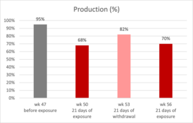
Giancarlo Bozzo’s team (39) reported and published a veterinary case regarding natural mycotoxin contamination in commercial egg production: up to week 47 of age, production parameters were on top of the genetic standards. However, a drop in egg production started at around week 47, and at week 50, egg production was only 68% (figure 1).

Figure
Figure 1: production of laying hens fed naturally contaminated feed with AFB1 and OTA
The house with the reduced performance received feed with linseed. In other houses of the same complex, which did not include linseed in the feed, production was unaffected. Therefore, this raw material was considered a possible cause of the issue. Linseed was removed from the formula, and three weeks after (53 weeks of age), egg production was at 84%. Afterward, linseed got back into the formulation, and the laying rate dropped again to 70% (week 56), this time accompanied by a significant increase in mortality.

Samples were collected at week 56, and AFB1 and OTA were detected in feed and the kidneys and livers of the hens consuming it (table 2). While the levels in the feed were not considered high risk, evidence from necropsy and histopathology suggested either a higher or a prolonged exposure; a synergistic effect of both mycotoxins on hen’s health and productivity can be inferred.

Table 2: mycotoxin analysis results for feed and organs

HPLC analysis results in samples of:
toxin Feed 1
(n=5)
Feed 2
(n=5)
Kidney

(n=10)

Liver

(n=10)

OTA 1.1 ± 0.1 ppb 31 ± 3 ppb 47 ± 3 ppb 24 ± 2 ppb
AFB1 ND 5.6 ± 0.3 ppb 1.4 ± 0.3 ppb 3.6 ± 0.4 ppb

The liver and kidneys were enlarged and showed signs of damage. Furthermore, urate crystals in the peritoneum and the abdominal air sac were observed, indicating renal failure. This limited the excretion of both toxins in the urine, increasing their half-life in the organism and enhancing the effects in target organs, contributing to the synergistic effect observed.

After using mycotoxin-free certified linseed, the problem receded. Though this is the best option to keep animals healthy and productive, it may not be practical in the long term due to the ubiquitous nature of the toxins and the cost and availability constraints of feed raw materials. Moreover, the mycotoxin levels present in the feed were relatively low and fell under recommended guidelines. For these reasons, in-feed toxin mitigation solutions must also be considered to reduce exposure for production animals.

In-feed intervention mitigates the effects of intermittent exposure to multiple mycotoxins

EW Nutrition conducted a study with Hy-Line W-36 layer-breeders intercalating three 10-day cycles of feed with 100ppb AFB1 + 100ppb OTA, with two 21-day cycles of non-challenged feed. An in-feed intervention (Solis Max 2.0, displayed as IFI) containing bentonite, yeast cell wall components, and a mixture of phytogenic components mitigated all effects.

Table 3: experimental groups and mycotoxin challenge

Treatment Group 100 ppb AFB1+ 100 ppb OTA IFI (2 kg/ton)
T-1 Control (C)
T-2 C+IFI X
T-3 Challenge (Ch) X
T-4 Ch+IFI X X

Trial design:

A total of 576 hens (18 replicates per diet, 8 hens each) and 58 roosters were randomly assigned to four diets at 28 weeks of age, as shown in Table 3. The 72-day experimental period included alternating 10-day challenge and 21-day non-challenge intervals (Figure 2). During the challenge intervals, the breeders in T-3 and T-4 were fed the mycotoxin-contaminated feed with and without the IFI.

FigureFigure 2: trial timeline showing challenge and non-challenge intervals and days of data collection and sampling.

Mitigated effects on egg production and egg quality

The challenge decreased overall egg production (Figure 3), egg mass, and shell thickness (Table 4). The first challenge interval did not affect production, but days later, from the first non-challenge period, all parameters were lower for the challenged group.

FigureDifferent letters indicate significant differences (p<0.05). Statistical tendencies (p<0.1) are indicated by (*).

Figure 3: Egg production of hens intermittently challenged with AFB1 and OTA, with and without in-feed Solis Max

The adverse effects on productivity and egg quality started after the first challenged feed was withdrawn and persisted through the following intervals until the end of the experiment. Similar effects in chronic mycotoxin challenges have been previously found (37, 39).

Table 4: Average egg quality parameters of hens intermittently challenged with AFB1+OTA, with and without an in-feed intervention (IFI)

Group Eggshell strength (N) Eggshell thickness (mm) Haugh Units
Control 21,02a 0,3661ab 70,88
IFI 21,16a 0,3702a 71,68
Challenge 20,05b 0,3630b   70,07*
Ch+IFI 21,06a 0,3698a 71,06

Different letters indicate significant differences (p<0.05). Statistical tendencies (p<0.1) are indicated by (*).

Mitigated effects on the progeny in incubation trials

Three incubation trials were performed: after the first challenge and non-challenge interval and at the end of the trial period after the third challenge interval. A significant decrease in fertility and hatchability was observed for the challenged group in all incubation trials. As mycotoxins affect egg quality (22-24) and can be transferred to the eggs (10, 14, 27), the effects were also shown in the case of hatchability and offspring performance. Fertility was affected from the first challenge interval onwards, continuing to be low for the challenge group until the end of the trial. However, the hatchability of fertile eggs dropped after the withdrawal of the contaminated feed and showed the lowest value during the third challenge interval.

The in-feed supplementation of Solis Max 2.0 (IFI) resulted in the consistent recovery of egg production and egg quality throughout the whole experimental period, achieving the same levels of productivity as the non-challenged control.

Figure
Letters indicate significant differences (p<0.05). Statistical tendencies (p<0.1), indicated by (*).

Figure 4: Hatchery parameters of eggs from breeders intermittently challenged with AFB1 and OTA, with and without an in-feed intervention (IFI).

Results in hatch of fertile can be related to egg quality, as the thickness of the eggshell influences the egg’s moisture loss and exchange with the environment during the incubation period. Thinner eggshells lead to higher embryo mortality (31, 32). The group having the challenge with Solis Max showed the same performance as the non-challenged control regarding hatchery performance.

Day-old chick weight was not affected. However, weight gain and mortality after ten days were hindered for the chicks from breeders taking the mycotoxin-contaminated feed (Table 5).

Table 5: Average day- and 10-day-old chick parameters from hens intermittently challenged with AFB1+OTA, with and without an in-feed intervention (IFI)

Parameter Control Challenge Ch + IFI
DOC body weight (g) 36,67 36,24 36,80
10-day body weight (g) 76,30a 75,94b 79,50a
10-day mortality (%) 0,94 1,26 0,97

Letters indicate significant differences (p<0.05). Statistical tendencies (p<0.1) indicated by (*)

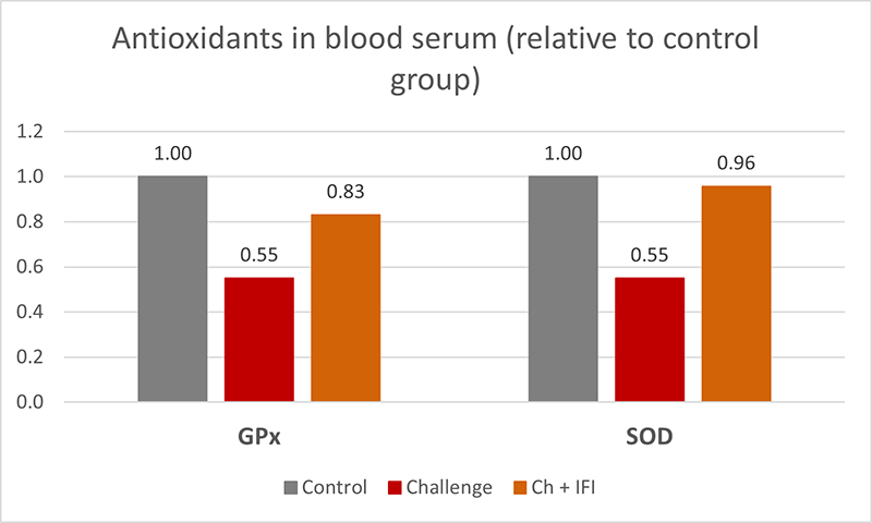
At the end of the experiment, oxidative stress biomarkers were measured in the blood serum of 15 hens per treatment, showing significantly lower GPx, and SOD (figure 5) in the challenged group, which indicates a depletion of the mechanisms to fight oxidative stress (40), the hens taking the in-feed product did not show this depletion.

FigureFigure 5: Antioxidants in blood serum, glutathione peroxidase (GPx), and superoxide dismutase (SOD) from breeders intermittently challenged with AFB1 and OTA, with and without an in-feed intervention (IFI).

Intermittent exposure to AFB1 and OTA negatively affected layer breeder productivity, egg quality, and hatchability and promoted oxidative stress in the birds. Intermittent mycotoxin challenges may affect animals even after the contamination is withdrawn. In-feed interventions showed effectiveness in mitigating these effects.

Climate changes bring new mycotoxin challenges – the right in-feed solutions can help

Today’s mycotoxin scenario shows increased frequency, quantity, and variety. Mainly long-living animals, such as laying hens and breeders, can be at more risk. Additionally, the contamination can be carried over to the eggs, potentially risking human health in the case of table eggs and hatchery performance and DOC quality in the case of breeders.

From case reports, we learn the consequences of real challenges and struggles in commercial production; from scientific trials based on possible commercial situations, we realize the advantages of interventions designed to tackle those challenges.

References

  1. Kos, Jovana, Mislav Anić, Bojana Radić, Manuela Zadravec, Elizabet Janić Hajnal, and Jelka Pleadin. “Climate Change—a Global Threat Resulting in Increasing Mycotoxin Occurrence.” Foods 12, no. 14 (July 14, 2023): 2704. https://doi.org/10.3390/foods12142704.
  2. Zingales, Veronica, Mercedes Taroncher, Piera Anna Martino, María-José Ruiz, and Francesca Caloni. “Climate Change and Effects on Molds and Mycotoxins.” Toxins 14, no. 7 (June 30, 2022): 445. https://doi.org/10.3390/toxins14070445.
  3. Loi, Martina, Antonio F. Logrieco, Tünde Pusztahelyi, Éva Leiter, László Hornok, and István Pócsi. “Advanced Mycotoxin Control and Decontamination Techniques in View of an Increased Aflatoxin Risk in Europe Due to Climate Change.” Frontiers in Microbiology 13 (January 10, 2023). https://doi.org/10.3389/fmicb.2022.1085891.
  4. Medina, Ángel, Jesús M González-Jartín, and María J Sainz. “Impact of Global Warming on Mycotoxins.” Current Opinion in Food Science 18 (December 2017): 76–81. https://doi.org/10.1016/j.cofs.2017.11.009.
  5. Grenier, Bertrand, and Todd Applegate. “Modulation of Intestinal Functions Following Mycotoxin Ingestion: Meta-Analysis of Published Experiments in Animals.” Toxins 5, no. 2 (February 21, 2013): 396–430. https://doi.org/10.3390/toxins5020396.
  6. Filazi, Ayhan, Begum Yurdakok-Dikmen, Ozgur Kuzukiran, and Ufuk Tansel Sireli. “Mycotoxins in Poultry.” Poultry Science, February 15, 2017. https://doi.org/10.5772/66302.
  7. Amirkhizi, Behzad, Seyed Rafie Arefhosseini, Masoud Ansarin, and Mahboob Nemati. “Aflatoxin B1in Eggs and Chicken Livers by Dispersive Liquid–Liquid Microextraction and HPLC.” Food Additives &amp; Contaminants: Part B, August 27, 2015, 1–5. https://doi.org/10.1080/19393210.2015.1067649.
  8. Emmanuel K, Tangni, Van Pamel Els, Huybrechts Bart, Delezie Evelyne, Van Hoeck Els, and Daeseleire Els. “Carry-over of Some Fusarium Mycotoxins in Tissues and Eggs of Chickens Fed Experimentally Mycotoxin-Contaminated Diets.” Food and Chemical Toxicology 145 (November 2020): 111715. https://doi.org/10.1016/j.fct.2020.111715.
  9. Ebrahem, Mohammad, Susanne Kersten, Hana Valenta, Gerhard Breves, and Sven Dänicke. “Residues of Deoxynivalenol (Don) and Its Metabolite de-Epoxy-Don in Eggs, Plasma and Bile of Laying Hens of Different Genetic Backgrounds.” Archives of Animal Nutrition 68, no. 5 (August 20, 2014): 412–22. https://doi.org/10.1080/1745039x.2014.949029.
  10. Salwa, A. Aly, and W. Anwer. “Effect of Naturally Contaminated Feed with Aflatoxins on Performance of Laying Hens and the Carryover of Aflatoxin B1 Residues in Table Eggs.” Pakistan Journal of Nutrition 8, no. 2 (January 15, 2009): 181–86. https://doi.org/10.3923/pjn.2009.181.186.
  11. Devreese, Mathias, Gunther Antonissen, Nathan Broekaert, Siegrid De Baere, Lynn Vanhaecke, Patrick De Backer, and Siska Croubels. “Comparative Toxicokinetics, Absolute Oral Bioavailability, and Biotransformation of Zearalenone in Different Poultry Species.” Journal of Agricultural and Food Chemistry 63, no. 20 (May 19, 2015): 5092–98. https://doi.org/10.1021/acs.jafc.5b01608.
  12. Galtier, P. “Biotransformation and Fate of Mycotoxins.” Toxin Reviews 18, no. 3 (August 1, 1999): 295–312. https://doi.org/10.3109/15569549909162648.
  13. Galtier, P., M. Alvinerie, and J.L. Charpenteau. “The Pharmacokinetic Profiles of Ochratoxin A in Pigs, Rabbits and Chickens.” Food and Cosmetics Toxicology 19 (January 1981): 735–38. https://doi.org/10.1016/0015-6264(81)90528-9.
  14. Hassan, Zahoor Ul, Muhammad Z Khan, Ahrar Khan, Ijaz Javed, and Zahid Hussain. “Effects of Individual and Combined Administration of Ochratoxin A and Aflatoxin B1 in Tissues and Eggs of White Leghorn Breeder Hens.” Journal of the Science of Food and Agriculture 92, no. 7 (December 16, 2011): 1540–44. https://doi.org/10.1002/jsfa.4740.
  15. Li, Shao-Ji, Guangzhi Zhang, Bin Xue, Qiaoling Ding, Lu Han, Jian-chu Huang, Fuhai Wu, Chonggao Li, and Chunmin Yang. “Toxicity and Detoxification of T-2 Toxin in Poultry.” Food and Chemical Toxicology 169 (November 2022): 113392. https://doi.org/10.1016/j.fct.2022.113392.
  16. Prelusky, D.B., R.M.G. Hamilton, and H.L. Trenholm. “Transmission of Residues to Eggs Following Long-Term Administration of 14 C-Labelled Deoxynivalenol to Laying Hens.” Poultry Science 68, no. 6 (June 1989): 744–48. https://doi.org/10.3382/ps.0680744.
  17. Ringot, Diana, Abalo Chango, Yves-Jacques Schneider, and Yvan Larondelle. “Toxicokinetics and Toxicodynamics of Ochratoxin A, an Update.” Chemico-Biological Interactions 159, no. 1 (January 2006): 18–46. https://doi.org/10.1016/j.cbi.2005.10.106.
  18. Osselaere, Ann, Mathias Devreese, Joline Goossens, Virginie Vandenbroucke, Siegrid De Baere, Patrick De Backer, and Siska Croubels. “Toxicokinetic Study and Absolute Oral Bioavailability of Deoxynivalenol, T-2 Toxin and Zearalenone in Broiler Chickens.” Food and Chemical Toxicology 51 (January 2013): 350–55. https://doi.org/10.1016/j.fct.2012.10.006.
  19. Sudhakar, BV. “A Study on Experimentally Induced Aflatoxicosis on the Carryover of Aflatoxin B1 into Eggs and Liver Tissue of White Leghorn Hens.” The Pharma Innovation Journal 11, no. 2S (2022): 213–17.
  20. Yiannikouris, Alexandros, and Jean-Pierre Jouany. “Mycotoxins in Feeds and Their Fate in Animals: A Review.” Animal Research 51, no. 2 (March 2002): 81–99. https://doi.org/10.1051/animres:2002012.
  21. Bouhet, Sandrine, and Isabelle P. Oswald. “The Intestine as a Possible Target for Fumonisin Toxicity.” Molecular Nutrition &amp; Food Research 51, no. 8 (August 2007): 925–31. https://doi.org/10.1002/mnfr.200600266.
  22. Fakhri, Yadolah, Mansour Sarafraz, Amene Nematollahi, Vahid Ranaei, Moussa Soleimani-Ahmadi, Van Nam Thai, and Amin Mousavi Khaneghah. “A Global Systematic Review and Meta-Analysis of Concentration and Prevalence of Mycotoxins in Birds’ Egg.” Environmental Science and Pollution Research 28, no. 42 (September 9, 2021): 59542–50. https://doi.org/10.1007/s11356-021-16136-y.
  23. Osaili, Tareq M., Akram R. Al-Abboodi, Mofleh AL. Awawdeh, and Samah Aref Jbour. “Assessment of Mycotoxins (Deoxynivalenol, Zearalenone, Aflatoxin B1 and Fumonisin B1) in Hen’s Eggs in Jordan.” Heliyon 8, no. 10 (October 2022). https://doi.org/10.1016/j.heliyon.2022.e11017.
  24. Wang, Lan, Qiaoyan Zhang, Zheng Yan, Yanglan Tan, Runyue Zhu, Dianzhen Yu, Hua Yang, and Aibo Wu. “Occurrence and Quantitative Risk Assessment of Twelve Mycotoxins in Eggs and Chicken Tissues in China.” Toxins 10, no. 11 (November 16, 2018): 477. https://doi.org/10.3390/toxins10110477.
  25. Tolosa, J., Y. Rodríguez-Carrasco, M.J. Ruiz, and P. Vila-Donat. “Multi-Mycotoxin Occurrence in Feed, Metabolism and Carry-over to Animal-Derived Food Products: A Review.” Food and Chemical Toxicology 158 (December 2021): 112661. https://doi.org/10.1016/j.fct.2021.112661.
  26. Völkel, Inger, Eva Schröer-Merker, and Claus-Peter Czerny. “The Carry-over of Mycotoxins in Products of Animal Origin with Special Regard to Its Implications for the European Food Safety Legislation.” Food and Nutrition Sciences 02, no. 08 (2011): 852–67. https://doi.org/10.4236/fns.2011.28117.
  27. Yuan, Tao, Junyi Li, Yanan Wang, Meiling Li, Ao Yang, Chenxi Ren, Desheng Qi, and Niya Zhang. “Effects of Zearalenone on Production Performance, Egg Quality, Ovarian Function and Gut Microbiota of Laying Hens.” Toxins 14, no. 10 (September 21, 2022): 653. https://doi.org/10.3390/toxins14100653.
  28. Song, Bin, Teng Ma, Damien P. Prévéraud, Keying Zhang, Jianping Wang, Xuemei Ding, Qiufeng Zeng, et al. “Research Note: Effects of Feeding Corn Naturally Contaminated with Aflatoxin B1, Deoxynivalenol, and Zearalenone on Reproductive Performance of Broiler Breeders and Growth Performance of Their Progeny Chicks.” Poultry Science 102, no. 11 (November 2023): 103024. https://doi.org/10.1016/j.psj.2023.103024.
  29. Qureshi, MA, J Brake, PB Hamilton, WM Hagler, and S Nesheim. “Dietary Exposure of Broiler Breeders to Aflatoxin Results in Immune Dysfunction in Progeny Chicks.” Poultry Science 77, no. 6 (June 1998): 812–19. https://doi.org/10.1093/ps/77.6.812.
  30. Ul-Hassan, Zahoor, Muhammad Zargham Khan, Ahrar Khan, and Ijaz Javed. “Immunological Status of the Progeny of Breeder Hens Kept on Ochratoxin a (OTA)- and Aflatoxin B1(Afb1)-Contaminated Feeds.” Journal of Immunotoxicology 9, no. 4 (April 24, 2012): 381–91. https://doi.org/10.3109/1547691x.2012.675365.
  31. Devegowda, G., and D. Ravikiran. “Mycotoxins and Eggshell Quality: Cracking the Problem.” World Mycotoxin Journal 1, no. 2 (May 1, 2008): 203–8. https://doi.org/10.3920/wmj2008.1037.
  32. Onagbesan, O., V. Bruggeman, L. De Smit, M. Debonne, A. Witters, K. Tona, N. Everaert, and E. Decuypere. “Gas Exchange during Storage and Incubation of Avian Eggs: Effects on Embryogenesis, Hatchability, Chick Quality and Post-Hatch Growth.” World’s Poultry Science Journal 63, no. 4 (December 1, 2007): 557–73. https://doi.org/10.1017/s0043933907001614.
  33. Ebrahem, Mohammad, Susanne Kersten, Hana Valenta, Gerhard Breves, Andreas Beineke, Kathrin Hermeyer, and Sven Dänicke. “Effects of Feeding Deoxynivalenol (Don)-Contaminated Wheat to Laying Hens and Roosters of Different Genetic Background on the Reproductive Performance and Health of the Newly Hatched Chicks.” Mycotoxin Research 30, no. 3 (April 11, 2014): 131–40. https://doi.org/10.1007/s12550-014-0197-z.
  34. Yegani, M., T.K. Smith, S. Leeson, and H.J. Boermans. “Effects of Feeding Grains Naturally Contaminated with Fusarium Mycotoxins on Performance and Metabolism of Broiler Breeders.” Poultry Science 85, no. 9 (September 2006): 1541–49. https://doi.org/10.1093/ps/85.9.1541.
  35. Calini, F, and F Sirri. “Breeder Nutrition and Offspring Performance.” Revista Brasileira de Ciência Avícola 9, no. 2 (June 2007): 77–83. https://doi.org/10.1590/s1516-635×2007000200001.
  36. Hassan, ZU, MZ Khan, A Khan, I Javed, U Sadique, and A Khatoon. “Ochratoxicosis in White Leghorn Breeder Hens: Production and Breeding Performance.” Vet. J. 32, no. 4 (2012): 557–61.
  37. Verma, J., T. S. Johri, and B. K. Swain. “Effect of Varying Levels of Aflatoxin, Ochratoxin and Their Combinations on the Performance and Egg Quality Characteristics in Laying Hens.” Asian-Australasian Journal of Animal Sciences 16, no. 7 (January 1, 2003): 1015–19. https://doi.org/10.5713/ajas.2003.1015.
  38. Johnson-Dahl, M.L., M.J. Zuidhof, and D.R. Korver. “The Effect of Maternal Canthaxanthin Supplementation and Hen Age on Breeder Performance, Early Chick Traits, and Indices of Innate Immune Function.” Poultry Science 96, no. 3 (March 2017): 634–46. https://doi.org/10.3382/ps/pew293.
  39. Bozzo, Giancarlo, Nicola Pugliese, Rossella Samarelli, Antonella Schiavone, Michela Maria Dimuccio, Elena Circella, Elisabetta Bonerba, Edmondo Ceci, and Antonio Camarda. “Ochratoxin A and Aflatoxin B1 Detection in Laying Hens for Omega 3-Enriched Eggs Production.” Agriculture 13, no. 1 (January 5, 2023): 138. https://doi.org/10.3390/agriculture13010138.
  40. Surai, Peter F., Ivan I. Kochish, Vladimir I. Fisinin, and Michael T. Kidd. “Antioxidant Defence Systems and Oxidative Stress in Poultry Biology: An Update.” Antioxidants 8, no. 7 (July 22, 2019): 235. https://doi.org/10.3390/antiox8070235.


Effects on Performance and Gut Health of Ventar D Supplementation in Broiler Diets

Summary of study by Necmettin Ceylan, Sait Koca, Nejla Kahraman, Ankara University, Faculty of Agriculture, Animal Science, 6110 Ankara/Türkiye

The study conducted by Dr. Celyn et al. in 2023 focused on the impact of Ventar D supplementation in broiler diets on growth performance and gut health. The trial was carried out over six weeks on Ross 308 broiler chicks, comparing a control group with an experimental group supplemented with Ventar D. The trial feed was based on corn, soybean meal, wheat, sunflower meal, and poultry oil.

Key Findings

Growth Performance: The study demonstrated that Ventar D supplementation significantly improved body weight gain, feed consumption, feed conversion ratio (FCR) and EPEF during the starter, grower, and finisher periods. The overall performance of chickens fed with Ventar D was notably better, showing a 6.5% higher body weight and 1.67% better FCR compared to the control group.

Treatments BWG, g FCR Corrected FCR2565 FI, g Mortality,% EPEF
Control 2520.6a±32,77 1.620a±0.006 1.629a±0.011 4082.2a±46.77 3.25±0.28 367.2a±5.18
Ventar D 2684.3b±23.65 1.593b±0.010 1.568b±0.015 4273.9b±19.89 2.75±0.53 399.8b±4.35

Different letters indicate significance; P ≤ 0.05

Liver Enzymes: The addition of Ventar D led to a significant decrease in serum Alanine aminotransferase (ALT) levels

Treatments ALP ALT
Control 286.70±54.98 1.505a±0.390
Ventar D 301.50±87.19 0.832b±0.181

Different letters indicate significance; P ≤ 0.05

Gut Health: Ventar D supplementation resulted in higher concentrations of short-chain volatile fatty acids (SCVFA) in the cecum.

  Acetate Propionate Butyrate Isobutyrate Valerate Isovalerate BCFA Total SCFA
Control 27.22a±1.26 8.21±0.38 7.24a±0.41 0.848±0.078 0.964±0.043 0.881±0.054 2.69a±0.12 45.36a±1.53
Ventar D 30.51b±0.80 9.36±0.56 8.86b±0.44 0.878±0.070 1.121±0.077 0.993±0.031 2.99b±0.08 51.73b±1.32

Different letters indicate significance; P ≤ 0.05

Conclusion

Considering the results summarized in the tables above according to the feeding phases and the overall study (0-41 days): Ventar D supplementation of broiler feeds at the level of 100 g/ton significantly improved growth performance parameters during the starter, grower and finisher periods (P ≤ 0.05), and in the final results was stable at 6.5% higher BW and 1.67% better FCR compared to the control group. European Production Efficiency Factor (EPEF) was also significantly better than the control group (P ≤ 0.05).

In the study, liver enzyme and the concentration of short-chain volatile fatty acids also improved significantly with the addition of Ventar D, which may be attributed to the gut health related mode of action for Ventar D.


Low Crude Protein Diets in Poultry: Understanding the Consequences

BROILER

Conference report

The concept of feeding poultry, specifically broilers and layers, with reduced crude protein (CP) diets is gaining traction among nutritionists. The economic implications of balancing amino acids currently dictate dietary CP levels. At the recent EW Nutrition Poultry Academy in Jakarta, Indonesia, Dr. Steve Leeson, Professor Emeritus at the University of Guelph, Canada, raised a crucial question: “What does ‘low CP’ really mean?” He states that it typically means a reduction of maximum 2-3% relative to current CP levels.

Low CP diets generally involve a decrease in soybean meal, compensated by higher grain content. This change increases dietary starch and decreases dietary lipid levels. To meet nutritional needs, these diets also include higher amounts of crystalline (synthetic) amino acids.

Dr. Leeson outlined the advantages and disadvantages of low CP diets. Positives include improved gut health due to reduced proteolytic bacteria, less environmental pollution, lower water intake (improving litter quality), improved sustainability indices, increased dietary net energy, and better performance during heat stress. Negatives encompass issues like lower pellet quality, altered dietary electrolyte balance, higher diet costs, reduced growth rate and feed efficiency, and increased abdominal fat deposition. There are also questions about the presumed complete utilization of crystalline amino acids, which can be as high as 25kg/MT in these diets.

Challenges with Low CP Diets

  • Protein vs. Amino Acids: Diets are typically formulated based on digestible amino acid content, though minimum CP levels remain common, to avoid reduced performance: Dr. Leeson noted that broiler diets with less than 19% CP in starter and 15% in finisher phases, and layer diets below 13% CP, often fail to deliver adequate performance, regardless of digestibly amino acid supply.
  • Utilization of Free Amino Acids: The crystalline amino acids are immediately absorbable in the small intestine, contrasting with protein-bound amino acids that are absorbed as di- and tri-peptides. Amino acids absorption dynamics and endogenous loss of amino acids are affected by (high) levels of  crystalline amino acids.
  • Non-Essential Amino Acids: The impact of reduced CP on animal performance might be related to the lower levels of presumed non-essential amino acids, e.g. glycine and serine.  This is an area for further exploration.
  • Energy Level Considerations: Dr. Leeson suggests maintaining specific ratios of digestible lysine to apparent metabolizable energy in broilers at different growth stages. The heat increment of CP is an essential factor, as it reduces net energy efficiency, possibly requiring an adjustment in amino acid to metabolizable energy ratios as poultry diets are not based on net energy values.
  • Gut Health: Lower CP levels can reduce the flow of undigested protein into the hindgut, reducing the risk of necrotic enteritis, and the production of harmful metabolites, like biogenic amines.
  • Role of Proteases: Protease use can lead to a further 2-4% reduction in dietary CP, with the response depending on the inherent protein digestibility of the diets.
  • Impacts on Pellet Quality: Due to the binding properties of protein, each 1% reduction in CP typically results in a 2% decrease in pellet durability (index).
  • Electrolyte Balance: Reduced CP can significantly lower dietary electrolyte balance, which has to be considered in feed formulation. Amongst the nutrients contributing to DEB value, Sodium and Potassium appear to be the most influential minerals to consider.

Conclusion

Dr. Leeson anticipates that low CP diets will become increasingly relevant. They have the potential to reduce environmental pollution and dependence on soybean meal, despite current challenges in reducing feed costs.


***

EW Nutrition’s Poultry Academy, featuring Dr. Leeson, took place in Jakarta and Manila in early September 2023. With nearly 50 years of industry experience, Dr. Leeson has made significant contributions to poultry nutrition and management, evidenced by his numerous awards and over 400 published papers.


Feed and water management strategies to mitigate heat stress in layers

LAYER IMGP

Dr Daniel Valbuena, Global Manager of Technical Services, Hy-Line International – Conference Report

Feed and water management strategies are essential to help mitigate the negative effects of heat stress on bird welfare, production, and profitability. In EW Nutrition’s Poultry Academy in September, the topic was approached in a comprehensive and practical presentation.

Feed management

Feed consumption of the flock should be closely monitored during hot weather. It is important to rebalance the diet for critical nutrients, particularly amino acids, calcium, sodium and phosphorous according to the birds’ productivity demand (i.e., stage of production) and the observed feed intake. Insufficient amino acid intake is the primary reason for productivity loss during hot weather. Several strategies may be employed to help to manage elevated temperatures and maintain higher levels of feed intake:

  • Withdrawing feed from birds 6 hours before peak hot temperatures in the afternoon can lower the risk of heat stress. Encourage as much consumption as possible in the early morning or evening. Using lighting for midnight feeding encourages feed intake.
  • One third of the daily feed ration should be given in the morning and two thirds in the late afternoon. An additional advantage is the availability of calcium in the digestive system during shell formation at night and in the early hours of the morning. This will improve shell quality and reduce the birds from depleting bone calcium.
  • Normally a maximum 1 hour for feeder clean-out time is recommended, but this can be extended to 3 hours when the temperature exceeds 36°C.
  • Consider adding a 1-2-hour midnight feeding.
  • Alter feed particle size, either by increasing it or by feeding a crumble diet. With crumble diets in laying flocks, a supplementary source or presentation of large particle limestone is recommended.
  • Formulate diets using highly digestible materials, particularly protein sources. Metabolism of excess protein is particularly heat-loading on the bird. Formulate to digestible amino acid targets and do not apply a high crude protein minimum in the formula. Synthetic amino acids can reduce crude protein in the diet without limiting amino acid levels.
  • Increasing the proportion of energy contribution from highly digestible lipid, rather than starches or proteins, will reduce the body heat production resulting from digestion. This is known as heat increment and is lowest with the digestion of dietary fat.
  • The bird’s metabolizable energy requirement decreases as ambient temperature increases to above 21°C, resulting from a reduction of energy requirements for maintenance. The energy requirement will decrease with the rise of temperature up to 27°C, above which it will start to increase again since the bird needs additional energy for panting to reduce body heat.

Stress Management Schedule

Management schedule during times of heat stress

Water management

During periods of high environmental temperature, birds have a high demand for drinking water. The water-to-feed consumption ratio is normally 2:1 at 21°C but increases to 8:1 at 38°C. Adequate drinking water must be available to heat-stressed flocks. Ensure that drinkers have sufficient water flow (>70 mL/minute/nipple drinker). If water flow is less the lines need to be checked for flow restriction. If there’s a build-up of iron and other minerals, it needs to be removed. Don’t forget to routinely check water filters and replace them as needed.

It’s easy to overlook a non-functioning drinker here and there; drinkers must be systematically checked to make sure they’re all working. For floor-reared flocks, providing additional drinkers can help accommodate the increased water consumption.

During hot weather, you need to ensure your water system can accommodate the bird’s increased water consumption, and the additional water demands for foggers, evaporative cooling systems and roof sprinklers. The availability of drinking water to a heat-stressed flock should never be compromised.

Cool water temperatures (<25oC) will encourage the birds to drink and reduces the birds’ core temperature. Flush water lines and waterers routinely to keep the water fresh and cool, increasing water consumption, and sustaining egg production. If available, ice can also be added to header tanks. When mechanical cooling systems fail, water flushing can serve as an emergency measure during heat stress.

Drinking water from overhead water tanks can become hot if exposed to direct sunlight. These water tanks should be a light color, insulated and covered to avoid direct sunlight. Water tanks are ideally placed inside the house or underground. Water pipes in the house should not be installed close to the roof to avoid heat from the roof warming up the water in the pipes.

Eggs

Water Cooler
Having the water tank inside the house (above) or light-colored and covered to avoid direct sunlight (below) keeps the water cooler

Use vitamin (A, D, E and B complex) and electrolyte supplements in the drinking water to replenish the loss of sodium, chloride, potassium, and bicarbonate in the urine. Electrolyte supplements are best used in anticipation of a heat stress period and can be added to drinking water for up to 3 days.


Coping with evolutions in the performance and nutritional requirements of layers

LAYERHennen Auf Hof

Dr. Vitor Arantes, Global Technical Services Manager and Global Nutritionist, Hy-Line International – Conference Report

The layer industry has gone through significant changes during the past decades and has a remarkable capacity to cope with new challenges. Dr Vitor Arantes, Global Technical Services Manager and Global Nutritionist, Hy-Line International, noted that increased egg production, improved feed efficiency, and adaptation of egg quality and bird welfare to consumer preferences have contributed significantly to the success of the egg industry. However, continuous improvement in egg production per hen housed is the most important selection criteria in layer breeding.

Egg producers needs include:

  • More saleable eggs,
  • Eggshell quality,
  • Easier behaviour
  • Housing systems
  • Egg size specifics
  • Sanitary / environmental challenges
  • Profits through productivity

Primary breeders can deliver these producer needs through:

  • Having the correct product for each country
  • Constant follow up
  • Local presence, trust relationship
  • Accurate data collection
  • Critical data analysis
  • Understand the company’s goals
  • Customized technical services according to each customer needs

How has genetics changed?

Examples of genetic progress in layers from 1984 to 2022 cited by Dr Arantes include:

  • Higher persistency (+30 weeks >90%)
  • Higher egg mass (+5.5 kg/hen housed)
  • Smaller hen (-21% mature body weight)

Dr Arantes states the record clutch size, defined as the unstopped length of individual egg production on a daily basis, was an amazing 474 days for a White Plymouth Rock hen. This genetic progress necessitates adjustments in nutrition and management.

Layers weeks

As shown below, growth and organ development occur at various ages. “There is no margin for mistakes – a lack of growth during a stage could have a detrimental impact on pullet quality and subsequent production,” stressed Dr Arantes.

Multi-phasic growth and development during rearing and start of lay

System Age (weeks) Consequence
Gastrointestinal 0-6 Shorter intestinal tract/reduced nutrient absorption
Immune 0-6 Flocks more susceptible to disease challenges
Skeleton 6-12 Shorter frames/less calcium reserves
Muscle 6-12 Impact in persistency of production
Fat >12 Excess can lead to fatty liver, prone to prolapse and mortality

 0-6 weeks of age

Most of the development of the organs of the digestive tract and the immune system occurs during the first 6 weeks of age. Problems that occur during this period can have negative effects on the function of these systems. Birds stressed during this period may have lifelong difficulties in digesting and absorbing feed nutrients. Immunosuppression may also result from problems during this period, leaving the bird more susceptible to diseases and less responsive to vaccinations.

6-12 weeks of age

Most of the adult structural components – muscles, bones and feathers are obtained during the period of rapid growth that occurs at 6-12 weeks of age. Growth deficiencies during this period will prevent the bird from obtaining sufficient bone and muscle reserves, which are necessary to sustain a high level of egg production and to maintain good eggshell quality. About 95% of the skeleton is developed at the end of the bird’s 13 weeks of life. At this time, the plates of the long bones become calcified and further growth in bone size cannot occur.

12-18 weeks of age

During this period, the growth rate slows, and the reproductive tract matures and prepares for egg production. Muscle development continues and the proliferation of fat cells takes place. Excessive weight gain during this period can result in an excessive amount of abdominal fat. Low body weight and stressful events at this time can delay the start of egg production. From 7-10 days before oviposition of the first egg, the medullary bone that is located within the cavities of the long bones can be increased by feeding the bird a pre-laying ration with higher levels of calcium than the development stage.

Bodyweight is a key factor for flock management as this will influence future performance of birds. Consequently, bodyweight should be controlled during the whole life of the layer flocks. Management, in particular nutrition and lighting programs, can help to control bodyweight so birds can achieve their genetic potential.

Uniformity

Uniformity is the most important KPI in our business. However, with the trend towards larger flocks, maintaining uniformity is becoming more challenging. With larger flocks, it is difficult to source one unique flock which thus usually comprises multiple breeding flocks of different ages. Inevitably, uniformity will be poor, hence the need for tools to address unexpected issues. Lack of uniformity becomes a self-perpetuating cycle – dominant versus dominated.

Many egg producers use average body weights compared to the breeder recommendations as a guide to flock status. However, knowing if you have good body weight uniformity is another valuable management tool. In any flock some birds are lighter or heavier than the average body weight. Poor uniformity makes management decisions, such as lighting, feed amounts or diet phase more difficult.

Ideally, the body weight coefficient of variation (CV) should be +/-10% of the mean, increasing the likelihood that your management decision will be appropriate for most of the flock. Inappropriate diet changes, bird handling, vaccination and transfer can reduce uniformity. Flocks should be at 90% uniformity at the time of transfer to the laying facility. Body weight at point of lay significantly affected egg production and eggshell quality.

Grading into 2 or 3 sub-populations of different average bodyweights may be necessary so that each group can be managed in a way that will achieve good whole flock uniformity at the point of lay. The best predictor of future laying performance is the pullet’s body weight and body type at the point of lay.

Vision egg

Vision Egg is a custom diagnostic tool used to analyze data and emphasize flock performance to achieve the highest genetic potential from Hy-Line layers with recommendations connected to customer profitability. This growing, robust database includes data from over 1 billion hens strengthens our flock performance diagnostic tool for improved profitability for Hy-Line customers.

Hy-Line customers can take advantage of this opportunity by sending flock data to their regional business manager or technical service specialist. The information shared with Hy-Line is kept completely confidential.

Summary

The challenge is not egg numbers, stated Dr. Arantes, but saleable eggs. Correct body weight and high uniformity of the flock at point of lay will result in good performance over the laying period, with high peak production and good persistency of production and the production of good quality eggs. Management is the key factor to regulation of body weight during rearing and at point of lay.


How to mitigate formulation costs when ingredient prices are high

IMG

Conference Report

The price of corn and soybeans dictates the price of all other ingredients, including to some extent amino acids, stated Dr Steve Leeson Professor Emeritus, University of Guelph, Canada at the recent EW Nutrition Poultry Academy in Jakarta, Indonesia.

The big question is, when times get tough, can we reduce safety margins and still get good performance?, asked Dr Leeson. “When we formulate diets, we build in some insurance. But so do the breeding companies in their recommendations.  For sure, reducing safety margins takes us out of our comfort zones, but we need to be nutritionists, not mathematicians,” he stressed.

Protein and energy are now expensive. As a result of this economic pressure, there is a focus on strategies to reduce feed costs and improving the production efficiency and profitability of poultry enterprises. Feed cost/kg body weight gain is not always at the lowest feed:gain.

To help achieve these targets, Dr Leeson discussed feeding and management strategies that take into account the cost mitigation requirement.

Optimize current digestibility/efficiency

With high feed prices, it is especially important to review the use of feed additives that optimize nutrient release and improve ‘digestibility’. The most obvious class of such additives are the various exogenous enzymes that improve the availability of phosphorus, energy, and amino acids. In most instances, these different classes of enzymes are additive in terms of nutrient release, since they have different target substrates or modes of action. All too often, the position is taken that “I take energy uplift from my amylase, so I can’t expect energy release from phytase or protease”.

The energy release from phytase is invariably net energy related to removal of the phytate molecule, which in effect is an ‘antigen’ and takes energy to counter its negative effects. The energy release from an amylase, however, is obviously related simply to the improved digestibility of carbohydrate complexes. Similarly, a protease enzyme will always provide energy, since all protein/amino acids are eventually used for energy during protein turnover, hence our use of the often forgotten ‘n’ in AMEn. We also have the choice of enzyme concentration, especially for phytase, which in the current economic solution is likely to be close to 2 – 2.5 doses, assuming a single dose is around 500-600 FTUs. The economics of super-dosing or mega-dosing is greatly impacted by the cost of the enzyme.

The response of phytase varies with individual amino acids, and with ingredients, with greater responses with ingredients of lower inherent digestibility. Generally, Dr Leeson suggests that a protease will capture 20% of indigestible amino acids. For example:

  • 70% digestibility = +6% uplift
  • 90% digestibility = +2% uplift

Relax ingredient constraint maximums

Probably the greatest current cost savings can be made from relaxing the maximum levels on ingredients. While corn and soybean meal levels are usually without restriction, we often impose limits on the upper levels of ‘alternative’ ingredients such as distillers grains, rice by-products and rapeseed/canola meals, etc. When the upper levels are reached in the formula, this suggests cost savings from using higher levels. Current restraints are based on past knowledge of perhaps variable nutrient composition and so the decision to use more of any ingredient must be based on past knowledge of on-going quality control assays. Although we can achieve considerable detail today in such QC assays, monitoring for (consistency of) crude fiber, crude protein, fat, and moisture alone, provide a sound basis for decisions on whether to use more of an individual ingredient.

Source alternate ingredients

Another option is to consider ‘new’ alternative ingredients. In reality, however, there are no new ingredients as such, since all monogastric nutritionists around the world have only around 19 ingredients available in sufficient quantities to sustain large-scale modern feed mills. There are certainly smaller quantities of specialised local by-products that can be used to advantage, yet these are becoming scarce. Therefore, an ingredient is only novel to you, since inevitably the same ingredient has been used for many years in other regions. As such, there is a wealth of information available on the nutritive value of these ‘new’ ingredients that can be simply transposed to our formulation matrices.

The bird is very adaptable to new ingredients, in fact it is more responsive to nutrients. Unless there are toxins, antinutritional factors, or other negative factors, it doesn’t matter to the bird. Knowing the ingredient composition is the critical feature regarding the success or failure with new ingredients.

Reduce nutrient density

Both layers and meat birds still eat quite precisely to their energy requirements. They are amazingly adaptable to a vast range of nutrient densities, assuming that they can eat enough feed as the lower levels of feed energy are approached. Success in using lower levels of nutrient density is invariably negatively impacted by factors such as high stocking density and a high environmental temperature. Conversely, reducing diet energy usually has the hidden advantage of improved pellet quality.

The key to successful use of lower energy diets lies in prediction of change in feed intake and corresponding adjustment to all other nutrients in the diet.

Flexible cost of Dietary electrolyte balance (DEB)

When first introduced in the 1970s, maintaining DEB around 250MEq was seen to optimize broiler performance, especially leg condition. There is now less emphasis on this, perhaps because of genetic selection for skeletal integrity. DEB, however, may be important during heat stress to stimulate water intake and control manure moisture. Formulating to fixed DEB levels always adds costs. Instead, Dr Leeson suggested to focus on sodium and chloride at a ratio of 1:1.3.

Optimize feed texture (pelleting)

The first consideration is to make a good quality pellet, then worry about pellet size, noted Dr Leeson. He also added he was “a big fan of sunflower meal – it’s great for pellet quality.”

When given a choice in particle sizes, birds invariably show a preference for the largest particles. This situation becomes obvious when ‘fines’ accumulate in the feeder pans over time. As shown below, as pellet size increases, so does the bird’s need to consume fewer pellets. As a result, they need to spend less time at the feeder. Naturally, this idealised pellet size must be balanced against the willingness of mill managers to accommodate the necessary changes in pellet die size. Matching pellet size to bird age becomes critical as stocking density increases.

Impact of pellet size on pellet number consumed by a 30-day-old broiler

Pellet size (diameter) 4 mm length 6 mm length
3 mm 580 390
4 mm 330 220
5 mm 210 140

In the end, cost mitigation should not require complex mathematics. Nutritionists should be able to play with several types of improvements without affecting health and performance.


***

EW Nutrition’s Poultry Academy took place in Jakarta and Manila in early September 2023. Dr. Steve Leeson, an expert in Poultry Nutrition & Production with nearly 50 years’ experience in the industry, was the distinguished keynote speaker.

Dr. Leeson had his Ph.D. in Poultry Nutrition in 1974 from the University of Nottingham. Over a span of 38 years, he was a Professor in the Department of Animal &Poultry Science at the University of Guelph, Canada. Since 2014, he has been Professor Emeritus at the same University. As an eminent author, he has more than 400 papers in refereed journals and 6 books on various aspects of Poultry Nutrition & Management. He also won the American Feed Manufacturer’s Association Nutrition Research Award (1981), the Canadian Society of Animal Science Fellowship Award (2001), and Novus Lifetime Achievement Award in Poultry Nutrition (2011).


Meat quality is a result of genetics, feeding, the microbiome, and the handling of animals and meat

Different Pieces Of Meat Shutterstock

by Dr. Inge Heinzl, Editor EW Nutrition

Nowadays, nutrition is no longer about pure nutrient intake; enjoyment is also a priority. Consumers attach great importance to the high quality of food and, therefore, also of meat. The genetic selection for faster growth and feeding high-energy diets made meat production more efficient and shortened the raising period. However, this selection may sometimes also result in challenges to meat quality, such as worse water holding capacity, less marbling, less flavor, and reduced storage & processing properties.

The following article will provide detailed information about what meat quality is, how the gut microbiota influences it, and how we can increase meat quality by feeding and modulating the intestinal microflora.

Which factors can contribute to meat quality?

Meat quality is a complex term. On the one hand, meat quality covers measurable parameters such as the content of nutrients, moisture, microbial contamination, etc. On the other hand, and to no small extent, the consumers’ preferences are significant. Since meat today is often sold as cuts or in parts (e.g., broiler drumsticks, breast), processing also affects the quality of meat and meat products.

Physical characteristics are objective determinants of meat quality

Physical characteristics are parameters that can be measured. For meat, the following measurable parameters determine meat quality:

1.  Fat content and fatty acid composition influence tenderness and taste

Some years ago, the majority of consumers asked for completely lean meat, which, fortunately, has now changed. Fat is a flavor carrier. Especially intramuscular fat (marbling) melts during the preparation, making the meat tender, juicy, and taste good. Fat also transports fat-soluble vitamins.

A further criterion is the composition of the fat, the fatty acids. Geese fat, e.g., is known for its high content of oleic, linoleic, linolenic, and arachidonic acid, all of them derivates of the enzymatic denaturation of stearic acid (Okruszek, 2012).

One exception is cholesterol. Although belonging to the lipids and improving the sensory quality of meat, consumers prefer meat with low cholesterol content.

2.  Protein and amino acid content influence the meat value

The content and the composition of protein are important factors in meat quality. Protein is essential for constructing and maintaining organs and muscles and for the functionality of enzymes. The human body needs 20 different amino acids for these tasks, eleven of which it can manufacture by itself. Nine amino acids, however, must be provided by food and are called essential amino acids. Meat is a highly valuable protein source, rich in protein and essential amino acids. The protein quality, therefore, includes the chemical and amino acid score, the index for essential amino acids, and the biological value.

In addition to the pure nutritional value, amino acids contribute to flavor and taste. These flavor amino acids directly influence meat’s freshness and flavor and include threonine, alanine, serine, lysine, proline, hydroxyproline, glutamic acid (glutamate is important for the umami taste), aspartic acid, and arginine.

3.  Vitamins and trace elements are essential nutrients

Meat is a primary source of B vitamins (B1-B9) and, together with other animal products such as eggs and milk, the only provider of Vitamin B12. Vitamin A is available in the innards, vitamin D in the liver and fat fish, and vitamin K in the flesh.

The most important mineral compounds in meat are zinc, selenium, and iron. Humans can utilize the iron from animal sources particularly well.

4.  pH and speed of pH decline decide if the meat is suited for cooking

Since broiler chicken meat nowadays is usually consumed as cut-up pieces or processed products, the appearance at the meat counter or in the plastic box is essential for being sold. The color, seen as an apparent measurement of the freshness and quality of the meat, is influenced by the pH. The muscle pH post-mortem plays an essential role in meat quality. Due to the glycolytic process, the pH post-mortem is a good indication for evaluating physiological meat quality. A rapid pH decline post-mortem to 5.8-6.0 in most cases leads to pale, soft, and exudative (PSE) meat with reduced water retention (Džinić et al., 2015), whereas a high ultimate pH results in dark, firm, and dry (DFD) meat with poor storage quality (Allen et al., 1997)

5.  Nobody wants meat like leather

The shear force is a measure of the tenderness of the meat. To determine the shear force, the meat undergoes the process of cooking and chilling. Afterward, standardized meat blocks, with fibers running along the length of the sample, are put into the Warner-Bratzler system. The blade used simulates teeth, and the system measures the force necessary to tear the piece of meat.

6.  Microbial contamination is a no-go

The microbial contamination of the meat often occurs during the slaughter process. Let’s take a look at salmonella or campylobacter in poultry. The chickens take up salmonella with contaminated feed or water. Campylobacter is transmitted by infected wild birds, inadequately cleaned and disinfected cages, or contaminated water. The bacteria proliferate in the intestine. At slaughter, the intestine’s microorganisms can spread onto the meat intended for human consumption.

7.  High water holding capacity is necessary to have tender meat

The moisture content contributes to the meat’s juiciness and tenderness and improves its quality. If the meat loses its moisture, it gets tough, and quality decreases. Additionally, drip loss reduces the nutritional value of meat and its flavor.

8.  Fat oxidation makes meat rancid, and oxidative stress can cause myopathies in broiler breasts

Rancidity of meat occurs when the fat in the flesh gets oxidized. There are different signs of meat rancidity: bad odor, changed color, and a sticky, slimy texture. Poultry meat is considered more susceptible to the development of oxidative rancidity than red meat. This can be explained by its higher content of phospholipids, PUFAs, especially in the thighs. The breast meat, however, has a relatively low level of intramuscular fat (up to 2 %) and, additionally, myoglobin is a natural antioxidant.

But oxidative stress in broiler breasts – and this more and more happens due to a selection of always bigger breasts – can lead to muscle myopathies such as white stripes or wooden breasts, making the meat only usable for processed products.

Sensory meat quality addresses the human senses

Besides physical quality, the sensory and chemical characteristics are essential to meat’s economic importance. All attributes of meat that stimulate the human senses (vision, smell, taste, and touch) belong to the sensory quality. It, therefore, is more subjective and hard to determine. The most important features for the consumer include color (attractive or unattractive), texture (tenderness, juiciness, marbling, drip loss), and taste/ flavor (Thorslund et al., 2016).

The appearance is the first impression

Nowadays, meat is often sold as cuts lying in polystyrene or clear plastic trays, over-wrapped with transparent plastic films, so the appearance is paramount. The meat must show an attractive color. Muscle myopathies, such as the ones occurring in chickens, would not meet consumers’ needs.

How does the flavor of meat develop?

There is a reaction between reducing sugars and amino acids when meat is cooked (Mottram, 1998). This Maillard reaction, along with the degradation of vitamins, lipid oxidation, and their interaction, is responsible for the production of the volatile flavor components forming the characteristic aroma and flavor of cooked meat (MacLeod, 1994). Werkhoff et al. (1990) consider cysteine and methionine the most significant contributors to meat flavor development. One factor deteriorating this quality characteristic is lipid peroxidation, which turns the taste to rancid.

Some sensory characteristics are related to physical ones

The parameters of sensory meat quality can be partly explained by measurable parameters. Water retention, e.g., influences the juiciness of the meat. The palatability increases with higher intramuscular fat or marbling (Stewart et al., 2021), the initial pH and the speed of decline decide if the flesh will be pale, soft, and exudative or normal, and lipid peroxidation is the leading cause of a decrease in meat quality (Pereira & Abreu, 2018).

Processing quality

For the processing quality, muscle structure, chemical ingredient interactions, and muscle post-mortem changes are decisive (Berri, 2000).

Does the microbiome influence the meat quality?

The gastrointestinal tract of monogastric animals disposes of a microbiome of primarily bacteria, mainly anaerobic Gram-positive ones (Richards et al., 2005). With its complex microbial community, the digestive tract is responsible for digesting feed and absorbing nutrients, but also for eliminating pathogens and developing immunity. Gut microbiotas play an essential role in digestion, are decisive concerning the synthesis of fatty acids, proteins, and vitamins, and, therefore, influence meat quality (Chen, 2022).

Intestinal microbiotas vary by species/breeds and age (Ma et al., 2022; Sun et al., 2018), and so does meat quality. For example, Duroc pigs with meat of high tenderness, good flavor, and excellent tastiness show different microbiota than other breeds (Xiao, 2017). Zhao et al.(2022) examined high- and low-fat Jinhua pigs, with the high-fat pigs showing more increased backfat thickness but also a higher fat content in the longissimus dorsi. They found low-fat pigs showed a higher abundance of Prevotella and Bacteroides, Ruminococcus sp. AF12-5, Faecalibacterium sp.OFO4-11AC und Oscillibacter sp. CAG:155, which are all involved in fiber fermentation and butyrate production. The high-fat animals showed a higher abundance of Firmicutes and Tenericutes, indicating that they are responsible for higher fat production of the organism in general but also a better fat disposition in the flesh. Lei et al. (2022) showed that abdominal fat was positively correlated with the occurrence of Lachnochlostridium and Christensenelleceae.

The intestinal microbiota-muscle axis enables us to improve meat quality by controlling intestinal microbiota (Lei, 2022). However, to develop strategies to enhance the quality of meat, understanding the composition of the microbiota, the functions of the key bacteria, and the interaction between the host and microbiota is of utmost importance (Chen et al., 2022).

Different factors influence the microbiome

Apart from that microbiotas are different in different breeds, they are additionally influenced by diseases, feeding (diets, medical treatments with, e.g., antibiotics), and the environment (climate, geographical position). This could be shown by different trials. The genetic influence on microbiota was impressively documented by Goodrich et al. (2014), who detected that the microbiomes of monozygotic twins differ less than the ones of dizygotic twins. Lei et al. (2022) compared the microbiota of two broiler breeds (Arbor Acres and Beijing-You, the last one with a higher abdominal fat rate) and found remarkable differences in their microbiota composition. When raising them in the same environment and with the same feed, the microbiotas became similar. Zhou et al. (2016) contrasted the cecal microbiota of five Tibetan chickens from five different geographic regions with Lohmann egg-laying hens and Daheng broiler chickens. Besides seeing a difference between the breeds, slightly distinct microbiota between the regions could also be noticed.

The intestinal microbiome can actively be changed by

  • promoting the wanted microbes by feeding the appropriate nutrients (e.g., prebiotics)
  • reducing the harmful ones by fighting them, for example, with organic acids or phytomolecules
  • directly applying probiotics and adding, therefore, desired microbes to the microbiome.

An increase in the abundance of Lactobacillus and Succiniclasticum could be achieved in pigs by feeding them a fermented diet, and Mitsuokella and Erysipelotrichaceae proliferated by adding a probiotic containing B. subtilis and E. faecalis to the diet (Wang et al., 2022).

How to change the intestinal microbiome to improve meat quality?

Before changing the microbiome, we must know which microbes are “responsible” for which characteristics. However, the microbiotas do not act individually but as consortia. The following table shows a selection of bacteria that, besides supporting the gut and its functions, influence meat quality in some way.

Metabolites Producing bacteria Biological functions and effects on pigs
Short-chain fatty acids (acetate, butyrate, and propionate) Ruminococcaceae

Ruminococcus

Lachnospiraceae

Blautia

Roseburia

Lactobacillaceae

Clostridium

Eubacterium

Faecalibacterium

Bifidobacterium

Bacteroides

Regulate lipid metabolism

Improve meat quality

Lactate Lactic acid bacteria

Bifidobacterium

Important metabolite for cross-feeding of SCFA-producing microbiota
Bile acids (primary and secondary bile acids) Clostridium species

Eubacterium

Parabacteroides

Lachnospiraceae

Regulate lipid metabolism
Ammonia Amino acid fermenting commensals

Helicobacter

By-product of amino acid fermentation

Inhibits short-chain fatty acid oxidation

B Vitamins and vitamin K Bacteroides

Lactobacillus

Serve as coenzymes in neurological processes (B vitamins)

• Essential vitamin for proper blood clotting (vitamin K)

Table 1: Bacteria influencing meat quality (according to Vasquez et al., 2022)

Fat for meat quality is intramuscular fat

If we talk about increasing fat to improve meat quality, we talk about increasing intramuscular fat or marbling, not depot fat. The fat in meat-producing animals is mostly a combination of triglycerides from the diet and fatty acids synthesized. Fat deposition and composition in non-ruminants reflect the fatty acid composition of the diet but are also closely related to the design of the microbiome; short-chain fatty acids in monogastric, e.g., are exclusively produced by the gut microbiome (Dinh et al., 2021; Vasquez et al., 2022). Intramuscular fat is mainly made of triglycerides but also disposes of phospholipids associated with proteins, such as lipoproteins or proteolipids, influencing meat flavor. The fermentation of indigestible polysaccharides or amino acids results in short-chain or branched-chain fatty acids, respectively. Lactate, produced by lactic acid bacteria, is utilized by SCFA-producing microbiota. An imbalance in the microbiome fosters lipid deposition, as shown by Kallus and Brandt (2012), who found a higher proportion of Firmicutes to Bacteroidetes (50% higher) in obese mice than in lean ones. In a trial described by Zhou et al. (2016), tiny Tibetian chickens with a low percentage of abdominal fat were compared to two breeds (Lohmann layers and Daheng broilers) being large and with a high percentage of abdominal fat. The Tibetan chickens showed a two to four-fold higher abundance of Christensenellacea in the cecal microbiome. Christensenellas belong to the bacterial strain of firmicutes. They are linked to slimness in human nutrition, which was already proven by Goodrich et al. (2014) and is the contrary stated by Lei et al. (2022).

Another example was provided by Wen et al. (2023). They compared two broiler enterotypes distinguished by Clostridia vadinB60 and Rikenellaceae_RC9_gut and saw that the type with an abundance of Clostridia_vadinBB60 showed higher intramuscular fat content but also more subcutaneous fat tissue. The scientists also found another bacterium especially responsible for intramuscular fat: A lower plethora of Clostridia vadimBE97 resulted in a higher intramuscular fat content in breast and thigh muscles but not adipose tissues. Similar results were achieved in a trial with pigs and mice: Jinhua pigs showed a significantly higher level of intramuscular fat than Landrace pigs. When transplanting the fecal microbiota of the two breeds in mice, the mice showed similar characteristics in fat metabolism as their donors of feces (Wu et al., 2021).

According to several studies (e.g., Chen et al., 2008; Liu et al., 2019), intramuscular fat in chicken has a low heritability but may be controlled by feeding up to a certain extent. In pigs, Lo et al. (1992) and Ding et al. (2019) found a moderate to low (0.16 – 0.23) heritability for intramuscular fat, but Cabling et al. (2015) calculated a heritability of 0.79 for the marbling score.

At least, especially the composition of fatty acids can easily be changed in monogastric (Aaslyng and Meinert, 2017). Zou et al. (2017) examined the effect of Lactobacillus brevis and tea polyphenol, each alone or combining both. Lactobacillus is probably involved in turning complex carbohydrates into metabolites lactose and ethanol, but also acetic acid and SCFA. SCFAs are mainly produced by Saccharolytic and anaerobic microbiota, aiding in the degradation of carbohydrates the host cannot digest (e.g., cellulose or resistant polysaccharides into monomeric and dimeric sugars and fermenting them subsequently into short-chain fatty acids). Including fibers and various oligosaccharides was shown to increase the gut microbiome’s fermentation capacity for producing short-chain fatty acids.

In a trial conducted by Jiao et al. (2020), they showed that SCFAs applied in the ileum modulate lipid metabolism and lead to higher meat quality in growing pigs. A plant polyphenol was used by Yu et al. (2021). The added resveratrol, a plant polyphenol in grapes and grape products, to the diet of Peking ducks and could significantly increase intramuscular fat.

Oxidation of lipids and proteins must be prevented

The composition of the fatty acids and occurring oxidative stress in adipose and muscle tissue influences or impacts meat quality in farm animals (Chen et al., 2022). During the last few years, the demand for healthier animal products containing higher levels of polyunsaturated fatty acids has increased. Consequently, the risk of lipoperoxidation has risen (Serra et al., 2021). Solutions are needed to counteract this deterioration of meat quality. As can be seen in table 1, ammonia produced by amino acid-fermenting commensals and Helicobacter inhibits the oxidation of SCFAs. Ma et al. (2022) changed the microbiome of sows by feeding a probiotic from mating till day 21 of lactation and achieved a decreased level of MDA, a sign of reduced oxidative stress. Similar results were achieved by He et al. (2022). In their trial, the supplementation of 200 mg yeast ß-glucan/kg of feed significantly decreased the abundance of the phylum WPS-2 as well as markedly increased catalase, superoxide dismutase (both p<0.05) and the total antioxidant activity (p<0.01) in skeletal muscle. Another approach was done by Wu et al. (2020) in broilers. They applied glucose oxidases (GOD) produced by Aspergillus niger and Penicillium amagasakiense. Both enzymes did not disturb but improved beneficial bacteria and microbiota. The GOD produced by A. niger reduced the content of malondialdehyde in the plasma.

Another alternative is antioxidant extracts from plants (Džinić, 2015). As consumers nowadays bet more on natural products, they would be good candidates. They are considered safe and, therefore, well-accepted by consumers and have beneficial effects on animal health, welfare, and production performance.

Hazrati et al. (2020) showed in a trial that the essential oils of ajwain and dill decreased the concentration of malondialdehyde (MDA) in quails’ breast meat and, therefore, lipid peroxidation and reduced cooking loss. The antioxidant effects of thymol and carvacrol were shown by Luna et al. (2010). The group receiving the essential oils showed lower TBARS in the thigh samples than the control group but similar TBARS to the butylated hydroxytoluene-provided group.

Protein quality is a question of essential amino acids

Protein with a high content of essential amino acids is one of the most critical components of meat. Alfaig et al. (2014) tested probiotics and thyme essential oil in broilers. They found out that the content of EAAs in breast and thigh muscles numerically increased gradually from the control over the probiotic and a combination of a probiotic up to the thyme essential oil group. A significant (p<0.05) increase in all tested amino acids (arginine, cysteine, phenylalanine, histidine, isoleucine, leucine, lysine, methionine, threonine, and valine) could be observed in the samples of the breast and the thigh muscles when comparing the thyme essential oil group with the control. Zou et al. (2017) provided similar results, showing a significant increase in leucine and glutamic acid as well as a numerical increase in lysin, valine, methionine, isoleucine, phenylalanine, threonine, asparagine, alanine, glycin, serin, and proline through the addition of a combination of Lactobacillus brevis and tea polyphenols. They also determined an increase in the beneficial bacteria Lactobacillus and Bacteroides. The experimental results led them to the assumption that both additives may also improve the taste of meat by increasing some of the essential and delicate flavors produced by amino acids.

Tenderness is closely related to drip loss

The already mentioned trial conducted by Lei et al. (2022) with two different broiler breeds (Arbor Acres and Beijing-You) having different microbiota showed a negative correlation between drip loss and the abundance of Lachnochlostridium. They remodeled the Arbor Acres’ microbiome by applying a bacterial suspension derived from the Beijing-You breed and decreased drip loss in their meat. He et al. (2022) changed the microbiome by adding yeast ß-glucan to the diet of finisher pigs. They achieved a reduced cooking loss (linear, p<0.05) and a lower drip loss (p<0.05), together indicating a better water-holding capacity, as well as a decreased lactate content. The addition of a multi-species probiotic to the diet of finishing pigs tended to result in lower cooking and drip loss(p<0.1) besides modulating the intestinal flora (higher lactobacilli and lower E. coli counts in the feces) (Balasubramanian et al., 2017) and the inclusion of Lactobacillus brevis and tea polyphenol individually or in a synergistic combination improved water holding capacity and decreased drip loss Zou et al. (2017).

Puvača et al. (2019) observed the lowest drip-loss values in breast meat and thigh with drumstick through feeding chickens 0.5 g or 1.0 g of hot red pepper per 100 g of feed, respectively, in the grower and finisher phase. The feeding of resveratrol reduced drip loss of Peking ducks’ leg muscles. SCFA infused into the ileum enlarged the longissimus dorsi area and alleviated drip loss (Jiao et al, 2021).

The decrease and increase of the pH after slaughtering determines meat quality

The pH in the muscles of a living animal is about 7.2. With slaughtering and bleeding, the energy supply of the muscles is interrupted. The stored glycogen gets degraded to lactic acid, lowering the pH. Usually, the lowest pH value of 5.4-5.7 in meat is reached after 18 to 24 hours. Afterward, it starts to rise again.

In stressed animals, the stress hormones adrenalin and noradrenalin provoke a rushly occurring and, due to a lack of oxygen, anaerobic metabolism and the quick production of lactic acid. This too rapid decrease in pH leads to the denaturation of proteins in the muscle cells and reduced water-holding capacity. The result is PSE (pale, soft, and exudative) meat.

On the contrary, DFD meat (dark, firm, and dry) occurs if the glycogen reserves, due to challenges, are already used up, and the lactic acid production is insufficient. Especially PSE meat is closely related to breeds – some are more susceptible to stress, others less. However, some trials show that influencing pH in meat is possible to a certain extent.

He et al., 2022 added yeast ß-glucan to the diets of finishing pigs and a higher pH45 min (linear and quadratic, p<0.01) and a higher redness (a*; linear, p<0.05) of the meat. Wu et al. (2020) achieved a significantly increased pH24h through the addition of Glucose oxidase produced by Aspergillus niger.

Sensory characteristics are very subjective

In general, the sensory characteristics of meat are seen very individually. Some prefer lean, others fatty meat, some like meat with a characteristic taste, and others with a neutral. However, the typical meat taste of umami is partly determined by the nucleotide inosine monophosphate (IMP), which is regarded as an essential index for evaluating meat flavor and the acceptability of meat products. IMP provides about 40-fold higher umami taste than sodium glutamate (Huang et al. 2022).IMP is the organophosphate of inosin. Inosine, however, according to Kroemer and Zitvogel (2020), is produced by Bifidobacterium pseudolongum, which possibly can be controlled by feeding. Sun et al. (2018) compared Caoke and Partridge Shank chickens and divided them into free-range and cage groups. They found out that, except for acids, the amounts of flavor components were higher in the free-range than in the cage groups. The two housing systems also modified the microbiota, and Sun et al. took it as an indication that meat flavor, as well as the composition and diversity of gut microbiota, are closely associated with the housing systems. Fu et al. (2023) examined the addition of a mixture containing Pulsatilla, Gentian, and Rhizoma coptidis and a mixture with Codonopsis pilosula, Atractylodes, Poria cocos, and Licorice to the feed of Hungarian white geese. They saw that in both groups, the total amino acid levels, especially Glu, Lys, and Asp, increased, with, according to Liu et al. (2018), Glu and Asp directly affecting meat’s freshness and flavor. Yu et al. (2021) achieved similar results by adding resveratrol to the diet of Peking ducks. The addition of the herbs additionally led to a higher Firmicutes/Bacteroidetes ratio and an increased level of lactobacilli (Fu et al., 2023).

How can EW Nutrition’s feed additives help to improve meat quality?

Meat quality is influenced by the microbiome. So, feed additives that stabilize the microbiome or promote certain beneficial bacterial strains are an opportunity.

Ventar D modulates the microbiome

Ventar D balances the microbiome by promoting beneficial bacteria such as lactobacilli and fighting harmful ones such as Clostridia, E. coli, and Salmonella. (Heinzl, 2022). In another trial with broilers, the addition of Ventar D to all feeds (100 g/t) showed an increase in short-chain fatty acids in the intestine:
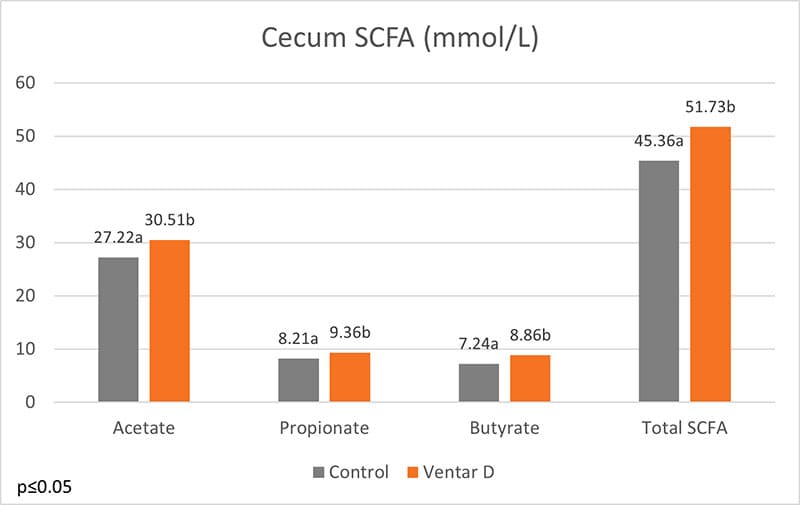
Figure Short Chain Fatty AcidsFigure 1: Short-chain fatty acids in the cecum of broilers

Santoquin countersteers oxidation

Another helpful product category is antioxidants. They can prevent the oxidation of lipids and proteins. For this purpose, EW Nutrition offers Santoquin M6*, a product tested by Kuttapan et al. (2021). Santoquin M6 was tested concerning its ability to minimize the oxidative damage caused by feeding oxidized fat. A control group receiving oxidized fat in feed was compared to one receiving oxidized fat plus 188 ppm Santoquin M6 (≙125 ppm ethoxyquin). The main parameters for this study were TBARS in the breast muscle, the incidence of wooden breast, and the live weight on day 48.

Results indicated that the inclusion of Santoquin M6 reduced the production of TBARS in the breast muscles, demonstrating a lower level of oxidative stress in the breast muscles.
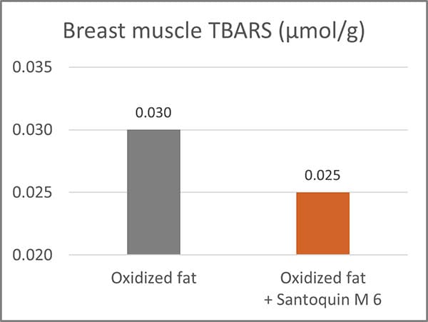
Figure Breast Muscle TBARSFigure 2: Thiobarbituric acid reactive substances (TBARS) in broiler breast muscles. TBARS are formed as a by-product of lipid peroxidation.

Additionally, it reduced the incidence of severe woody breasts (Score 3) by almost half and helped mitigate the impact of breast muscle degradation due to increased oxidative stress.
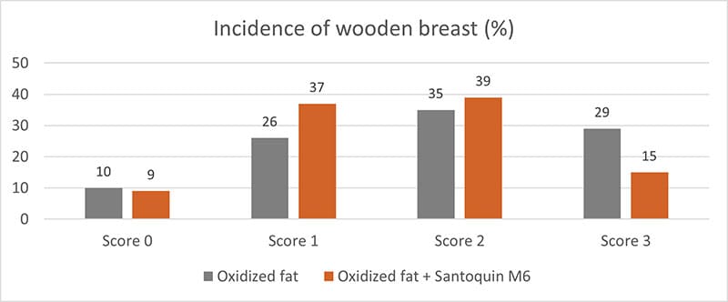
Figure Incidence Of Wooden BreastFigure 3: Incidence of wooden breast in broilers

*Usage of ethoxyquin is dependent on country regulations.

Feed hygiene with Acidomix products minimizes harmful pathogens

The Acidomix product line offers liquid, powdery, and micro-granulated products to be added to feed and water. The organic acids in Acidomix directly act against pathogens in the feed and the water and help keep the intestinal flora in balance.

A trial evaluating the effect of different Acidomix products against diverse pathogens showed lower MICs for most Acidomix products than for single organic acids. The trial was conducted with decreasing concentrations of the Acidomix products (2 – 0.015625 %) and 105 CFU of the respective microorganisms (microtiter plates; 50 µl bacterial solution and 50 µl diluted product).
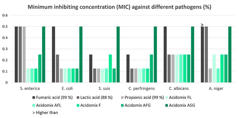
Figure Minimum Inhibiting Concentration
Feeding is the one side, slaughtering the other one

With feeding, the microbiota and some meat characteristics can be changed; however, the last step, handling the animals before and the meat after slaughtering also significantly contributes to a good quality of meat. Stress due to the transport and the slaughterhouse atmosphere, combined with stress-sensible breeds, can lead to PSE meat. Incorrect handling at the slaughterhouse can lead to meat contaminated with pathogens.

Combining feeding measures with professional and calm handling of the animals is the best strategy to achieve high-quality meat.


References

Aaslyng, Margit D., and Lene Meinert. “Meat Flavour in Pork and Beef – from Animal to Meal.” Meat Science 132 (2017): 112–17. https://doi.org/10.1016/j.meatsci.2017.04.012.

Alfaig, Ebrahim, Maria Angelovičova, Martin Kral, and Ondrej Bučko. “EFF ECT of Probiotics and Thyme Essential Oil on the Essential Amino Acid Content of the Broiler Chicken Meat.” Acta Scientiarum Polonorum Technologia Alimentaria 13, no. 4 (2014): 425–32. https://doi.org/10.17306/j.afs.2014.4.9.

Allen, CD, SM Russell, and DL Fletcher. “The Relationship of Broiler Breast Meat Color and Ph to Shelf-Life and Odor Development.” Poultry Science 76, no. 7 (1997): 1042–46. https://doi.org/10.1093/ps/76.7.1042.

Balasubramanian, Balamuralikrishnan, Sang In Lee, and In-Ho Kim. “Inclusion of Dietary Multi-Species Probiotic on Growth Performance, Nutrient Digestibility, Meat Quality Traits, Faecal Microbiota, and Diarrhoea Score in Growing–Finishing Pigs.” Italian Journal of Animal Science 17, no. 1 (2017): 100–106. https://doi.org/10.1080/1828051x.2017.1340097.

Berri, Céile. “Variability of Sensory and Processing Qualities of Poultry Meat.” World’s Poultry Science Journal 56, no. 3 (2000): 209–24. https://doi.org/10.1079/wps20000016.

Cabling, M. M., H. S. Kang, B. M. Lopez, M. Jang, H. S. Kim, K. C. Nam, J. G. Choi, and K. S. Seo. “Estimation of Genetic Associations between Production and Meat Quality Traits in Duroc Pigs.” Asian-Australasian Journal of Animal Sciences 28, no. 8 (2015): 1061–65. https://doi.org/10.5713/ajas.14.0783.

Chen, Binlong, Diyan Li, Dong Leng, Hua Kui, Xue Bai, and Tao Wang. “Gut Microbiota and Meat Quality.” Frontiers in Microbiology 13 (2022). https://doi.org/10.3389/fmicb.2022.951726.

Chen, J.L., G.P. Zhao, M.Q. Zheng, J. Wen, and N. Yang. “Estimation of Genetic Parameters for Contents of Intramuscular Fat and Inosine-5′-Monophosphate and Carcass Traits in Chinese Beijing-You Chickens.” Poultry Science 87, no. 6 (2008): 1098–1104. https://doi.org/10.3382/ps.2007-00504.

Ding, Rongrong, Ming Yang, Jianping Quan, Shaoyun Li, Zhanwei Zhuang, Shenping Zhou, Enqin Zheng, et al. “Single-Locus and Multi-Locus Genome-Wide Association Studies for Intramuscular Fat in Duroc Pigs.” Frontiers in Genetics 10 (2019). https://doi.org/10.3389/fgene.2019.00619.

Dinh, Thu T., K. Virellia To, and M. Wes Schilling. “Fatty Acid Composition of Meat Animals as Flavor Precursors.” Meat and Muscle Biology 5, no. 1 (2021). https://doi.org/10.22175/mmb.12251.

Džinić, N., N. Puvača, T. Tasić, P. Ikonić, and Okanović. “How Meat Quality and Sensory Perception Is Influenced by Feeding Poultry Plant Extracts.” World’s Poultry Science Journal 71, no. 4 (2015): 673–82. https://doi.org/10.1017/s0043933915002378.

Džinić, N., N. Puvača, T. Tasić, P. Ikonić, and Okanović. “How Meat Quality and Sensory Perception Is Influenced by Feeding Poultry Plant Extracts.” World’s Poultry Science Journal 71, no. 4 (2015): 673–82. https://doi.org/10.1017/s0043933915002378.

Fu, Guilin, Yuxuan Zhou, Yupu Song, Chang Liu, Manjie Hu, Qiuyu Xie, Jingbo Wang, et al. “The Effect of Combined Dietary Supplementation of Herbal Additives on Carcass Traits, Meat Quality, Immunity and Cecal Microbiota Composition in Hungarian White Geese (v0.2)”.” Peer J.; 11:e15316, May 8, 2023. https://doi.org/10.7287/peerj.15316v0.2/reviews/3.

Fu, Guilin, Yuxuan Zhou, Yupu Song, Chang Liu, Manjie Hu, Qiuyu Xie, Jingbo Wang, et al. “The Effect of Combined Dietary Supplementation of Herbal Additives on Carcass Traits, Meat Quality, Immunity and Cecal Microbiota Composition in Hungarian White Geese.” PeerJ 11 (2023). https://doi.org/10.7717/peerj.15316.

Goodrich, Julia K., Jillian L. Waters, Angela C. Poole, Jessica L. Sutter, Omry Koren, Ran Blekhman, Michelle Beaumont, et al. “Human Genetics Shape the Gut Microbiome.” Cell 159, no. 4 (2014): 789–99. https://doi.org/10.1016/j.cell.2014.09.053.

Hazrati, S., V. Rezaeipour, and S. Asadzadeh. “Effects of Phytogenic Feed Additives, Probiotic and Mannan-Oligosaccharides on Performance, Blood Metabolites, Meat Quality, Intestinal Morphology, and Microbial Population of Japanese Quail.” British Poultry Science 61, no. 2 (2019): 132–39. https://doi.org/10.1080/00071668.2019.1686122.

He, Linjuan, Jianxin Guo, Yubo Wang, Lu Wang, Doudou Xu, Enfa Yan, Xin Zhang, and Jingdong Yin. “Effects of Dietary Yeast β-Glucan Supplementation on Meat Quality, Antioxidant Capacity and Gut Microbiota of Finishing Pigs.” Antioxidants 11, no. 7 (2022): 1340. https://doi.org/10.3390/antiox11071340.

Heinzl, Inge. “Efficient Microbiome Modulation with Phytomolecules.” EW Nutrition, July 6, 2022. https://ew-nutrition.com/pushing-microbiome-in-right-direction-phytomolecules/.

Huang, Zengwen, Juan Zhang, Yaling Gu, Zhengyun Cai, Dawei Wei, Xiaofang Feng, and Chaoyun Yang. “Analysis of the Molecular Mechanism of Inosine Monophosphate Deposition in Jingyuan Chicken Muscles Using a Proteomic Approach.” Poultry Science 101, no. 4 (2022): 101741. https://doi.org/10.1016/j.psj.2022.101741.

Jiao, Anran, Hui Diao, Bing Yu, Jun He, Jie Yu, Ping Zheng, Yuheng Luo, et al. “Infusion of Short Chain Fatty Acids in the Ileum Improves the Carcass Traits, Meat Quality and Lipid Metabolism of Growing Pigs.” Animal Nutrition 7, no. 1 (2021): 94–100. https://doi.org/10.1016/j.aninu.2020.05.009.

Kallus, Samuel J., and Lawrence J. Brandt. “The Intestinal Microbiota and Obesity.” Journal of Clinical Gastroenterology 46, no. 1 (2012): 16–24. https://doi.org/10.1097/mcg.0b013e31823711fd.

Khan, Muhammad Issa, Cheorun Jo, and Muhammad Rizwan Tariq. “Meat Flavor Precursors and Factors Influencing Flavor Precursors—a Systematic Review.” Meat Science 110 (2015): 278–84. https://doi.org/10.1016/j.meatsci.2015.08.002.

Kroemer, Guido, and Laurence Zitvogel. “Inosine: Novel Microbiota-Derived Immunostimulatory Metabolite.” Cell Research 30, no. 11 (2020): 942–43. https://doi.org/10.1038/s41422-020-00417-1.

Kuttappan, Vivek A., Megharaja Manangi, Matthew Bekker, Juxing Chen, and Mercedes Vazquez-Anon. “Nutritional Intervention Strategies Using Dietary Antioxidants and Organic Trace Minerals to Reduce the Incidence of Wooden Breast and Other Carcass Quality Defects in Broiler Birds.” Frontiers in Physiology 12 (2021). https://doi.org/10.3389/fphys.2021.663409

Lei, Jiaqi, Yuanyang Dong, Qihang Hou, Yang He, Yujiao Lai, Chaoyong Liao, Yoichiro Kawamura, Junyou Li, and Bingkun Zhang. “Intestinal Microbiota Regulate Certain Meat Quality Parameters in Chicken.” Frontiers in Nutrition 9 (2022). https://doi.org/10.3389/fnut.2022.747705.

Liu, R., M. Zheng, J. Wang, H. Cui, Q. Li, J. Liu, G. Zhao, and J. Wen. “Effects of Genomic Selection for Intramuscular Fat Content in Breast Muscle in Chinese Local Chickens.” Animal Genetics 50, no. 1 (2018): 87–91. https://doi.org/10.1111/age.12744.

Lo, L. L., D. G. McLaren, F. K. McKeith, R. L. Fernando, and J. Novakofski. “Genetic Analyses of Growth, Real-Time Ultrasound, Carcass, and Pork Quality Traits in Duroc and Landrace Pigs: II. Heritabilities and Correlations.” Journal of Animal Science 70, no. 8 (1992): 2387–96. https://doi.org/10.2527/1992.7082387x.

Luna, A., M.C. Lábaque, J.A. Zygadlo, and R.H. Marin. “Effects of Thymol and Carvacrol Feed Supplementation on Lipid Oxidation in Broiler Meat.” Poultry Science 89, no. 2 (2010): 366–70. https://doi.org/10.3382/ps.2009-00130.

Ma, Cui, Md. Abul Azad, Wu Tang, Qian Zhu, Wei Wang, Qiankun Gao, and Xiangfeng Kong. “Maternal Probiotics Supplementation Improves Immune and Antioxidant Function in Suckling Piglets via Modifying Gut Microbiota.” Journal of Applied Microbiology 133, no. 2 (2022): 515–28. https://doi.org/10.1111/jam.15572.

Ma, Jianfeng, Jingyun Chen, Mailin Gan, Lei Chen, Ye Zhao, Yan Zhu, Lili Niu, Shunhua Zhang, Li Zhu, and Linyuan Shen. “Gut Microbiota Composition and Diversity in Different Commercial Swine Breeds in Early and Finishing Growth Stages.” Animals 12, no. 13 (2022): 1607. https://doi.org/10.3390/ani12131607.

MacLeod, G. “The Flavour of Beef.” Essay. In Shahidi, F. (Eds) Flavor of Meat and Meat Products, 4–37. Boston, MA: Springer, 1994.

Mottram, Donald. “Flavour Formation in Meat and Meat Products: A Review.” Food Chemistry 62, no. 4 (1998): 415–24. https://doi.org/10.1016/s0308-8146(98)00076-4.

Okruszek, A. “Fatty Acid Composition of Muscle and Adipose Tissue of Indigenous Polish Geese Breeds.” Archives Animal Breeding 55, no. 3 (2012): 294–302. https://doi.org/10.5194/aab-55-294-2012.

Pereira, Ana Lúcia F., and Virgínia Kelly G. Abreu. “Lipid Peroxidation in Meat and Meat Products.” Essay. In Lipid Peroxidation Research. London: IntechOpen, 2020.

Puvača, Nikola, Tatjana Peulić, Predrag Ikonić, Sanja Popović, Jasmina Lazarević, Olivera Đuragić, Magdalena Cara, and Nedeljka Nikolova. “Effects of Medicinal Plants in Broiler Chicken Nutrition on  Selected Parameters of Meat Quality.” Macedonian Journal of Animal Science 9, no. 2 (2019): 45–51. https://doi.org/10.54865/mjas1992045p.

Richards, J. D., J. Gong, and C. F. de Lange. “The Gastrointestinal Microbiota and Its Role in Monogastric Nutrition and Health with an Emphasis on Pigs: Current Understanding, Possible Modulations, and New Technologies for Ecological Studies.” Canadian Journal of Animal Science 85, no. 4 (2005): 421–35. https://doi.org/10.4141/a05-049.

Serra, Valentina, Giancarlo Salvatori, and Grazia Pastorelli. “Dietary Polyphenol Supplementation in Food Producing Animals: Effects on the Quality of Derived Products.” Animals 11, no. 2 (2021): 401. https://doi.org/10.3390/ani11020401.

Stewart, S.M., G.E. Gardner, P. McGilchrist, D.W. Pethick, R. Polkinghorne, J.M. Thompson, and G. Tarr. “Prediction of Consumer Palatability in Beef Using Visual Marbling Scores and Chemical Intramuscular Fat Percentage.” Meat Science 181 (2021): 108322. https://doi.org/10.1016/j.meatsci.2020.108322.

Sun, Jing, Yan Wang, Nianzhen Li, Hang Zhong, Hengyong Xu, Qing Zhu, and Yiping Liu. “Comparative Analysis of the Gut Microbial Composition and Meat Flavor of Two Chicken Breeds in Different Rearing Patterns.” BioMed Research International 2018 (2018): 1–13. https://doi.org/10.1155/2018/4343196.

Thorslund, Cecilie A.H., Peter Sandøe, Margit Dall Aaslyng, and Jesper Lassen. “A Good Taste in the Meat, a Good Taste in the Mouth – Animal Welfare as an Aspect of Pork Quality in Three European Countries.” Livestock Science 193 (2016): 58–65. https://doi.org/10.1016/j.livsci.2016.09.007.

Vasquez, Robie, Ju Kyoung Oh, Ji Hoon Song, and Dae-Kyung Kang. “Gut Microbiome-Produced Metabolites in Pigs: A Review on Their Biological Functions and the Influence of Probiotics.” Journal of Animal Science and Technology 64, no. 4 (2022): 671–95. https://doi.org/10.5187/jast.2022.e58.

Wang, Cheng, Siyu Wei, Bojing Liu, Fengqin Wang, Zeqing Lu, Mingliang Jin, and Yizhen Wang. “Maternal Consumption of a Fermented Diet Protects Offspring against Intestinal Inflammation by Regulating the Gut Microbiota.” Gut Microbes 14, no. 1 (2022). https://doi.org/10.1080/19490976.2022.2057779.

Wen, Chaoliang, Qinli Gou, Shuang Gu, Qiang Huang, Congjiao Sun, Jiangxia Zheng, and Ning Yang. “The Cecal Ecosystem Is a Great Contributor to Intramuscular Fat Deposition in Broilers.” Poultry Science 102, no. 4 (2023): 102568. https://doi.org/10.1016/j.psj.2023.102568.

Werkhoff, Peter, Juergen Bruening, Roland Emberger, Matthias Guentert, Manfred Koepsel, Walter Kuhn, and Horst Surburg. “Isolation and Characterization of Volatile Sulfur-Containing Meat Flavor Components in Model Systems.” Journal of Agricultural and Food Chemistry 38, no. 3 (1990): 777–91. https://doi.org/10.1021/jf00093a041.

Wu, Choufei, Wentao Lyu, Qihua Hong, Xiaojun Zhang, Hua Yang, and Yingping Xiao. “Gut Microbiota Influence Lipid Metabolism of Skeletal Muscle in Pigs.” Frontiers in Nutrition 8 (2021). https://doi.org/10.3389/fnut.2021.675445.

Xiao, Yingping, Kaifeng Li, Yun Xiang, Weidong Zhou, Guohong Gui, and Hua Yang. “The Fecal Microbiota Composition of Boar Duroc, Yorkshire, Landrace and Hampshire Pigs.” Asian-Australasian Journal of Animal Sciences 30, no. 10 (2017): 1456–63. https://doi.org/10.5713/ajas.16.0746.

Yu, Qifang, Chengkun Fang, Yujing Ma, Shaoping He, Kolapo Matthew Ajuwon, and Jianhua He. “Dietary Resveratrol Supplement Improves Carcass Traits and Meat Quality of Pekin Ducks.” Poultry Science 100, no. 3 (2021): 100802. https://doi.org/10.1016/j.psj.2020.10.056.

Zhao, Guangmin, Yun Xiang, Xiaoli Wang, Bing Dai, Xiaojun Zhang, Lingyan Ma, Hua Yang, and Wentao Lyu. “Exploring the Possible Link between the Gut Microbiome and Fat Deposition in Pigs.” Oxidative Medicine and Cellular Longevity 2022 (2022): 1–13. https://doi.org/10.1155/2022/1098892.

Zhou, Xueyan, Xiaosong Jiang, Chaowu Yang, Bingcun Ma, Changwei Lei, Changwen Xu, Anyun Zhang, et al. “Cecal Microbiota of Tibetan Chickens from Five Geographic Regions Were Determined by 16s Rrna Sequencing.” MicrobiologyOpen 5, no. 5 (2016): 753–62. https://doi.org/10.1002/mbo3.367.

Zou, Xiaozhuo, Rong Xiao, Huali Li, Ting Liu, Yong Liao, Yuanliang Wang, Shusong Wu, and Zongjun Li. “Effect of a Novel Strain of Lactobacillus Brevis M8 and Tea Polyphenol Diets on Performance, Meat Quality and Intestinal Microbiota in Broilers.” Italian Journal of Animal Science 17, no. 2 (2017): 396–407. https://doi.org/10.1080/1828051x.2017.1365260.