The big challenge: Keeping sows healthy and productive – Part 1 General aspects to be observed

HEADER DSC

Dr. Inge Heinzl – Editor of EW Nutrition and
Dr. Merideth Parke – Global Application Manager for Swine, EW Nutrition

Sow mortality critically impacts herd performance and efficiency in modern pig production. Keeping the sows healthy is, therefore, the best strategy to keep them alive and productive and the farm’s profitability high.

Rising mortality rates are alarming

In recent years, sow mortality has increased across pig-raising regions in many countries. Eckberg’s (2022) findings from the MetaFarms Ag Platform (including farms across the United States, Canada, Australia, and the Philippines) determined an increase of 66.2% between 2012 and 2021.

Sow mortality rates from 2012 to 2021
Figure 1: Sow mortality rates from 2012 to 2021 (Eckberg, 2022)

What can be done to decrease mortality rates?

Several measures can be taken to reach a particular stock of healthy and high-performing sows. In the following, the main remedial actions will be explained.

1. Determination of the cause of death

If a sow is dead, it must first be clarified why it has died. If the sow is culled, the reason for this decision is usually apparent. If the sow suddenly dies, investigations, including a thorough postmortem, are extremely valuable to determine the cause of death. Kikuti et al. (2022) provided a collection of the most-occurring causes of death in the years 2009 to 2018. As often, no necropsy is conducted, and the causes of death remain unclear, as shown by the high numbers of “other”. Locomotory (e.g., lameness) and reproductive (e.g., prolapse, endotoxemic shock from retained fetuses) incidents account for approximately half of the recorded sow mortalities (Kikuti et al., 2022), especially during the first three parities. (Marco, 2024).

Removal reasons and their frequency from 2009 to 2018
Figure 2: Removal reasons and their frequency from 2009 to 2018 (Kikuti et al., 2022)

Evaluating detailed breeding history together with the cause of death will provide perspective and assist veterinary, nutritionist, and husbandry teams with interventions to prevent similar events and early sow mortality.

Selection of the gilts

After selecting the best genetics and rearing the gilts under the best conditions, further selection must focus on physical traits such as structure, weight, height, leg, and hoof integrity.

Additionally, as we have more and more group housing for sows, the selection for stress resilience can positively impact piglet performance (Luttmann and Ernst, 2024). The following table compares stress-resilient and stress-vulnerable sows concerning piglet performance and shows the piglets of the vulnerable sows with worse performance.

Table 1: Influence of stress resilience on performance (Luttmann and Ernst, 2024)

Trait SR SV p-Value
Birth weight (kg) 1.350 ± 0.039 1.246 ± 0.041 0.083
Wean weight(kg) 6.299 ± 0.185 5.639 ± 0.202 0.033*
Suckling ADG (kg/d) 0.191 ± 0.005 0.165 ± 0.005 0.004**

Least square means and standard error of stress resilient (SR) and stress vulnerable (SV) for each trait; significance threshold of p<0.05 with * indicating 0.01<p<0.05, ** indicating 0.001<p<0.01

How to manage the gilts best

The management of the gilts must consider the following:

  1. Age at first estrus should be <195 days:
    Gilts having their first estrus earlier show higher daily gain and usually higher lifetime productivity. In a study conducted by Roongsitthichai et al. (2013), sows culled at parity 0 or 1 exhibited first estrus at 204.4±0.7 days of age, while those culled at parity ≥5 exhibited first estrus at 198.9±2.1 days of age (P=0.015).
  2. Age at first breeding should lay between 200 and 225 days:
    If the sows are bred at a higher age, they have the risk of being overweight, leading to smaller second-parity litters, longer wean-to-service intervals, and shorter production life.
  3. The body weight at first mating should be between 135 and 160 kg:
    To reach this target within 200-225 days, the gilts must have 600-800 g of average daily gain. Breeding underweight gilts reduces first-litter size and lactation performance. Overweight gilts (>160 kg) face higher maintenance costs and locomotion issues.
  4. The number of estruses at first mating should be 2 or 3:
    Accurately track estrus and breed on the second estrus. Research shows that delaying breeding to the second estrus positively affects litter size. Only delay breeding to the third estrus to meet minimum weight targets.
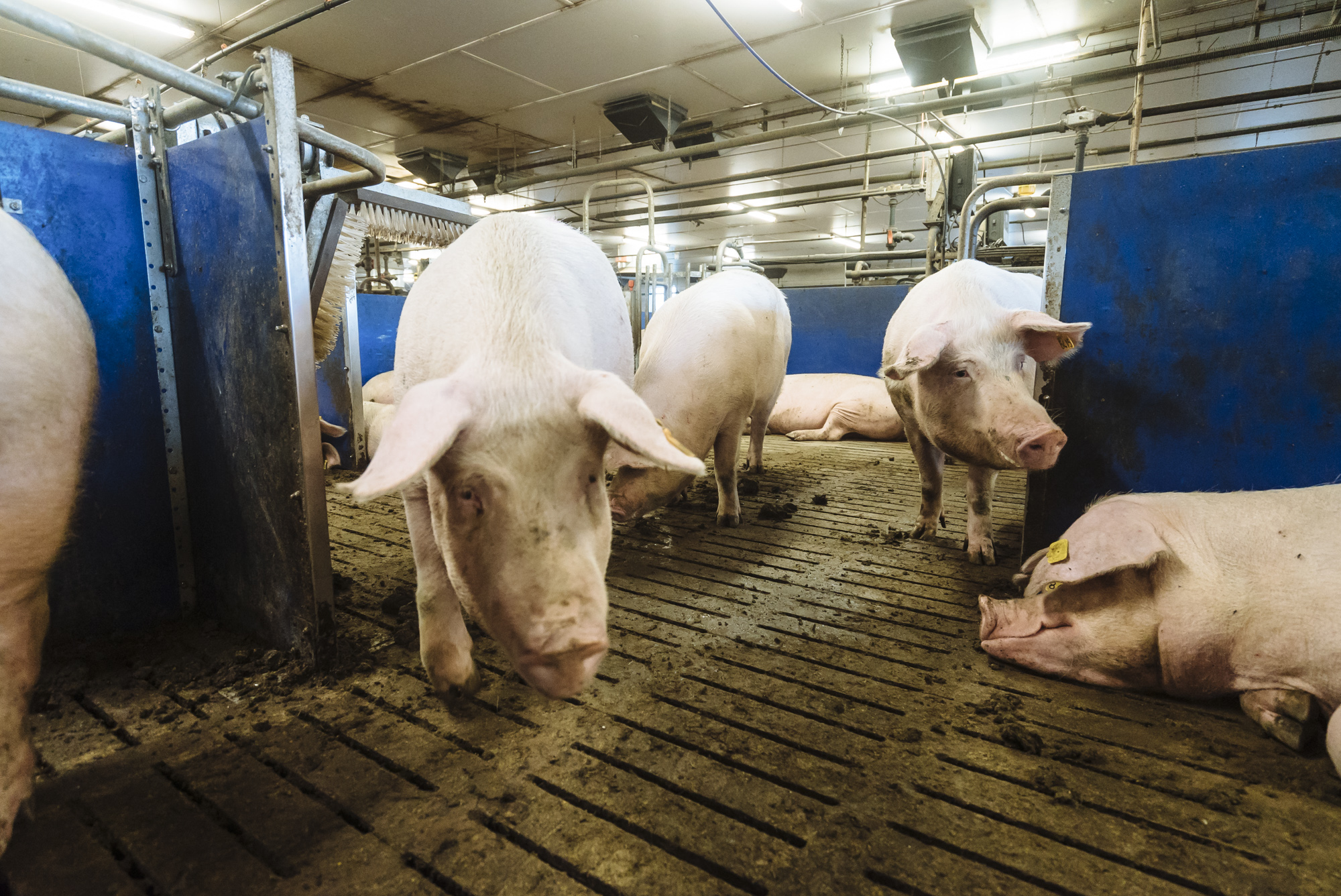
Housing

Gestating sows are more and more held in groups. Understanding the process of group housing is essential for success. The following graphic shows factors impacting successful grouping.

 Factors influencing group housing
Figure 3: Factors influencing group housing

If the groups are not well-established yet, the stress levels among sows are higher, leading to

  • More leg injuries due to aggressive behavior or fighting for resources
  • Higher rates of abortions and returns to service
  • Reduced sow performance, including decreased productivity, lower milk yield, and poor piglet growth due to compromised immune function and overall health

To mitigate stress in group housing, it is crucial to implement proper group management practices, which include gradual introductions, maintaining stable social structures, and ensuring adequate space and resources. This helps promote a calmer environment, improving animal welfare and herd performance.

Responsible on-farm pig care

Caregivers must be well-trained and equipped to provide high-quality care. Insufficient or unskilled pig caregivers can significantly affect the growth and development of prospective replacement gilts, ultimately influencing their suitability for the breeding herd:

  • Growth Rates: Suboptimal nutrition and health management result in slower growth rates and poor body condition.
  • Health Issues: Unskilled handling may increase the risk of disease transmission, injuries, and stress, all of which can adversely affect growth and overall development.
  • Behavioral Problems: Poorly managed environments can increase aggression and competition among animals, hindering growth and health.
  • Selection Criteria: Ineffective growth and health monitoring can result in misjudging the potential of gilts, leading to the selection of less suitable candidates for the breeding herd.

Table 2: Influence of handling on growth performance and corticosteroid concentration of female grower pigs from 7-13 weeks of age (Hemsworth et al., 1987)

Unpleasant Pleasant Inconsistent Minimal
ADG (g) 404a 455b 420ab 4.58b
FCR (F:G) 2.62b 2.46a 2.56b 2.42a
Corticosteroid conc (ng/mL) 2.5a 1.6b 2.6a 1.7b

Responsible on-farm pig care is crucial to keep sows healthy and performing. Poor sow observations (e.g., failure to identify stressed, anorexic, or heat-stressed sows) or inappropriate farrowing interventions can directly influence sow health and potentially reduce subsequent performance or mortality. On the contrary, rapid and proactive identification of sows needing intervention can save many animals that would otherwise die or need to be culled.

Keeping sows healthy and performing is manageable

The maintenance of sows’ health is a challenge but manageable. Observing all the points mentioned, from selecting the right genetics over rearing the piglets under the best conditions to managing the young gilts, can help prevent disease and performance drops. For all these tasks, farmers and farm workers who do their jobs responsibly and passionately are needed. The following article will show nutritional interventions supporting the sow’s gut and overall health.

References:

Eckberg, Bradley. “2021 Sow Mortality Analysis.” National Hog Farmer, February 3, 2022. https://www.nationalhogfarmer.com/hog-health/2021-sow-mortality-analysis.

Hemsworth, P.H., J.L. Barnett, and C. Hansen. “The Influence of Inconsistent Handling by Humans on the Behaviour, Growth and Corticosteroids of Young Pigs.” Applied Animal Behaviour Science 17, no. 3–4 (June 1987): 245–52. https://doi.org/10.1016/0168-1591(87)90149-3.

Kikuti, Mariana, Guilherme Milanez Preis, John Deen, Juan Carlos Pinilla, and Cesar A. Corzo. “Sow Mortality in a Pig Production System in the Midwestern USA: Reasons for Removal and Factors Associated with Increased Mortality.” Veterinary Record 192, no. 7 (December 22, 2022). https://doi.org/10.1002/vetr.2539.

Marco, E. “Sow Mortality: How and Who? (1/2).” Pig333.com Professional Pig Community, March 18, 2024. https://www.pig333.com/articles/sow-mortality-how-are-sows-dying-which-sows-are-dying_20105/.

Luttmann, A. M., and C. W. Ernst. “Classifying Maternal Resilience for Improved Sow Welfare, Offspring Performance.” National Hog Farmer, September 2024. https://informamarkets.turtl.co/story/national-hog-farmer-septemberoctober-2024/page/5.

Roongsitthichai, A., P. Cheuchuchart, S. Chatwijitkul, O. Chantarothai, and P. Tummaruk. “Influence of Age at First Estrus, Body Weight, and Average Daily Gain of Replacement Gilts on Their Subsequent Reproductive Performance as Sows.” Livestock Science 151, no. 2–3 (February 2013): 238–45. https://doi.org/10.1016/j.livsci.2012.11.004.


Nutritional strategies to maximize the health and productivity of sows

Muttersau Mit Ferkel

Conference Report

During lactation, the focus should be on maximizing milk production to promote litter growth while reducing weight loss of the sow, stated Dr. Jan Fledderus during the recent EW Nutrition Swine Academies in Ho Chi Minh City and Bangkok. A high body weight loss during lactation negatively affects the sow’s performance in the next cycle and impairs her longevity.

Milk production – ‘push’ or ‘pull’?

“Is a sow’s milk production driven by “push” – the sow is primarily responsible for milk production, or “pull” – suckling stimulates the sow to produce milk?” asked Dr. Jan Fledderus at the beginning of his presentation. The answer is that it is primarily a pull mechanism: piglets that suckle effectively and frequently can activate all compartments of the udder, leading to increased milk production. Therefore, the focus should be optimizing piglet suckling behavior (pull) to enhance milk production. This highlights the importance of piglet vitality and access to the udder in maximizing milk yield.”

Modern sows are lean

Modern sows have been genetically selected for increased growth rates and leanness, which alters their body composition. This makes traditional body condition scoring (BCS) methods, which rely on subjective visual assessment and palpation of backfat thickness, less effective. This may not accurately represent a sow’s true condition, especially in leaner breeds where muscle mass is more prominent than fat. Technology, such as ultrasound measurements of backfat and loin muscle depth, provide more accurate assessments of body condition and can help quantify metabolic reserves more effectively than visual scoring.

Due to their increased lean body mass, we must consider adjusted requirements for amino acids, energy, digestible phosphorus, and calcium. Their dietary crude protein and amino acid requirements have increased drastically.

Importance of high feed intake for milk production

Sows typically catabolize body fat and protein to meet the demands of milk production. Adequate feed intake helps reduce this catabolism, allowing sows to maintain body condition while supporting their piglets’ nutritional needs.

Feeding about 2.5kg on the day of farrowing ensures that sows receive adequate energy to support the farrowing process and subsequent milk production. Sows that are well-fed before farrowing tend to have shorter farrowing durations due to better energy availability during labor.

A short interval between the last feed and the onset of farrowing (3 hours) has been shown to significantly reduce the probability of both assisted farrowing and stillbirths without increasing the risk of constipation. To enhance total feed intake, feeding lactating sows at least three times a day is helpful.

Dr. Fledderus recommended a gradual increase in feed intake during lactation, then from day 12 of lactation to weaning, feeding 1% of sow’s bodyweight at farrowing + 0.5 kg/piglet. For example, for a 220kg sow with 12 piglets:

(220 kg x 0.01) + (12 x 0.5 kg) = 2.2 +6 = 8.2 kg total daily feed intake

Energy source – starch versus fat

The choice between starch and fat as an energy source in sow diets has substantial implications for body composition and milk production.

Starch digestion leads to glucose release, stimulating insulin secretion from the pancreas. Insulin is essential for glucose uptake and utilization by tissues. Enhanced insulin response can help manage body weight loss by promoting nutrient storage and reducing the mobilization of the sow’s body reserves.

Sows fed diets with a higher fat supplementation had an increased milk fat, which is crucial for the growth and development of piglets.

Table 1: Effect of energy source (starch vs. fat) on sows’ body composition and milk yield (Schothorst Feed Research)

.ewn-art-241-table-energy-source, .ewn-art-241-table-energy-source th, .ewn-art-241-table-energy-source td {border: 1px solid black;} .ewn-art-241-table-energy-source th {background-color: var(--color-content-primary);} .ewn-art-241-table-energy-source tr:nth-child(odd) {background-color: #ecedaf;)}
Diet 1 Diet 2 Diet 3
Energy value (kcal/kg) 2,290 2,290 2,290
Starch (g/kg) 300 340 380
Fat (g/kg) 80 68 55
Feed intake (kg/day) 6.7 6.7 6.8
Weight loss (kg) 15 11 10
Weight loss (kg) 3.1 2.7 2.3
Milk fat (%) 7.5 7.2 7.0
Milk fat (%) 260 280 270

Heat stress impacts performance

The optimum temperature for lactating sows is 18oC. A meta-analysis concluded that each 1°C above the thermal comfort range (from 15° to 25°C) leads to a decrease in sows’ feed intake and milk production and weaning weight of piglets, as shown below.

Effect Of Heat Stress
Effect of heat stress on lactating sows (according to Ribeiro et. al., 2018 Based on 2,222 lactating sows, the duration of lactation was corrected to 21 days)

To mitigate the effects of heat stress, which reduces feed intake, it is beneficial to schedule feeding during cooler times of the day. This strategy helps maintain appetite and ensures that sows consume sufficient nutrients for milk production. Continuous access to cool, clean water can also enhance feed consumption.

Pigs produce much heat, which must be “excreted”. Increased respiratory rate (>50 breaths/minute) has been shown to be an efficient parameter for evaluating the intensity of heat stress in lactating sows.

When sows resort to panting as a mechanism to dissipate heat, this rapid breathing increases the loss of carbon dioxide, resulting in respiratory alkalosis. To prevent a rise in blood pH level, HCO3 is excreted via urine, and positively charged minerals (calcium, phosphorous, magnesium, and potassium) are needed to facilitate this excretion. However, these minerals are crucial for various physiological functions. As their loss can lead to deficiencies that affect overall health and productivity, this mineral loss must be compensated for.

Implications for management

Implementing effective nutritional strategies together with robust management practices is crucial for maximizing the health and productivity of sows. The priority is to stimulate the sow to eat more. This not only enhances milk production and litter growth but also supports the overall well-being of the sow. Regularly assessing sow performance metrics – such as body condition score, feed intake, and litter growth – can help identify areas for improvement in nutritional management.

EW Nutrition’s Swine Academy took place in Ho Chi Minh City and Bangkok in October 2024. Dr. Jan Fledderus, Product Manager and Consultant at the S&C team at Schothorst Feed Research, with a strong focus on continuously improving the price/quality ratio of the diets for a competitive pig sector and one of the founders of the Advanced Feed Package, was a reputable guest speaker in these events.


Rearing pigs without antibiotics

HIGH Shutterstock Header

Conference Report

Holistic management is essential for successfully rearing pigs, particularly in systems that aim to minimize antibiotics. The method emphasizes the interconnectedness of various factors contributing to sustainable pig health and productivity. Some of the key components of this holistic management were discussed by Dr. Seksom.

Sow lifetime productivity

Suggested targets for sow lifetime productivity are

  • >70 marketed fattening pigs
  • At least 6 parities with at least 10.5 pigs marketed per parity
  • 25 fattening pigs/sow/year (2.4 parities/year x 10.5 fattening pigs)

To achieve these targets, we need 29.2 born alive piglets/sow/year (or 12.2 born alive piglets/parity), and it is essential to control losses during each production period: <10% pre-weaning, <3% during nursery, and <2% in fattening.

Since the occurrence of African swine fever (ASF), with improved genetics, we can now produce pigs with 120 kg+ bodyweight at slaughter without carcass problems and reach about 3 tons of bodyweight/sow/year, compared to around 2 tons before.

Modern pig genetics and subsequent problems

Despite the advancements in modern pig genetics leading to improved production and bigger litters, several ensuing problems have emerged:

  • Less average body weight of piglets at birth
  • Large number of piglets born with less than 1.0 kg (target <5%)
  • High pre-weaning mortality
  • High post-weaning mortality and morbidity

Dr. Seksom highlighted that birthweights decrease with increasing sow prolificacy. He stated that “piglets should be divided into groups with similar body weights at weaning” and that “a key objective for successful weaning is a piglet that weighs a minimum of 6-6.5 kg at three weeks of age, and that less than 25% of the piglets have a weight of ≤5.9 kg.”

Sow body condition

Sows should be fed to feed to body condition score (BCS), not a fixed amount of feed. Ideally, the sows have a BCS of 2.75 (the sow’s backbone is visible, and the tips of the short ribs can be felt but are smooth) or 3.0 (well-rounded appearance, hips, and spine can only be felt with firm pressure) at 12 weeks of pregnancy, so we can feed more in the last month to achieve a BCS of 3-3.25 at farrowing. This is essential to ensure that sows have sufficient energy reserves for lactation and overall health.

Target body condition score – 2.75 at three months of gestation

Screenshot


Feed intake must be increased gradually during the last month of gestation as most fetal growth and mammary gland development occur during this period. This may involve raising energy-dense feeds or adjusting protein levels as needed.

Dr. Seksom stressed that “nutrition is not just the feed; it’s about feeding as well. To feed sows to BCS, assessments of BCS should be done regularly throughout gestation, ideally every 2-4 weeks. This allows for timely adjustments in feeding based on individual sow’s needs. Ensure that staff are trained one-on-one to accurately assess the body condition of sows. This includes recognizing the visual and tactile indicators of different scores and understanding how BCS impacts reproductive performance, longevity, and overall farm profitability.”

After farrowing, the sows must be monitored closely for any signs of excessive weight loss and feeding strategies adjusted accordingly to support recovery and lactation needs.

Piglet diarrhea

Many factors cause diarrhea and must be thoroughly investigated. For bacteria-caused diarrhea, Dr. Seksom advised a good hygiene program, whereas for viral causes, a vaccination program is essential. However, he emphasized that “for a vaccination program, you can’t just copy from another farm; it needs to be created specifically using the titers for diseases on your farm.”

Swine influenza is an often-overlooked cause of diarrhea in piglets. While it is primarily recognized for causing respiratory issues, the virus can also lead to scours in the first two weeks of piglets’ life. So, sows should be checked for symptoms of swine influenza (such as nasal discharge, sneezing and coughing, and inappetence) before farrowing. If necessary, they must be treated with paracetamol to reduce fever and symptoms.

Main disease causes of pre-weaning diarrhea

.ewn_art_238_table_main_disease_cause {border: 1px solid black !important;}.ewn_art_238_table_main_disease_cause td, .ewn_art_238_table_main_disease_cause th {border: 1px solid black !important;}.ewn_art_238_table_main_disease_cause td.text-center, .ewn_art_238_table_main_disease_cause th.text-center {text-align: center;}.ewn_art_238_table_main_disease_cause th {background-color: var(--color-content-primary);}.ewn_art_238_table_main_disease_cause tr:nth-child(even) {background-color: #ecedaf;}
Nursery period Mortality level
Days 1-3 Days 3-7 Days 7-14 Days 14-21
Agalactia ü ü ü ü Moderate
Clostridia ü ü ü High
Coccidiosis ü ü ü Low
E. coli ü ü ü Moderate
PED ü ü ü Variable
PRRS ü ü ü ü Variable
Rotavirus ü ü Low
TGE ü ü ü ü High
Influenza ü ü Low

Ensuring colostrum intake

The intake of an adequate quantity of colostrum is crucial for piglets to be protected during the first days of life. Best practices to ensure that piglets get 250 mL of colostrum include:

  • Teat access – if a sow has a large litter or is unable to nurse all her piglets effectively, consider split suckling by separating larger, more vigorous piglets from the litter for a couple of hours after birth. This allows smaller or weaker piglets better access to the udder without competition. Syringe-feeding colostrum to smaller piglets is also effective.
  • Early access – six hours after farrowing, the quality of colostrum begins to decline significantly. Additionally, the piglet can only absorb intact large IgG molecules, the major source of passive immunity, during the first 24 h after birth, prior to gut closure. In any case, by this time, the sow will start producing milk and not colostrum.
  • Sow behavior – if a sow experiences pain or discomfort from injuries caused by her piglets’ teeth, she may become less willing to allow them to nurse, leading to delays in colostrum intake. Genetic background influences maternal behavior significantly. For example, some breeds exhibit stronger maternal instincts and better nursing
    behaviors than others. Selecting sows with proven good maternal traits can lead to improved outcomes in piglet survival and growth.
  • Drafts – newborn piglets are born with low fat reserves and are highly susceptible to hypothermia. Drafts significantly impact the effective temperature experienced by piglets.
  • Staff training – Staff must be trained to recognize signs of distress in both sows and piglets; training in techniques enables them to assist with nursing and feeding, which is crucial for timely interventions.

PIGLET Shutterstock Cmyk Smaller

Weaning is a process, not just a one-time event

Research has shown that heavier piglets at weaning have better lifetime performance than lighter ones. Weaning weight is a more accurate indication of post-weaning growth than either birth weight or age. It is, therefore, important to establish the weaner immediately post-weaning to maintain growth rates, reduce pen variation, and lessen the amount of ‘tail-enders’ at the point of sale.

Dr. Seksom emphasized that “viewing weaning merely as a single event, rather than a process, overlooks the complexities involved in ensuring a smooth transition for the animals. He advocated for a comprehensive approach to weaning that includes the shown well-planned steps to support piglets during this critical phase. If the weaning process is managed effectively, you can significantly reduce the need for antibiotics.”

Conclusion

“By integrating these holistic management strategies, pig producers can effectively raise pigs without antibiotics while promoting animal health, improving productivity, and addressing consumer concerns about antibiotic use in livestock production,” summarized Dr. Seksom.

EW Nutrition’s Swine Academy took place in Ho Chi Minh City and Bangkok in October 2024. Dr. Seksom Attamangkune, a leading expert in the nutrition and management of pigs in tropical conditions and former Head of the Animal Science Department and Dean of the Faculty of Agriculture at Kamphaeng Saen, Kasetsart University, was a reputable guest speaker at this event.


Building and boosting the immunity shield of pigs

ART IMAGE

Conference report

A well-functioning immune system is vital for the survival and performance of animals. It helps piglets cope with challenging periods, such as their first days of life or weaning. Measures can be taken around farrowing to support the piglets during their first days by enhancing the quality and quantity of colostrum and helping them develop their own immune system as fast as possible.

Adequate feeding of the sow before and around farrowing

Feeding of both the sow and the piglet has an important influence on farrowing, the health of the sow, colostrum and milk production, piglets’ development of immunity, and their later performance. A well-functioning immune system is crucial for the piglets to withstand upcoming challenges such as weaning.

Colostrum quality can be influenced by feeding

Newborn piglets have no functioning immunity system. They rely entirely on immunoglobulin G (IgG) absorption from colostrum within the first few hours after birth to establish their immunity shield. Dr. Megan Edwards, Animal Nutrition Consultant from Integral Nutrition (S) Pte Ltd, highlighted the payback of adequate colostrum quality and intake: Adequate colostrum intake can positively affect whole-of-life immunity and, ultimately, growth performance. The contained IgG is essential for providing passive immunity to piglets, protecting them from infections during their early days of life when their immune systems are still developing. There is a positive correlation between the amount of IgG they absorb from colostrum and their performance. This benefit of colostrum intake is independent of birth weight.

We have a 3-week window to influence colostrogenesis. However, the fat content of colostrum is determined in the last 48 hours before farrowing. According to Dr. Edwards, influencing colostrum quality is generally easier than affecting quantity. She identified several compounds that can serve as immunomodulators, such as MCFAs, yeast extracts, and butyrate. However, by moving IgG to colostrum and milk in late gestation and lactation, the sow compromises her immunity status by depleting her own reserves for about two weeks.

Feeding at farrowing

Sow body condition has been shown to have more impact on colostrum yield than feeding level. The highest colostrum yield was achieved when sows entered the farrowing unit with a moderate body condition (3-3.25 – the ribs, spine, and hip bones can only be felt with firm pressure but are not visibly prominent). Overfeeding should be avoided to prevent sows from becoming excessively fat pre-farrowing.

Sows experience increased energy demands during farrowing due to the physical demands of parturition and the physiological changes occurring in their bodies. Dr. Edwards does not encourage withholding feed on the day of farrowing and suggests offering up to 3kg if the sow has the appetite. Feeding just below the energy requirement helps the sow to mobilize her own body fat.

Many producers mistakenly withhold feed on the day of the farrow to reduce the incidence of constipation. Feeding, however, stimulates gut motility. Withholding feed can slow down gut transit time and actually increase the likelihood of constipation.

Piglet feeding for developing intestinal tract and immune system

In piglet feeding, two strategies are decisive: the early intake of immunoglobulins via colostrum to protect the piglets against pathogens during their first days of life and the offering of creep feed to stimulate their intestinal development.

High-quality colostrum as much and as soon as possible

When the piglets are born, it is of the highest importance that they ingest colostrum as much and as soon as possible. The piglet can only absorb intact large IgG molecules, the primary source of passive immunity, before gut closure, which begins about 6–12 hours after birth and progresses rapidly to completion in about 24 hours. In any case, the sow will start producing milk by this time and no more colostrum. The concentration of colostrum IgG decreases by 50% within 6 hours after the birth of the first piglet. The target is for piglets to consume 250 g of colostrum within the first 24 hours, ideally within the first 6 hours. However, about 30% of sows produce insufficient colostrum.

Figure 1: Mortality of piglets until 42 days of age
Figure 1: Mortality of piglets until 42 days of age according to intervals of birthweight and colostrum intake
(Hasan et al. 2019; the numbers of piglets are shown in parenthesis)

Split suckling jump-starts weak piglets

Split suckling is an effective management strategy to improve piglets’ access to colostrum and milk, particularly in increasingly common situations where sows give birth to large litters. This involves temporarily separating the more vigorous piglets from the sow to allow smaller or weaker piglets better access to the teats. This method helps ensure that all piglets receive adequate nutrition during the critical early hours after birth.

Large litters provoke energy deficiency in piglets

Piglets are born with limited energy reserves (glycogen and brown fat tissue). Ingestion of colostrum is associated with a considerable increase in the metabolic rate, contributing to maintaining body temperature. About 70% of the piglets’ energy requirement in the first 72 hours is provided by colostrum. “Most piglets that die within this period do so primarily due to energy deficiencies rather than immune-related issues. The trend towards larger litter sizes has exacerbated the issue of energy deficiency,” stated Dr. Edwards.

Creep feeding

The primary role of creep feed is to accelerate the development of the piglets, their digestive and immune systems, and their gut microbiome, not for weight gain. Creep feeding helps evolve digestive enzymes and acid secretion necessary for breaking down complex carbohydrates and proteins. This early feeding supports piglets in adapting to solid diets, mitigating stress during weaning.
Creep feeding also helps piglets develop an oral tolerance to avoid transient hypersensitivity due to various dietary ingredients. This process is essential for preventing allergic reactions and hypersensitivity, which can occur when the immune system mistakenly identifies harmless substances as threats. It takes about two weeks for the piglet to recognize an ingredient as a nutrient, not a pathogen. To facilitate this process, she recommends that creep diets contain a broad range of ingredients at low doses. This approach gradually exposes piglets to various nutrients, allowing their immune systems to adapt without overwhelming them with high concentrations of any single ingredient.

Mycotoxins must be managed – even in piglets

The significance of mycotoxins in piglets is often underestimated due to their relatively small feed intake. However, there is substantial evidence that mycotoxins can be transferred from sows to piglets through colostrum and milk, which can have profound health implications.

Dr. Edwards is convinced that managing mycotoxins is managing immunity. Mycotoxins are transferrable via the placenta, colostrum, and milk. There is a positive correlation between the mycotoxin levels in feed and colostrum. For example, adverse effects seen in piglets consuming colostrum with low doses of deoxynivalenol (DON) include:
• Decreased villus height
• Reduced mucosal integrity
• Increased inflammation
• Alternated immune response
The bottom line is that mycotoxins are a real and everyday risk to the immune quality of your piglets.

Nutrition influences piglets’ immune development

Dr. Edwards summarized that adequate nutrition is fundamental for developing a strong immune system in pigs, which is the basis for high performance. By focusing on the appropriate nutrition of the sow, ensuring an adequate intake of high-quality colostrum intake in piglets, and implementing creep feeding strategies, producers can significantly enhance the lifetime health and productivity of their piglets from an early age.

EW Nutrition’s Swine Academy took place in Ho Chi Minh City and Bangkok in October 2024. Dr. Megan Edwards, an Australian animal nutrition consultant with global research and praxis experience and a keen interest in immuno-nutrition and functional nutrients, was an esteemed guest speaker at this event.


Mycotoxins pose a threat to the horse’s digestive system

Eye Of Arabian Bay Horse

Author: Judith Schmidt, Product Manager On Farm Solutions

Alarm in the gut! Horses have a susceptible digestive system that can quickly become unbalanced. Intestinal disorders in horses are usually associated with colic. Many factors can be responsible for intestinal issues. Have you ever thought about mycotoxins? What can horse owners do to support their horse´s gut health?
The equine stomach is not robust at all. Depending on their age and use, more than half of all horses suffer from stomach pain. Their digestive system is very sensitive and very different from that of other mammals: Horses cannot vomit and often suffer from severe abdominal pain, diarrhea, or cramps if they overeat or ingest spoiled feed.

The horse´s digestive system is complex and sensitive

The horse´s stomach has a relatively small capacity of around twelve to fifteen liters. Depending on the feed’s consistency and composition, it remains in the stomach for around one to five hours before it is pressed through the stomach outlet (pylorus) into the small intestine. The horse´s entire intestine is about ten times its body length.

Figure Digestive TractFigure 1: The horse’s digestive tract

The horse´s gastrointestinal tract is a complex network, reacting extremely sensitively to changes and, therefore, highly susceptible to disorders. It essentially consists of the head intestine (lips, oral cavity, teeth, and esophagus), stomach (blind pouch, fundus, and stomach outlet), small intestine (duodenum, jejunum, and ileum), and large intestine (caecum, colon and rectum). Each section plays a crucial role in the digestive process; any disruption can lead to health issues. Understanding this structure is key to maintaining a horse’s digestive health.

Digestive disorders can have various reasons

Intestinal problems in horses can stem from diverse causes, often a complex interplay of multiple factors. By understanding these causes more deeply, horse owners can be better equipped to prevent and manage these issues. In the following, we delve into several of these causes.

1.   Too long time between the feedings

Usually, a feeding break should be at most four to six hours, as, in nature, a horse is busy eating for at least 18 hours a day. In contrast to humans, who produce stomach acid only after food intake, the horse’s stomach produces gastric acid around the clock. The continuous intake of roughage, intensive chewing, and high saliva production (a horse produces 5 to 10 L of saliva per day) is, therefore, essential to protect the stomach mucosa by neutralizing excess gastric acid.

A too-long time between feedings and, therefore, no saliva production leads to an accumulation of gastric acid in the stomach. Four hours without roughage can already cause inflammation of the mucosa and probably ulcers.

2.   Excessive amounts of concentrated feed

Excessive amounts of concentrates such as wheat or rye, conditioned by less chewing activity, increase gastric acid and histamine production, and the stomach lining can be attacked. Also, in this case, the development of stomach ulcers is possible.

Furthermore, the possibly resulting hyperacidity of the organism can lead to malfunctions of the organs, the skin, and the hooves.

3.   Stress

Stress can also lead to a higher production of gastric acid and, therefore, to gastric ulcers. The horse is a flight animal.  When it is under stress, it prepares for the impending escape, and the muscles are preferably supplied with blood, resulting in a lower blood flow to the mucous membranes. Furthermore, the rising cortisone level reduces the hydrochloric acid-suppressing prostaglandin E. As a result, more stomach acid is produced, irritating the gastric mucosa.

Stress can be triggered, e.g., by transportation, competitions, training, a change of house, a new rider, unsuitable equipment, or poor posture.

4.   Dental diseases

The teeth are essential for digestion. When feed is chewed, it is broken down and mixed with saliva. Chipped teeth cannot chew well, and the feed is not sufficiently salivated or crushed, which has a detrimental effect on digestion.

For this reason, an expert vet should check the horse´s teeth at least once a year.

5.   Administration of painkillers/medication

As with humans, long-term medication administration can promote the formation of stomach ulcers. For this reason, it is essential to ensure that horses are fed a gentle diet on the stomach, especially when using oral pain therapy, and to add stomach protection if necessary.

6. Endotoxins

If pathogens such as E. coli or clostridia proliferate extremely or are killed by an antibiotic, endotoxins can be released. These toxins can cause transformation or inflammation of the gut mucosa. In drastic cases, whole areas of the mucosa can die off. 

7. Mycotoxins – the hidden danger in horse feed

Mycotoxins in plants and horse feed are a common but often unnoticed danger to horses’ health. Mycotoxins are natural, secondary metabolites of molds that have a toxic effect on humans and animals and can trigger mycotoxicosis. Contaminated feed can severely affect the horse’s health and, in the worst case, lead to death.

Over 90 % of the world´s feed production is estimated to be contaminated with at least one mycotoxin (see also Global Mycotoxin Report 2023, EW Nutrition. The intake of mycotoxins via hay, grain, silage, or compound feed can hardly be avoided. Mycotoxins are an increasing problem for all horse owners. Scientific studies show that the mycotoxins DON and ZEA are most frequently found in horse feed and, therefore, are also frequently detected in sports horses’ urine and blood samples.

Due to the highly toxic metabolic products, feed contaminated with molds can lead to severe liver and kidney diseases in horses, affect fertility, trigger colic, and promote digestive issues (diarrhea and watery stools).

Pictures ART
Mycotoxins Horses

Figure 2: Mycotoxins and their impact on horses

How to protect the horse from mycotoxins?

The first measure against the ingestion of mycotoxins is prevention. Correct pasture management and adequate barn and feed hygiene can contribute to preventing the ingestion of toxins.

However, despite the best prophylactic measures, it is impossible to prevent mycotoxin contamination of feed completely. As mycotoxins are not visible, analyzing the feed regarding mycotoxin contamination is recommended.

To protect your horse from mycotoxins, EW Nutrition developed MasterRisk, a tool for evaluating the risk of mycotoxins. Additionally, EW Nutrition has developed a complementary feed specifically for your horse´s needs in the form of granules. The sophisticated formulation of “Toxi-Pearls” is designed to bind mycotoxins and mitigate the adverse effects of mycotoxin contamination.

The pearls contain a mixture of mycotoxin binder, brewer’s yeast, and herbs:

  • The contained mycotoxin binder effectively controls the most important feed myco- and endotoxins. It additionally supports the liver and immune system and strengthens the intestinal barrier.
  • Brewer´s yeast supports the natural strength of the gastrointestinal tract. Due to its high natural content of beta-glucans and mannan-oligosaccharides (MOS), unique surface structure, and the associated high adsorption power, brewer´s yeast has a prebiotic effect on the intestinal microbiome.
  • The additional unique herbal mixture consists of the typical gastrointestinal herbs oregano, rosemary, aniseed, fennel, and cinnamon. The processed beetroot is a true all-rounder. Literature shows that it has an antioxidant effect and strengthens the immune system. It also promotes bile secretion and, therefore, supports fat digestion.

Conclusion

The horse’s digestive tract is highly sensitive and must be supported by all means. Besides failures in management, such as too long breaks between feedings or too high amounts of feed concentrate, mycotoxins present a high risk in horse nutrition. To prevent horses from intestinal issues, feed and stress management, dental care, and medication in the case of disease must be optimized. Particular attention should be paid to possible mycotoxin contamination. Effective toxin risk management, which consists of analysis, risk evaluation, and adequate toxin risk-managing products, should be implemented.


Respiratory disease – one of the biggest problems in horses

Houser Home Bg

By Judith Schmidt, Product Manager On-Farm Solutions

The respiratory tract in horses is prone to various problems, ranging from allergic reactions and inflammation to severe infections. Respiratory diseases are a constant topic of suffering and irritation in horse breeding and keeping. According to a study published in 2005, respiratory diseases account for about 40 % of all equine internal diseases recorded worldwide (Thein 2005). Through early diagnosis, appropriate treatment, and preventive measures, horse owners can help maintain the respiratory health of their horses and promote their well-being and performance.

The horse’s lung – a high-performance organ

The respiratory tract of our horses is a high-performance system with a large surface, allowing the exchange between the inside of the body and the environment. The lungs enable the gas exchange, i.e., the transfer of oxygen from the air into the horse’s bloodstream and the discharge of CO2. A functioning gas exchange is crucial for the horse to supply its muscles with sufficient oxygen and perform.

Even when resting, a 600-kg horse breathes about 50 to 80 liters of air per minute into its lungs. With increasing load, this value can rise to 2.000 liters per minute at maximum load. If a horse is healthy, it breathes calmly and slowly and takes eight to sixteen deep breaths per minute.

A special mucous membrane covering the entire respiratory tract protects the lungs from harmful influences. When irritated by pathogens or foreign bodies, this mucous membrane generates higher amounts of mucous and transports it toward the mouth cavity with the help of the finest cilia. In this way, most harmful particles are usually trapped quickly, reliably, and, above all, effectively and, if necessary, coughed up before they can even reach the alveoli and cause damage there.

The most common respiratory diseases in horses

Chronic obstructive bronchitis

Chronic obstructive bronchitis is better known as COB or equine asthma. COB is more common in horses regularly kept in dusty or poorly ventilated environments, such as cramped stables or pastures with high mold levels. Inhalation of dust particles and allergens can cause respiratory tract inflammation, leading to coughing, increased mucus expectoration, and breathing difficulties. The clinical picture of COB can vary greatly. From occasional poor performance in show horses to chronic coughing with purulent nasal discharge or significant weight loss.

Tracheitis

Another common respiratory disease in horses is tracheitis, often caused by bacterial or viral infections. Young and older horses and those with a weakened immune system are particularly susceptible to tracheitis. Besides infections, factors such as dust, smoke, or chemicals can also irritate the mucous membrane of the trachea and trigger inflammation.

Hay fever

Hay fever, also known as allergic respiratory disease or rhinitis, is a common condition affecting horses. Known to humans, it is an allergic reaction to certain pollen, molds, or other environmental allergens that are present in the air. Common signs include sneezing, a runny nose, and itchy eyes. However, some horses may also suffer from coughing or respiratory symptoms. Hay fever in horses can occur seasonally, depending on the pollen emerging, and the symptoms may be more severe during spring, summer, or autumn.

Asthma

Asthma in horses, also known as equine asthma or heaves, is a chronic respiratory disease similar to asthma in humans in many ways. The main cause of this disease is hypersensitivity of the respiratory tract to dust, allergens, or mold spores in the horses’ environment.

How to differentiate between respiratory distress and harmless rattling?

Hourser Side Bg

Horse owners know it – the four-legged friends have an impressive range of breathing sounds. But which are harmless, such as the exited trumpeting through the nostrils during a fright, and which could be respiratory disease symptoms?

Diagnosing respiratory problems in horses can be challenging because symptoms are often non-specific signs and similar to several diseases.

Snorting: When horses snort, it is a sign of relaxation. There is usually no cause for concern—quite the opposite.
Snorting at a gallop: Many horses snort rhythmically at a gallop, which is also considered harmless. Snorting is particularly common in thoroughbreds.
Coughing during, e.g., trotting: Occurs so frequently that it is often perceived as usual. But it is not. Coughing is always an alarm signal and can indicate an allergy, asthma, or a viral or bacterial infection.
Whistling when inhaling: In this case, to be on the safe side, a veterinarian should be consulted.

What are the consequences of respiratory disease?

Respiratory disease in horses can have significant economic consequences. If a horse suffers from chronic obstructive bronchitis or another respiratory illness, this can lead to various problems:

  • Veterinary costs increase: Diagnosing and treating respiratory diseases often require veterinary visits, medication, and possibly further examinations such as x-rays or endoscopy.
  • Performance decreases: A horse with respiratory problems may have severely limited performance. It may have difficulty breathing, negatively affecting its athletic performance, equestrian work, or other activities.
  • Downtime: During the treatment or recovery, horses may have to take a break or be taken out of training, resulting in loss of income, especially if the horse was intended for competition or show.
  • Decrease in value: A horse with chronic respiratory problems may lose its value as a sport or breeding horse. The demand for that horse and, therefore, the selling price might decrease.

Early diagnosis and treatment are crucial for containing the economic impact. However, the best strategy is to minimize the risk of respiratory disease by appropriate preventive measures.

Prevention

Preventing cough in horses is considerably important to reduce the incidence and severity of respiratory disease. Several measures can be taken to achieve this goal:

  1. A clean horse stable is crucial: Dust is a common trigger of respiratory symptoms in horses. Removing dust, dirt, and mold spores regularly from the stable and horse boxes can help improve air quality and reduce respiratory stress.
  2. Allow horses to breathe fresh air with efficient pasture management: When possible, horses should have access to fresh pastures. The natural outdoor environment helps horses breathe cleaner air and inhale fewer harmful particles.
  3. Hay feeding should not increase exposure to allergens: The exposure to allergens can be reduced by choosing high-quality, low-dust hay. Moist soaking of the hay before feeding can also help reduce dust levels.
  4. Ventilation ensures air exchange: Appropriate ventilation in the stable is essential to avoid stagnant air and dust accumulation. The use of fans or natural ventilation systems can improve air circulation.
  5. Feed management: High-quality feed free of molds and allergens can reduce the risk of respiratory problems. It is vital to adjust feed rations to the individual needs of each horse.
  6. Supplements support hygiene measures: Supplements can play a positive role in preventing respiratory problems in horses if used selectively and with expert advice.
    • Immune system support: Supplements such as vitamins, minerals, and antioxidants can strengthen the immune system. A healthy immune system helps the horse to better defend itself against infections and inflammation of the respiratory tract.
    • Certain supplements contain ingredients with anti-inflammatory properties, such as omega-3 fatty acids or herbal extracts. They can help alleviate inflammation in the respiratory tract and thus reduce the risk of respiratory problems.
    • Supporting respiratory health: Some supplements on the market have been specially designed to support respiratory function. They help regulate mucus production, improve respiratory protection, and facilitate the expectoration of mucus.
    • Strengthening lung capacity: Certain ingredients in supplements can support the horse’s lung capacity and promote better oxygen uptake, which is essential for performance and respiratory health.

Conclusion

Respiratory health is essential for horses. So, you should consult the vet in case of noticeable breathing sounds, coughing, fever, or a drop in performance. Respiratory diseases tend to become chronic and long-term problems if they are not treated appropriately. Fresh air and species-appropriate husbandry, feeding dust- and mold-free feed are the first steps to support the normal function of your horse’s respiratory tract. A holistic approach to equine health, including proper stable and feed hygiene, sufficient exercise, and good air quality in the stable is crucial. Appropriate feed supplements can be an excellent tool to round this approach off.

References:

Handbuch Pferd: Dr. med. vet. Peter Thein, 2005

Tierklink Kaufungen (2016): Chronische Obstruktive Bronchitis (COB), Barbara Liese & Dr. Kristian Sander


Ketosis: the most critical metabolic disease in dairy cows

Kuehe Stal

Judith Schmidt, Product Manager On-Farm Solutions

Improvements in genetics, nutrition, and management continue to enhance dairy cows’ performance. However, being high-performance athletes comes at a cost, putting an extremely high burden on the animals’ energy metabolism. Especially around calving and during the first eight weeks of lactation, dairy cows can experience many stress factors: subclinical hypocalcemia, abomasum displacements, herd composition changes, or lameness. The more stress factors put the cows’ organism under pressure, the more likely they will become sick. A common consequence of stress is the occurrence of metabolic diseases, especially ketosis.

Both in terms of animal health and economic aspects, ketosis is probably the most critical dairy cow disease when also considering the correlated diseases. In this article, we explore the causes and consequences of ketosis and highlight prevention strategies that keep this issue under control.

Ketosis: causes and consequences

How ketosis develops

A restricted feed intake capacity and/or reduced energy concentration in the ration lead to a deficit in the animal’s energy balance. This situation occurs, for instance, at calving when the mother animal focuses her resources on the calf and its care. To compensate for the energy deficit, body fat is broken down for energy production. This process creates free fatty acids that accumulate in the liver and are partially converted into ketone bodies. These ketone bodies are a “transport medium” for energy, which various organs can use as an alternative energy source.

The problem arises when the deficiency lasts too long: more and more body fat is broken down, more and more fatty acids reach the liver, which leads to a fatty liver, and too high an amount of ketone bodies is formed and released into the blood. The ketone bodies in the blood inhibit appetite, resulting in less feed consumption and an energy deficit – the vicious cycle of ketosis begins.

Subclinical ketosis

Subclinical ketosis is defined as the stage of the disease at which an increased level of ketone bodies can be detected in the blood, urine, and milk. Furthermore, signs of hypoglycemia, increased levels of non-esterified fatty acid, and decreased hepatic gluconeogenesis can be seen in the blood. These conditions are typically not detected because there are no clinical signs.

Subclinical ketosis is a problem as it does not cause visible symptoms but leads to an increased incidence of subsequent diseases such as lab stomach displacement, clinical ketosis, and uterine inflammation. In addition, there may be loss of milk and fertility problems. Subclinically ill animals cannot be identified by the farmer by observation alone. Therefore, subclinical ketosis must be detected at an early stage to be able to act at the right time: prophylaxis instead of therapy.

There are several test possibilities to find out if an animal suffers from ketosis:

  1. Milk: Milk test for ketosis detection has been available for many years. The results are to be obtained based on a color gamut. In contrast to blood analysis, the milk test does not evaluate exact values but shows a color change of the contained indicator. However, an increased milk cell content of the feeding of poorly fermented silages with a high butyric acid content significantly influences the result. The test often does not adequately reflect the actual conditions.
  2. Urine: Another possibility is the examination of urine samples. Urine can be obtained spontaneously or with the help of a catheter. The results can also be read on a color scale of the urine test stripes. Like the milk test, the urine test only distinguishes different concentration ranges, but these are more finely graded than in the milk tests.
  3. Blood: The most accurate but also most complex and expensive method is a blood test. It has the advantage that not only ketone bodies but also other parameters such as free fatty acids, minerals, and liver enzymes can be analyzed. In addition, the blood analysis results are evaluated in numbers and are more comparable than the color changes of test stripes. A good alternative is a rapid test by using a rapid test device, which is also used for measuring human blood sugar. A result is displayed with a drop of blood on a test strip within a few seconds.

Clinical ketosis

Depending on why there are elevated ketone body levels in the blood, we distinguish between primary and secondary clinical ketosis. For the primary form of clinical ketosis, the energy deficit itself (due to high performance and/or incorrect feeding) causes the condition. This form mainly occurs in susceptible, high-yielding dairy cows between the second and seventh weeks of lactation (Vicente et al., 2014). Secondary ketosis is caused indirectly by other diseases disease. A cow suffering from, for example, a claw disease might no longer consume a performance-based feed ration, leading to an energy deficit.

Typical symptoms

Typical of metabolic diseases, ketosis leads to a broad spectrum of symptoms. The classic symptoms at the beginning of the disease are a loss of appetite and decreased milk performance. As the disease develops, motor skills may be affected, and the excrement’s consistency becomes firmer and darker in color. The respiratory rate of sick animals increases, and they show dyspnea. Dyspnea is the medical description for breathing difficulties. Affected animals suffer from air shortage, which can occur in different situations. Due to the excretion of ketone bodies via the mucous membranes, the animals’ breath smells more or less strongly of acetone (Robinson and Williamson, 1977).

In addition, the animals undergo rapid and severe weight loss, and their general body conditions deteriorate noticeably. Furthermore, cows suffering from ketosis show increased milk fat content or an increased milk fat/protein quotient. Clinical symptoms include reduced general well-being, apathy, blindness, staggering, persistent “absent-minded” licking of the environment or overexcitability, muscle tremors, and aggressiveness (Andersson, 1984).

Effects on animal health and performance

Even in its subclinical form – if untreated – ketosis will engender health risks and reduced performance, negatively impacting milk yield and cows’ fertility. For clinical cases, typical effects include infertility, udder and hoof problems, and a fatty liver. Ketosis during early lactation is usually associated with fatty liver disease. In severe cases, the liver becomes enlarged and more fragile. It then no longer performs its detoxification function, toxic compounds increase, and the central nervous system is damaged. Anorexia or even a total loss of consciousness, the so-called hepatic coma, might ensue, ending in a complete liver function failure.

Direct economic costs range from high veterinary costs to the total loss of the dairy cow, i.e., approximately € 600 to € 1.000 per cow. Moreover, producers face indirect costs from secondary diseases such as fatty liver disease, increased postpartum behavior such as uterine infections, abomasum dislocations, or claw diseases.

­­Ketosis prevention: feeding and targeted supplementation

Feeding strategy

As part of the preparatory feeding, both dry and pregnant cows should receive rations that lead to an optimal (and not maximum) body condition at the time of calving. Animals with a poorer nutritional status do not have enough body fat reserves to compensate for lack of energy in the first phase of lactation. In more cases, animals have a too high BCS, leading to a risk of difficult births, and the cows have too little appetite at the beginning of lactation. These cows tend to show an excessive mobilization of fat reserves and develop a fatty liver. So prevention of ketosis of the current lactation starts with preventing a too-high BCS in the middle of the previous lactation.

The aim of feeding measures is to keep the lactating cow’s discrepancy between nutrient requirements and nutrient uptake as low as possible when the genetically determined performance potential is exhausted. For this reason, the ration must have a certain minimum energy density (high-quality forage and appropriate concentrate supplements). Also, anything that prevents the cows from ingesting the maximum amount of dry matter should be avoided.

Ket-o-Vital bolus for metabolic support

Another important preventive measure is the specific support of the calving cow’s liver, rumen, and immune system. EW Nutrition’s Ket-o-Vital Bolus was explicitly designed to reduce the risk of ketosis. It contains fast-available glucogenic substances, positively influencing the cow’s energy metabolism. Another advantage the bolus offers is the slow release of the contained cobalt, selenium, niacin, and active yeast:

  • Cobalt is a trace element important to form cobalamin, the so-called vitamin B12. It is essential for blood formation and the functioning of the nervous system.
  • Selenium protects cells from oxidative damage and ensures an intact immune defense;
  • Niacin is a B vitamin that intervenes in energy metabolism and prevents fatty liver syndrome;
  • And active yeast supports rumen health, preventing rumen acidosis and increasing feed intake.

The application of the Ket-o-Vital Bolus is profitable and straightforward. Only one bolus per application is required.

Ketosis control: be one step ahead

High-performance dairy cows are at risk of ketosis, which results in involuntary culling, poor health, and performance losses. Advanced feed management practices combined with the targeted use of the Ket-o-Vital bolus offer a solution for preventing this debilitating disease. The bolus protects the cows from clinical and subclinical ketosis, reduces metabolic disorders, increases appetite, and improves health – leading to a quick recovery and ensuring profitable production.

References

Vicente, Fernando, María Luisa Rodríguez, Adela Martínez-Fernández, Ana Soldado, Alejandro Argamentería, Mario Peláez, and Begoña de la Roza-Delgado. “Subclinical ketosis on dairy cows in transition period in farms with contrasting butyric acid contents in silages.” The Scientific World Journal 2014 (November 25, 2014): 1–4. https://doi.org/10.1155/2014/279614.

Andersson, L. “Concentrations of blood and milk ketone bodies, blood isopropanol and plasma glucose in dairy cows in relation to the degree of hyperketonaemia and clinical signs*.” Zentralblatt für Veterinärmedizin Reihe A 31, no. 1-10 (1984): 683–93. https://doi.org/10.1111/j.1439-0442.1984.tb01327.x.

Robinson, A. M., and D. H. Williamson. “Effects of acetoacetate administration on glucose metabolism in mammary gland of fed lactating rats.” Biochemical Journal 164, no. 3 (1977): 749–52. https://doi.org/10.1042/bj1640749.


Respiratory disease – the biggest problem in horses

MG

Author: Judith Schmidt, Product Manager On-Farm Solutions

The respiratory tract in horses is prone to various problems, ranging from allergic reactions and inflammation to infections. Through early diagnosis, appropriate treatment, and preventive measures, horse owners can help maintain the respiratory health of their horses and promote their well-being and performance.

Respiratory diseases are a constant topic of suffering and irritation among horse owners. According to a study published in 2005, respiratory diseases account for about 40 % of all equine internal diseases recorded worldwide (Thein 2005).

The high-performance organ: the horse’s lung

The respiratory tract of our horses is a high-performance system with a large exchange surface between the inside of the body and the environment. The lungs enable the so-called gas exchange, i.e., the transfer of oxygen from the air into the horse´s bloodstream. Only when this gas exchange functions properly can the horse supply its muscles with sufficient oxygen.

Even at rest, about 50 to 80 liters of air per minute enter the lungs of a 600 kg horse. With increasing load, this value can rise up to 2.000 liters per minute at maximum load. If a horse is healthy, it breathes calmly and slowly and takes eight to sixteen deep breaths per minute.

In order to protect the lungs as best as possible from harmful influences, the entire respiratory tract is equipped with a special mucous membrane. When irritated by pathogens or foreign bodies, for example, this mucous membrane forms more mucous and transports it towards the mouth cavity with the help of the finest cilia. In this way, most harmful particles are usually intercepted quickly, reliably and, above all, effectively and, if necessary, coughed up before they can even reach the alveoli and cause damage there.

Horse Pixabay

The most common causes of respiratory diseases in horses

Chronic obstructive bronchitis

Chronic obstructive bronchitis is better known as COB or equine asthma. COB is more common in horses that are regularly kept in dusty or poorly ventilated environments, such as cramped stables or pastures with high levels of mold. Inhalation of dust particles and allergens can cause inflammation of the respiratory tract, resulting in coughing, increased mucus expectoration and breathing difficulties. The clinical picture of COB can vary greatly. From occasional poor performance in show horses to chronic coughing with purulent nasal discharge or significant weight loss.

Tracheitis

Another common respiratory disease in horses is tracheitis. This disease is often caused by bacterial or viral infections. Young horses, older horses or those with a weakened immune system are particularly susceptible to tracheitis. Besides infections, irritating factors such as dust, smoke or chemicals can also irritate the mucous membrane of the trachea and trigger inflammation.

Hay fever

Hay fever, also know as allergic respiratory disease or allergic rhinitis, is a common condition that can also affect horses. Like humans, it is an allergic reaction to certain pollens, molds or other environmental allergens that are suspended in the air. Common signs include sneezing, a runny nose and itchy eyes. However, some horses may also suffer from coughing or respiratory symptoms. Hay fever in horses can occur seasonally, depending on the pollen seasons. Depending on the region and season, the symptoms may be more serve during spring, summer or autumn.

Asthma

Asthma in horses, also known as equine asthma or heaves, is a chronic respiratory disease that occurs mainly in horses. It is similar to in many ways to asthma in humans. The main cause of this disease is hypersensitivity of the respiratory tract to dust, allergens or mold spores in the horse´s environment.


Respiratory distress or harmless rattling?

Horse owners know it – the four-legged friends have an impressive range of breathing sounds. But which are harmless, such as the exited trumpeting through the nostrils during a fright? And which ones could be symptoms of a respiratory disease?

Diagnosing respiratory problems in horses can be challenging because symptoms can often be non-specific and/or show signs similar to several diseases.

Snorting: When horses snort, it is a sign of relaxation. There is usually no cause for concern. Quite the opposite.

Snorting at gallop: Many horses snort rhythmically at a gallop. This is also considered harmless. Snorting is particularly common in thoroughbreds.

Coughing, for example when trotting:  Occurs so often that it is often perceived as normal. But it is not. Coughing is always an alarm sign and can indicate an allergy, asthma or a viral or bacterial infection.

Whistling when inhaling: To be on the safe side, a veterinarian should be consulted.

Consequences of respiratory disease

Respiratory disease in horses can have significant economic consequences. If a horse suffers from chronic obstructive bronchitis or another respiratory disease, this can lead to various problems:

  • Veterinary costs: The diagnosis and treatment of respiratory diseases often require veterinary visits, medication, and possibly further examinations such as x-rays or endoscopy.
  • Reduced performance: A horse with respiratory problems may be severely limited in its performance. It may have difficulty breathing, which can have a negative effect on its athletic performance, equestrian work, or other activities.
  • Downtime: During the treatment or recovery period, horses may have to take a break or be taken out of training. This may result in loss of income, especially if the horse was intended for competition or showing.
  • Decrease in value: A horse with chronic respiratory problems may lose its value as a sport or breeding horse. Selling price might decrease and the demand for such a horse might decrease too.

To minimize economic impact, early diagnosis and treatment is important, as the implementation of appropriate preventive measures to reduce the risk of respiratory disease.

Prevention

Prevention of equine cough is of big importance to reduce the incidence and severity of the disease.

Clean stable environment

Dust is a common trigger of respiratory symptoms in horses. Regular removal of dust, dirt and mold spores from the stable and horse boxes can help to improve air quality and reduce respiratory stress.

Pasture management

When possible, horses should be allowed access to fresh pastures. The natural outdoor environment helps horses breathe cleaner air and inhale fewer harmful particles.

Hay feeding

Choosing high quality, low dust hay can reduce exposure to allergens. Moist soaking of hay before feeding can also help reduce dust levels.

Ventilation in the stable

Good ventilation in stables is essential to avoid stagnant air and dust accumulation. The use of fans or natural ventilation systems can improve air circulation.

Feed management

Feeding high quality feed that is free of mold and allergens can reduce the risk of respiratory problems. It is important to adjust feed rations to the individual needs of each horse.

Supplements

Supplements can play a positive role in the prevention of respiratory problems in horses if they are used selectively and with expert advice.

  • Immune system support: Supplements such as vitamins, minerals and antioxidants can strengthen the immune system. A healthy immune system helps the horse to better defend itself against infections and inflammation of the respiratory tract.
  • Certain supplements contain ingredients with anti-inflammatory properties, such as omega-3-fatty acids or herbal extracts. These can help reduce inflammation in the respiratory tract and thus reduce the risk of respiratory problems.
  • Supporting respiratory health: Some supplements on the market have been specially designed to support respiratory function. They can help to regulate mucus production, improve respiratory protection, and facilitate the expectoration of mucus.
  • Strengthening lung capacity: Certain ingredients in supplements can support the horse´s lung capacity and promote better oxygen uptake, which is important for performance and respiratory health.

Conclusion

If there are noticeable breathing sounds, coughing, fever or a drop in performance, the vet should come quickly. A respiratory disease tends to develop into a long-term problem if it is not treated appropriately. Without treatment, it can become chronic in some cases. Fresh air and species-appropriate husbandry, as well as feed that is free of mold und dust, are the first steps to supporting the normal function of your horse´s respiratory tract. Supplements can be an excellent tool for prevention. A holistic approach to equine health is crucial. This includes proper stable and feed hygiene, sufficient exercise, and good air quality in stables.

References:

Handbuch Pferd: Dr. med. vet. Peter Thein, 2005

Tierklink Kaufungen (2016): Chronische Obstruktive Bronchitis (COB), Barbara Liese & Dr. Kristian Sander


Fighting antimicrobial resistance with immunoglobulins

OLYMPUS DIGITAL CAMERA

By Lea Poppe, Regional Technical Manager On-Farm Solutions Europe, and Dr. Inge Heinzl, Editor

One of the ten global public health threats is antimicrobial resistance (AMR). Jim O’Neill predicted 10 million people dying from AMR annually by 2050 (O’Neill, 2016). The following article will show the causes of antimicrobial resistance and how antibodies from the egg could help mitigate the problem of AMR.

Global problem of AMR results from the incorrect use of antimicrobials

Antimicrobial substances are used to prevent and cure diseases in humans, animals, and plants and include antibiotics, antivirals, antiparasitics, and antifungals. The use of these medicines does not always happen consciously, partially due to ignorance and partially for economic reasons.

There are various possibilities for the wrong therapy

  1. The use of antibiotics against diseases that household remedies could cure. A recently published German study (Merle et al., 2023) confirmed the linear relationship between treatment frequency and resistant scores in calves younger than eight months.
  2. The use of antibiotics against viral diseases: antibiotics only act against bacteria and not against viruses. Flu, e.g., is caused by a virus, but doctors often prescribe an antibiotic.
  3. Using broad-spectrum antibiotics instead of determining an antibiogram and applying a specific antibiotic.
  4. A too-long treatment with antimicrobials so that the microorganisms have the time to adapt. For a long time, the only mistake you could make was to stop the antibiotic therapy too early. Today, the motto is “as short as possible”.

Let’s take the example of neonatal calf diarrhea, one of the most common diseases with a high economic impact. Calf diarrhea can be caused by a wide range of bacteria, viruses, or parasites. This infectious form can be a complication of non-infectious diarrhea caused by dietary, psychological, and environmental stress (Uetake, 2012). The pathogens causing diarrhea in calves can vary with the region. In Switzerland and the UK, e.g., rotaviruses and cryptosporidia are the most common pathogens, whereas, in Germany, E. coli is also one of the leading causes. To minimize the occurrence of AMR, it is always crucial to know which pathogen is behind the disease.

Prophylactic use of antibiotics is still a problem

  1. The use of low doses of antibiotics to promote growth. This use has been banned in the EU now for 17 years now, but in other parts of the world, it is still common practice. Especially in countries with low hygienic standards, antibiotics show high efficacy.
  2. The preventive use of antibiotics to help, e.g., piglets overcome the critical step of weaning or to support purchased animals for the first time in their new environment. Antibiotics reduce pathogenic pressure, decrease the incidence of diarrhea, and ensure the maintenance of growth.
  3. Within the scope of prophylactic use of antimicrobials, also group treatment must be mentioned. In veal calves, group treatments are far more common than individual treatments (97.9% of all treatments), as reported in a study documenting medication in veal calf production in Belgium and the Netherlands. Treatment indications were respiratory diseases (53%), arrival prophylaxis (13%), and diarrhea (12%). On top, the study found that nearly half of the antimicrobial group treatment was underdosed (43.7%), and a large part (37.1%) was overdosed.

However, in several countries, consumers request reduced or even no usage of antibiotics (“No Antibiotics Ever” – NAE), and animal producers must react.

Today’s mobility enables the spreading of AMR worldwide

Bacteria, viruses, parasites, and fungi that no longer respond to antimicrobial therapy are classified as resistant. The drugs become ineffective and, therefore, the treatment of disease inefficient or even impossible. All the different usages mentioned before offer the possibility that resistant bacteria/microorganisms will occur and proliferate. Due to global trade and the mobility of people, drug-resistant pathogens are spreading rapidly throughout the world, and common diseases cannot be treated anymore with existing antimicrobial medicines like antibiotics. Standard surgeries can become a risk, and, in the worst case, humans die from diseases once considered treatable. If new antibiotics are developed, their long-term efficacy again depends on their correct and limited use.

Different approaches are taken to fight AMR

There have already been different approaches to fighting AMR. As examples, the annually published MARAN Report compiled in the Netherlands, the EU ban on antibiotic growth promoters in 2006, “No antibiotics ever (NAE) programs” in the US, or the annually published “Antimicrobial resistance surveillance in Europe” can be mentioned. One of the latest approaches is an advisory “One Health High-Level Expert Panel” (OHHLEP) founded by the Food and Agriculture Organization of the United Nations (FAO), the World Organization for Animal Health (OIE), the United Nations Environment Program (UNEP), and the World Health Organization (WHO) in May 2021. As AMR has many causes and, consequently, many players are involved in its reduction, the OHHLEP wants to improve communication and collaboration between all sectors and stakeholders. The goal is to design and implement programs, policies, legislations, and research to improve human, animal, and environmental health, which are closely linked. Approaches like those mentioned help reduce the spread of resistant pathogens and, with this, remain able to treat diseases in humans, animals, and plants.

On top of the pure health benefits, reducing AMR improves food security and safety and contributes to achieving the Sustainable Development Goals (e.g., zero hunger, good health and well-being, and clean water).

Prevention is better than treatment

Young animals like calves, lambs, and piglets do not receive immunological equipment in the womb and need a passive immune transfer by maternal colostrum. Accordingly, optimal colostrum management is the first way to protect newborn animals from infection, confirmed by the general discussion on the Failure of Passive Transfer: various studies suggest that calves with poor immunoglobulin supply suffer from diarrhea more frequently than calves with adequate supply.

Especially during the immunological gap when the maternal immunoglobulins are decreasing and the own immunocompetence is still not fully developed, it is crucial to have a look at housing, stress triggers, biosecurity, and the diet to reduce the risk of infectious diseases and the need for treatments.

Immunoglobulins from eggs additionally support young animals

Also, if newborn animals receive enough colostrum in time and if everything goes optimally, the animals suffer from two immunity gaps: the first one occurs just after birth before the first intake of colostrum, and the second one occurs when the maternal antibodies decrease, and the immune system of the young animal is still not developed completely. These immunity gaps raise the question of whether something else can be done to support newborns during their first days of life.

The answer was provided by Felix Klemperer (1893), a German internist researching immunity. He found that hens coming in contact with pathogens produce antibodies against these agents and transfer them to the egg. It is unimportant if the pathogens are relevant for chickens or other animals. In the egg, the immunoglobulins usually serve as an immune starter kit for the chick.

Technology enables us today to produce a high-value product based on egg powder containing natural egg immunoglobulins (IgY – immunoglobulins from the yolk). These egg antibodies mainly act in the gut. There, they recognize and tie up, for example, diarrhea-causing pathogens and, in this way, render them ineffective.

The efficacy of egg antibodies was demonstrated in different studies (Kellner et al., 1994; Erhard et al., 1996; Ikemori et al., 1997; Yokoyama et al., 1992; Marquart, 1999; Yokoyama et al., 1997) for piglets and calves.

Trial proves high efficacy of egg immunoglobulins in piglets

One trial conducted in Germany showed promising results concerning the reduction of mortality in the farrowing unit. For the trial, 96 sows and their litters were divided into three groups with 32 sows each. Two of the groups orally received a product containing egg immunoglobulins, the EP -1 + 3 group on days 1 and 3 and the EP – 1 + 2 + 3 group on the first three days. The third group served as a control. Regardless of the frequency of application, the egg powder product was very supportive and significantly reduced mortality compared to the control group. The measure resulted in 2 additionally weaned piglets than in the control group.

Figure

Egg immunoglobulins support young dairy calves

IgY-based products were also tested in calves to demonstrate their efficacy. In a field trial conducted on a Portuguese dairy farm with 12 calves per group, an IgY-containing oral application was compared to a control group without supplementation. The test product was applied on the day of birth and the two consecutive days. Key observation parameters during a two-week observation period were diarrhea incidence, onset, duration, and antibiotic treatments, the standard procedure on the trial farm in case of diarrhea. On-farm tests to check for the pathogenic cause of diarrhea were not part of the farm’s standards.

Figure

In this trial, 10 of 12 calves in the control group suffered from diarrhea, but in the trial group, only 5 calves. Total diarrhea and antibiotic treatment duration in the control group was 37 days (average 3.08 days/animal), and in the trial group, only 7 days (average 0.58 days/animal). Additionally, diarrhea in calves of the Globigen Calf Paste group started later, so the animals already had the chance to develop an at least minimally working immune system.

The supplement served as an effective tool to support calves during their first days of life and to reduce antibiotic treatments dramatically.

Conclusion

Antimicrobial reduction is one of the biggest tasks for global animal production. It must be done without impacting animal health and parameters like growth performance and general cost-efficacy. This overall demand can be supported with a holistic approach considering biosecurity, stress reduction, and nutritional support. Feed supplements such as egg immunoglobulins are commercial options showing great results and benefits in the field and making global animal production take the right direction in the future.


References upon request.


Cryptosporidia in calves – chickens can help

Header Calf Standing Fotolia L

By Lea Poppe, Regional Technical Manager, EW Nutrition

Diarrhea due to infestation with cryptosporidia is one of the most pressing problems in calf rearing. These protozoa, along with rotaviruses, are now considered the most common pathogens in infectious calf diarrhea. Due to their high resistance and thus limited possible control and prevention measures, they have now overtaken other pathogens such as coronaviruses, salmonellae, and E. coli.

Cryptosporidia show complex development

Cryptosporidia are single-celled intestinal parasites. In calves, Cryptosporidium parvum and Cryptosporidium bovis are most commonly found. C. bovis is normally considered nonpathogenic. Accordingly, the disease known as cryptosporidiosis is caused by C. parvum. The rapid tests for determining the diarrheal pathogens, which are increasingly widespread, are usually unsuitable for distinguishing between the individual strains, which can lead to false positive results.

Resistant in the environment, active in the animal

In the environment, cryptosporidia are distributed as oocysts. The oocysts are only about 5 µm in size and have a very resistant shell. They can remain infectious for up to 6 months in high humidity and moderate temperatures. Drought and extreme temperatures (below -18°C and above 65°C) cause the oocysts to die.

After oral ingestion, the oocysts are reactivated by conditions in the gastrointestinal tract (low pH and body temperature): As sporozoites, the parasites attach to the posterior small intestine, causing diarrhea symptomatology. There, they surround themselves with a special protective membrane, and the complex life cycle continues. Only a few days after infection, reproductive forms are detectable in the calf’s intestine, and excretion of infectious oocysts in the feces begins.

Header Calf En
Figure 1 (Olias et al., 2018): Life cycle of cryptosporidia: ingested oocysts release four sporozoites that invade host enterocytes (intestinal epithelial cells). There, they develop into trophozoites before asexual and sexual reproduction ensues, and thin- and thick-walled oocysts are formed. Thick-walled oocysts are excreted through the intestine. Thin-walled oocysts may break apart, and the sporozoites may infect other enterocytes, resulting in relapse or prolonged diarrhea. Infestation of the cells leads to their destruction, resulting in villi atrophy or fusion.

Oocysts bring the disease to the animal

Cryptosporidiosis is transmitted either by direct contact of calves with feces from infected animals or indirectly by ingesting contaminated feed, bedding, or water. Each gram of feces excreted by calves showing symptoms may contain up to 100 million oocysts. According to experimental studies, as few as 17 orally ingested oocysts are sufficient to trigger infection. In addition, some multiplication forms can infect other intestinal cells directly within the intestine and thus further advance the disease by autoinfection.

Cryptosporidiosis caused by cryptosporidia often presents with typical diarrhea symptoms and occurs primarily in calves up to 3 weeks of age. Older calves may also be infected with cryptosporidia but usually show no symptoms. Pathogen excretion and, thus, the spread of disease within the herd is nevertheless likely due to the minimal infectious dose.

Damage to the intestinal wall leads to retardation of growth

Attachment of cryptosporidia to the intestinal wall is associated with an inflammatory reaction, regression and fusion of the intestinal villi, and damage to the microvilli. As a result, nutrient absorption in the small intestine is impaired, and more undigested nutrients enter the colon. The microflora starts a fermentation process with lactose and starch, leading to increased lactate levels in the blood and, thus, hyperacidity in the calf. Faintness, unwillingness to drink, recumbency, and growth disorders are the consequences.

Diarrhea often occurs late or not at all and, accordingly, is not considered the main symptom of cryptosporidiosis. When diarrhea occurs, it lasts about 1-2 weeks. The feces are typically watery, greenish-yellow, and are often described as foul-smelling. Due to diarrhea, there is a loss of electrolytes and dehydration.

Studies show: Cryptosporidia are the most prevalent diarrheal pathogens

Several studies in different regions, which examined calf diarrhea and its triggers in more detail, came to a similar conclusion: Cryptosporidia are one of the most common causes of calf diarrhea. In addition, mixed infections often occur.


Country or region Number Age/Health status % Crypto-sporidia % Rota viruses Combined infections with crypto-sporidia Others (%) Source
Switzerland 2 – 21 DL

Ill and healthy

43 46 1 case of E. coli Luginbühl et al., 2012
Switzerland 63 1 – 4 DL

Ill and healthy

————–

7 – 20 DL

————–

26 – 49 DL

34.4

—————

54.0

—————

33.3

3.1

—————-

28.6

—————-

13.3

2 EP – 1.6

4 EP – 3.2

—————-

2 EP – 19

3 EP – 3.2

4 EP – 0

—————-

2 EP – 30

3 EP – 11.7

4 EP – 6.7

Corona 4.7

E. coli 4.7

Giardia 1.6

—————

Corona 0

E. coli 3.2

Giardia 6.3

—————

Corona 0

E. coli 15

Giardia 35


Weber et al., 2016


Weber et al., 2016 EN

Switzerland 147 Up to 3rd WL;

Diarrhea

55 58.7 5.5 % Rota

7.8 % BCV

Lanz Uhde et al., 2014
Sweden 782 1 – 7 DL

Diarrhea

25.3 Detected with Giardia, E. coli, Rota, Eimeria Silverlås et al., 2012
USA (East coast) 503 Pre-weaning 50.3 Santin et al., 2004
USA 30 2 weeks old

1-8 weeks old

3-12 months

12-24 months

96.7

45.8

18.5

2.2

Santin et al., 2008
Germany 521 32 9 Losand et al., 2021
Ethiopia 360 18.6 Ayele et al., 2018
Argentina 1073 n.m. / Ill and healthy 25.5 Lombardelli et al., 2019
UK n.m. Ill ?? 37 25 20 Coccidia 8

E. coli 4

Corona 3

Co infections not including Crypto-sporidia 3

APHA, SRUC, Veterinary investigation diagnosis analysis (VIDA) report (2014)

DL = days of life WL = weeks of life n.m. = not mentioned  EP = enteropathogen

Cryptosporidia reduces profit

Infection with cryptosporidia and sometimes subsequent diarrhea entails treatment of the animals and generates costs (veterinarian, medication, electrolyte drinks). In addition, poorer feed conversion, lower growth, and animal losses result in lower production efficiency.

A Scottish study shows 34 kg less gain in the first six months of life compared to healthy calves in beef calves that experienced severe cryptosporidiosis in the first three weeks of life. Similar results are described in lambs, also a susceptible species to cryptosporidia. These studies suggest a long-term negative effect of cryptosporidia on growth performance and production efficiency.

Here’s how you can support your calves against cryptosporidia

High resistance of the pathogens to environmental influences, a very low necessary infection dose combined with an elevated excretion of infectious oocysts, and the possibility of autoinfection make cryptosporidia tough opponents. This is also reflected in their worldwide distribution.

What is the treatment?

Suitable drugs for the treatment of cryptosporidiosis are currently unavailable on the market. The only medicine that can be used in case of cryptosporidiosis infestation may only be administered to calves that have had diarrhea symptoms for 24 hours or less. Accordingly, this agent is usually used only for prevention. Scientific studies on its effectiveness are contradictory; some suggest that it merely delays the onset of the disease. In addition, it is not always easy to use due to the exact dosage that must be followed. Doubling the dose (sometimes happening already due to incorrectly observed intervals between doses) can lead to a toxic overdose.

Accordingly, only the symptoms of the disease – diarrhea with its accompanying symptoms – can be treated. Electrolyte and water losses must be continuously compensated with the help of a high-quality electrolyte drink. The buffer substances contained also reduce the hyperacidity of the blood caused by faulty fermentation in the intestines. For successful treatment, the electrolyte drink should be given in addition to the milk drink. Under no circumstances should the feeding of milk or milk replacer be discontinued because the sick calf urgently needs energy and nutrients. Opinions to the contrary are outdated.

As always: prevention is better than treatment

To make it more difficult for cryptosporidiosis to spread from the outset, it is worth looking at the risk factors. These include direct contact with other calves and general herd size. Furthermore, organic farms seem to have more problems with cryptosporidia. Weather also influences calves born during warmer and, at the same time, wetter weather periods (temperature-humidity index) often get sick.

Due to the limited possibilities for treatment, prevention is of greater importance. For other diarrheal pathogens such as rotavirus, coronavirus, and E. coli, it has become established practice to vaccinate dams to achieve better passive immunization of the calf. However, commercial vaccination against cryptosporidia is not currently available, making dam vaccination as unavailable as calf vaccination.

Accordingly, optimal colostrum management is the first way to protect the calf from cryptosporidia infection. This also confirms the general discussion on the Failure of Passive Transfer: various studies suggest that calves with poor immunoglobulin supply suffer from diarrhea more frequently than calves with good supply, although a concrete link to cryptosporidia itself cannot always be established with certainty.

Furthermore, it is essential to break the chain of infection within farms. In addition to the separate housing of the calves, it is necessary to ensure consistent hygiene. One should take advantage of the pathogen’s weakness as well as its sensitivity to high temperatures and ensure that the water temperature is sufficiently high when cleaning the calf pens and calving area. When disinfecting afterward, it is crucial to consider the spectrum of activity of the agent used, as not all are effective against cryptosporidia.

Egg immunoglobulins support animals against cryptosporidia

Egg immunoglobulins were initially designed to help chicks get started. In this process, hens form antibodies against pathogens they are confronted with. As studies have shown, this also works with cryptosporidia. Cama and Sterling (1991) tested their produced antibodies in the neonatal mouse model and achieved a significant (P≤0.001) reduction in parasites there. Kobayashi et al. (2004) registered decreased binding of sporozoites to the intestinal cell model and their decreased viability in addition to oocyst reduction.

In the IRIG Research Institute (2009, unpublished), feeding egg powder with immunoglobulins against cryptosporidia (10 g/day) to 15 calves reduced oocyst excretion. Before administration, calves excreted an average of 106.42 oocysts/g of feces. After administration of egg powder, only two calves still showed 103.21 oocysts/g feces, and the other 13 of the 15 calves showed no oocyst excretion. All these results are confirmed by positive customer feedback on IgY-based feed supplements.

Egg immunoglobulins and optimal colostrum management as a key solution

Since there are no effective drugs against cryptosporidia, animals must be prophylactically protected against this disease as much as possible. In addition to optimal colostrum management, which means feeding high-quality colostrum (IgG≥50g/L) to the calf as soon as possible after birth, we have products with egg immunoglobulins available to support the calf as a prophylactic against cryptosporidia infestation and thus prevent significant performance losses, especially during rearing.

References

Brainard, J., Hooper, L., McFariane, S., Hammer, C. C., Hunter, P. R., & Tyler, K. (2020). Systemic review of modifiable risk factors shows little evidential support for most current practices in Cryptosporidium management in bovine calves. Parasitology research 119, 3572-3584.

Cama, V. A., and C. R. Sterling. “Hyperimmune Hens as a Novel Source of Anti-Cryptosporidium Antibodies Suitable for Passive Immune Transfer.” University of Arizona. Wiley-Blackwell, January 1, 1991. https://experts.arizona.edu/en/publications/hyperimmune-hens-as-a-novel-source-of-anti-cryptosporidium-antibo.

Kobayashi, C, H Yokoyama, S Nguyen, Y Kodama, T Kimata, and M Izeki. “Effect of Egg Yolk Antibody on Experimental Infection in Mice.” Vaccine 23, no. 2 (2004): 232–35. https://doi.org/10.1016/j.vaccine.2004.05.034.

Lamp, D. O. (25. Januar 2020). Rinder aktuell: Kälberdurchfall durch Kryptosporidien – Hartnäckig und weitverbreitet. BAUERNBLATT, S. 52-53.

Losand, B., Falkenberg, U., Krömker, V., Konow, M., & Flor, J. (2. März 2021). Kälberaufzucht in MV – Alles im grünen Bereich? 30. Milchrindtag Mecklemburg-Vorpommern.

Luginbühl, A., K. Reitt, A. Metzler, M. Kollbrunner, L. Corboz, and P. Deplazes. “Feldstudie Zu Prävalenz Und Diagnostik Von Durchfallerregern Beim Neonaten Kalb Im Einzugsgebiet Einer Schweizerischen Nutztierpraxis.” Schweizer Archiv für Tierheilkunde 147, no. 6 (2005): 245–52. https://doi.org/10.1024/0036-7281.147.6.245.

Olias, P., Dettwiler, I., Hemphill, A., Deplazes, P., Steiner, A., & Meylan, M. (2018). Die Bedeutung der Cryptosporidiose für die Kälbergesundheit in der Schweiz. Schweiz Arch Tierheilkd, Band 160, Heft 6, Juni 2018, 363-374.

Santín, M., Trout, J. M., Xiao, L., Zhou, L., Greiner, E., & Fayer, R. (2004). Prevalence and age-related variation of Cryptosporidium species and genotypes in dairy calves. Veterinary Parasitology 122, 103-117.

Shaw, H. J., Innes, E. A., Marrison, L. J., Katzer, F., & Wells, B. (2020). Long-term production effects of clinical cryptosporidiosis in neonatal calves. International Journal for Parasitology 50, 371-376.

Silverlås, C., H. Bosaeus-Reineck, K. Näslund, and C. Björkman. “Is There a Need for Improved Cryptosporidium Diagnostics in Swedish Calves?” International Journal for Parasitology 43, no. 2 (2013): 155–61. https://doi.org/10.1016/j.ijpara.2012.10.009.

Thomson, Sarah, Carly A. Hamilton, Jayne C. Hope, Frank Katzer, Neil A. Mabbott, Liam J. Morrison, and Elisabeth A. Innes. “Bovine Cryptosporidiosis: Impact, Host-Parasite Interaction, and Control Strategies.” Veterinary Research 48, no. 1 (2017). https://doi.org/10.1186/s13567-017-0447-0.

Uhde, F., Kaufmann, T., Sager, H., Albini, S., Zanoni, R., & Schelling, E. (2008). Prevalence of four enteropathogens in the feces of young diarrhoeic dairy calves in Switzerland. Veterinary Record (163), 362-366.