Hens support calves in their critical first phase of life
By Dr. Inge Heinzl, EW Nutrition
Calves are particularly susceptible to disease because ruminants have a multilayered placenta (placenta epitheliochorialis), and transplacental transfer of immunoglobulins is either absent or occurs only to a very limited extent. This means that the animals must build up their own defenses during the first weeks of life and depend on the immunological support provided by maternal colostrum. Its intake, therefore, is vital. Several studies evaluating the immunoglobulin Y (IgY) supply in calves have shown that not all calves have sufficient immunoglobulins to withstand disease challenges, especially diarrhea.
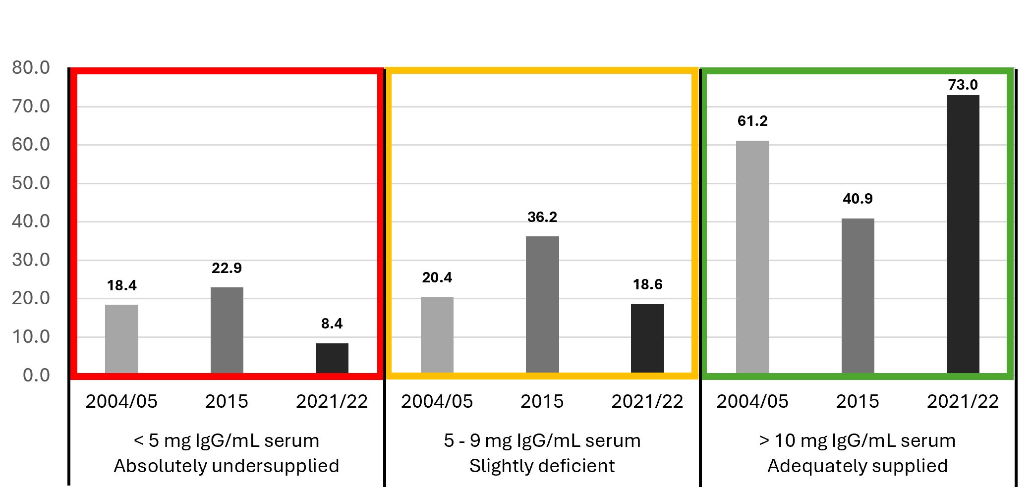
Insufficient intake of colostrum or colostrum with an inadequate content of relevant antibodies leads to failure of passive transfer in calves (Figure 1).
Figure 1: IgG supply of calves – Comparison 2004/05, 2015, and 2021/22 (2004/05: E.K. McMorran, 2006; 2015: K. Stemme et al., 2016; 2021/22: K. Stemme et al., 2022)
Old method – newly applied
To generally support young animals, and especially runts, it was common practice in “the good old days” to feed them raw chicken eggs. Back then, people may not have always known why, but it helped. Dr. Felix Klemperer (1893) provided the explanation. He discovered that chickens form antibodies against a wide variety of germs – not just chicken-relevant ones – they come into contact with and then transfer these antibodies into the egg.
Since, in the past, all animals were kept under one roof, chickens were also exposed to the diarrhea-causing pathogens of all animals living on the farm. Without becoming sick themselves, they formed immunoglobulins (IgY = immunoglobulins from the egg yolk) against these pathogens and transferred them to the egg – the only way to provide their offspring with an immunological head start.
Advantages of egg immunoglobulins: They work not only in birds but can also support mammals immunologically against diarrheal diseases (Yokoyama et al., 1992; Yokoyama et al., 1993; Ikemori et al., 1992; Kuroki et al., 1993; Kellner et al., 1994; Ehrhard et al., 1993; Kuroki, 1999), and they can be produced in high quantities without wounding or killing the hens.
Modern technology enables the provision of complementary dietary feeds containing egg immunoglobulins, supporting calves…and also other young animals suffering from failure of passive transfer (FPT) to overcome critical phases of life.
Egg immunoglobulins mainly act in the gut. There, they bind to pathogens such as E. coli, Cryptosporidia, rotaviruses, and coronaviruses.
Field trials prove the efficacy of egg immunoglobulins
Several trials were conducted in the field to evaluate the efficacy of egg immunoglobulins in calf rearing.
1. Lower number of treatments and decreased mortality rate
A trial conducted in the Netherlands with 300 veal calves (Holstein Friesian and Brown Swiss) examined the effect of egg immunoglobulins on the number of treatments and mortality rate. Both groups were fed standard milk replacer. The IgY group additionally received decreasing amounts of an IgY-containing product (Globigen Life Start).
Days 1-3: 16 g
Days 4-7: 8 g
Days 8-14: 4 g, and
Weeks 3-28: 0.4 g /animal/day.
Results:
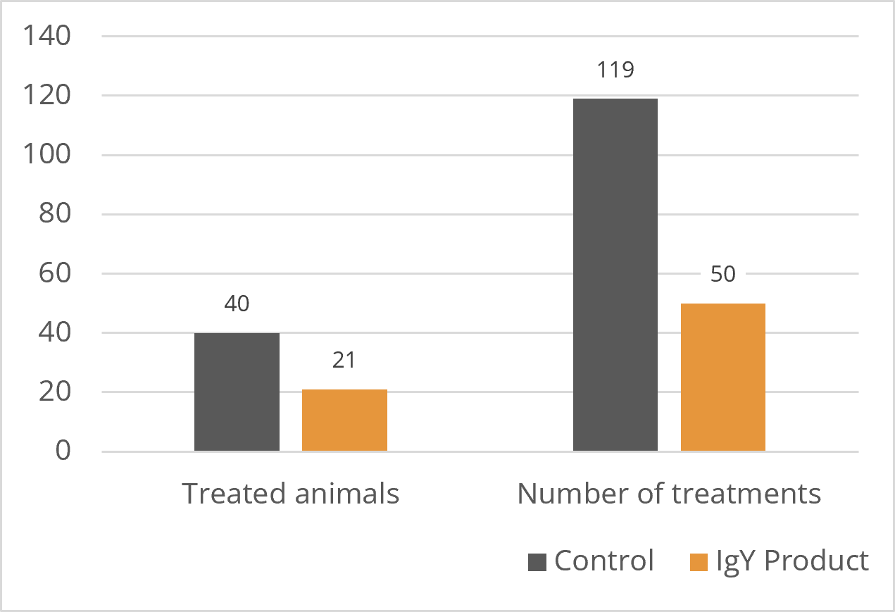
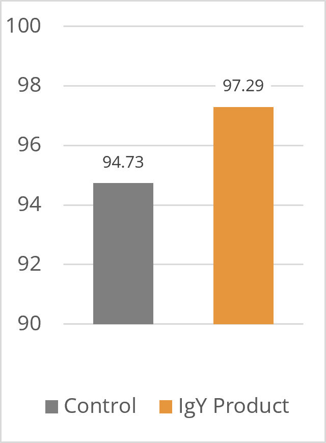
In the group fed the IgY product, nearly 50% fewer animals (21 vs. 40) required medical treatment, and the number of treatments also decreased significantly, from 119 in the control to 50 in the Globigen Life Start group. Better health status resulted in a 2.5% higher livability rate in the group receiving the IgY Product (97.29% vs. 94.73% in the control).
Figure 2: Treated animals and number of treatments (n)
Figure 3: Livability (%)
2. Improved weight development
Two trials were conducted in Germany to evaluate the effect of an IgY-containing product (Globigen Life Start) on the weight development of calves. In both trials, the trial group was fed 4 g of Globigen Life Start/animal per day, in trial 1 on days 2 to 30 and in trial 2 from day 2 until weaning.
Results:
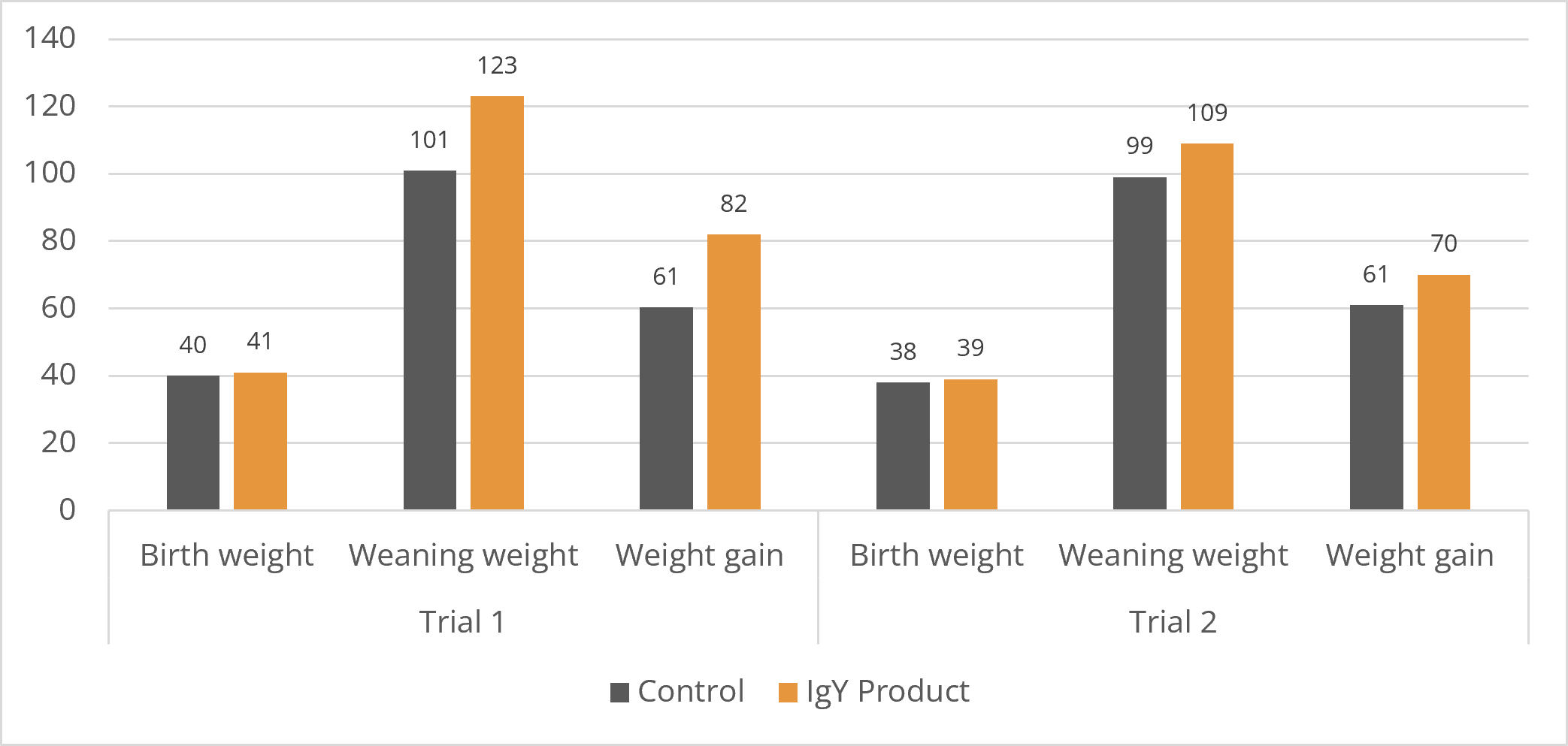
The trial results are presented in Figure 4.
FigureFigure 4: Weight development in calves with and without IgY Product
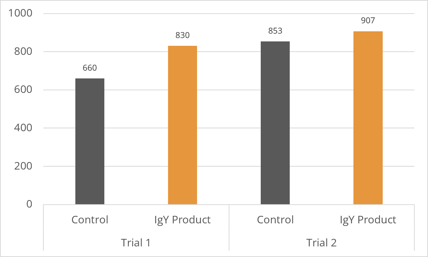
The IgY groups finished the trial with weaning weights more than 20 kg (trial 1) and 10 kg (trial 2) higher than their respective control groups, resulting in 9 kg and 21 kg higher weight gains, respectively. Daily gain is shown in Figure 5.
Figure 5: Daily gain with and without Globigen Life Start
Egg immunoglobulins improve animal health, welfare, and performance
Egg immunoglobulins support young animals such as calves during their first weeks of life by binding pathogens in the intestine. Reducing the pathogenic pressure has two positive consequences:
Decreased occurrence of diarrhea and
Fewer competitors for nutrients
The result, as shown in several trials, is fewer medical treatments, improved livability, and better performance, thereby reducing antibiotic use in calf rearing.
References:
Erhard, M.H., J. Kellner, J. Eichelberger, and U. Lösch. “Neue Möglichkeiten in Der Oralen Immunprophylaxe Der Neugeborenendiarrhoe Des Kalbes- Ein Feldversuch Mit Spezifischen Eiantikörpern.” Berl Münch Tierärztl Wochenschr 106 (1993): 383–87.
Ikemori, Yutaka, Masahiko Kuroki, Robert C. Peralta, Hideaki Yokoyama, and Yoshikatsu Kodama. “Protection of Neonatal Calves against Fatal Enteric Colibacillosis by Administration of Egg Yolk Powder from Hens Immunized with K99-Piliated Enterotoxigenic Escherichia Coli.” American Journal of Veterinary Research 53, no. 11 (November 1, 1992): 2005–8. https://doi.org/10.2460/ajvr.1992.53.11.2005.
Ikemori, Yutaka, Masahiko Kuroki, Robert C. Peralta, Hideaki Yokoyama, and Yoshikatsu Kodama. “Protection of Neonatal Calves against Fatal Enteric Colibacillosis by Administration of Egg Yolk Powder from Hens Immunized with K99-Piliated Enterotoxigenic Escherichia Coli.” American Journal of Veterinary Research 53, no. 11 (November 1, 1992): 2005–8. https://doi.org/10.2460/ajvr.1992.53.11.2005.
Kellner, J., M.H. Erhard, M. Renner, and U. Lösch. “Therapeutischer Einsatz Von Spezifischen Eiantikörpern Bei Saugferkeldurchfall – Ein Feldversuch.” Tierärztliche Umschau 49, no. 1 (January 1994): 31–34.
Klemperer, Felix. “Ueber Natürliche Immunität Und Ihre Verwerthung Für Die Immunisirungstherapie.” Archiv für Experimentelle Pathologie und Pharmakologie 31, no. 4–5 (June 1893): 356–82. https://doi.org/10.1007/bf01832882.
Kuroki, M. “Oral Passive Immunization Using Chicken Egg Yolk Immunoglobulins against Bovine Rotavirus and Coronavirus Infections.” Recent Res. Devel. Virol. 1 (1999): 95–106.
Kuroki, Masahiko, Yutaka Ikemori, Hideaki Yokoyama, Robert C. Peralta, Faustino C. Icatlo, and Yoshikatsu Kodama. “Passive Protection against Bovine Rotavirus-Induced Diarrhea in Murine Model by Specific Immunoglobulins from Chicken Egg Yolk.” Veterinary Microbiology 37, no. 1–2 (October 1993): 135–46. https://doi.org/10.1016/0378-1135(93)90188-d.
McMorran, Elizabeth Kay. “Bundesweite Untersuchung Zur Kolostralen Versorgung von Neugeborenen Kälbern von Elizabeth Kay McMorran.” Bundesweite Untersuchung Zur Kolostralen Versorgung von Neugeborenen Kälbern von Elizabeth Kay McMorran. Dissertation, LMU, 2006.
Stemme, K., E. Rauch, N. Franz, S. Reese, and M. Erhard. “11. Berlin-Brandenburgischer Rindertag.” In Das Gesunde Kalb – Eine Deutschlandweite Erhebung Zur Kolostralversorgung Neugeborener Kälber, 41–42. Berlin: Cuvillier, n.d.
Stemme, K., M. Erhard, D. Klaus-Halla, S. Reese, and E. Rauch. “DVG-Vet-Congress – Fachgruppenübergreifende Buiatrik-Tagung,” 49–53. Berlin: DVG e.V., 2022.
Yokoyama, H, R C Peralta, R Diaz, S Sendo, Y Ikemori, and Y Kodama. “Passive Protective Effect of Chicken Egg Yolk Immunoglobulins against Experimental Enterotoxigenic Escherichia Coli Infection in Neonatal Piglets.” Infection and Immunity 60, no. 3 (March 1992): 998–1007. https://doi.org/10.1128/iai.60.3.998-1007.1992.
Yokoyama, Hideaki, Robert C. Peralta, Kouji Umeda, Tomomi Hashi, Faustino C. Icatlo, Masahiko Kuroki, Yutaka Ikemori, and Yoshikatsu Kodama. “Prevention of Fatal Salmonellosis in Neonatal Calves, Using Orally Administered Chicken Egg Yolk Salmonella-Specific Antibodies.” American Journal of Veterinary Research 59, no. 4 (April 1, 1998): 416–20. https://doi.org/10.2460/ajvr.1998.59.04.416.
Yokoyama, Hideaki, Robert C. Peralta, Sadako Sendo, Yutaka Ikemori, and Yoshikatsu Kodama. “Detection of Passage and Absorption of Chicken Egg Yolk Immunoglobulins in the Gastrointestinal Tract of Pigs by Use of Enzyme-Linked Immunosorbent Assay and Fluorescent Antibody Testing.” American Journal of Veterinary Research 54, no. 6 (June 1, 1993): 867–72. https://doi.org/10.2460/ajvr.1993.54.06.867.
The Gut: A Main Component of Poultry’s Immune System
By Dr. Inge Heinzl, Editor EW Nutrition
Gut health is a critical factor in poultry production, influencing growth performance, feed efficiency, and overall bird health. A well-functioning digestive system ensures optimal nutrient absorption and ultimately contributes to economic sustainability in poultry farming.
However, another essential function of the gut is its significant role in immune defense, as evidenced by the fact that 80% of all active immune cells are in the gut. It is essential for the organism to keep a sensitive balance by eliminating invading pathogens while maintaining self-tolerance to avoid autoimmunity. Being 1.5 to 2.3 m long and with a big contact area to the external environment, the gut is the first line of defense when pathogens have orally entered the organism. For this purpose, the intestine has several specialized cells and a plethora of diverse microorganisms – the microbiome.
A balanced gut environment, therefore, enhances resistance to diseases, helps prevent infections, and reduces the need for antibiotics.
Which tools are available in the gut to counteract pathogenic attacks?
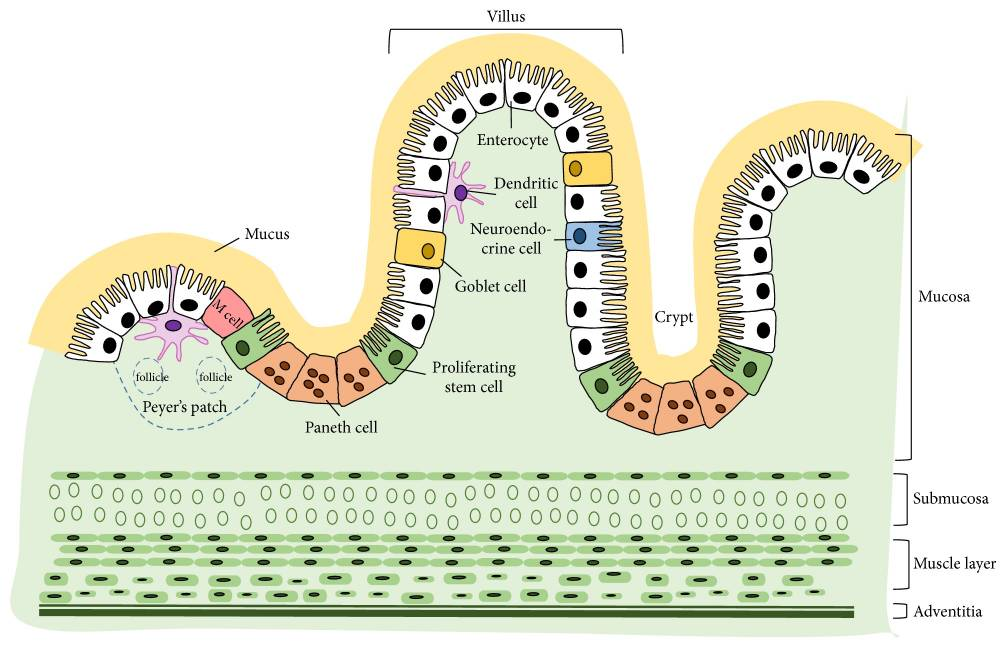
The gut wall, per se, has several fixed tools to fight pathogenic offenses, such as the mucus layers and the epithelium with highly specialized cells. Figure 1 shows in detail the different parts of the gut immune system.
Figure 1: Structure of the intestinal wall with its specialized cells (Kong et al., 2018)
1. Mucus layers
The mucus layers form the first host-derived line of defense. They help trap invasive bacteria and facilitate their removal via luminal flow. The protective properties may depend on whether the mucin is neutral or acidic, sialylated or sulfated (Broom and Kogut, 2018). The glycoprotein mucins forming the mucus layer (mainly MUC2 in the small and large intestine and MUC5ac in the proventriculus) are produced by the goblet cells, part of the intestinal epithelium just beneath.
2. Intestinal epithelium
The one-layered intestinal epithelium represents a physical barrier and consists of normal enterocytes, as well as specialized cells. All the cells are closely linked by tight junctions, consisting of claudin, occludin, and junctional adhesion molecules (JAM).
The following diverse specialized cells protect the organism from pathogenic attacks:
2.1 Proliferating stem cells
These cells are ready to replace damaged epithelial cells in the case of inflammation.
2.2 Paneth cells
Paneth cells are situated at the bottom of the Lieberkühn crypts, neighboring the stem cells in the jejunum and the ileum. Paneth cells have different tasks:
In normal conditions, they maintain homeostasis by regulating the microbiome’s composition via the secretion of antimicrobial peptides, which are accumulated in apically oriented secretory granules, performing phagocytosis and efferocytosis. Additionally, the Paneth cells provide niche factors for the intestinal stem cell compartment, absorb heavy metals, and preserve the integrity of the intestinal barrier. If one or more of these functions are impaired, intestinal and systemic inflammations or infections can develop (Wallaeys et al., 2022). The number of Paneth cells and their diameter can be enhanced via feeding. Agarwal et al. (2022) noticed a significant increase in the number and diameter of Paneth cells after feeding quinoa soluble fiber and/or quercetin 3-glucoside.
2.3 M cells
M cells (M coming from microfold and indicating the structure) are specialized epithelial cells localized along the antimesenteric border in the epithelium of the ileum. They are crucial for the immune system and an essential part of the gut-associated lymphoid tissue (GALT), a sub-system of the mucosa-associated lymphoid tissue (MALT).
M cells play an important role in the function of the immune system. They act as a transport system for antigens. They sample antigens (macromolecules, bacteria, viruses, small parasites) via the apical membrane. After the phagocytosis of the foreign organism/substance, the antigen gets through the cell and is consigned to cells of the adaptive immune system (e.g., the B-cells) at the basal side. The exact transport and the handover to the cells of the adaptive immune system are still unclear. It is also not clarified whether the antigens are processed inside the cells.
2.4 Dendritic cells
Dendritic cells are a kind of leucocyte derived from the bone marrow. Immature dendritic cells have a star-like shape. They are specialized to identify, uptake, transport, process, and present antigens to other immune system cells on their surface. To identify and uptake harmful substances/microbes, they carry receptors on their surface that recognize the attributes often occurring in pathogenic viruses, bacteria, and fungi. After contact with the antigen, the cell moves to secondary lymphoid tissue, and in the intestine, this is predominantly Mucosa-Associated Lymphoid Tissue (MALT). Arriving as mature and not phagocytizing dendritic cells, they present the antigens of the pathogens to the T-lymphocytes. For this purpose, they use cell surface proteins (MHC proteins). This presentation, together with co-stimulators and cytokines, activates naïve T-lymphocytes to develop into the relevant T-cell (fighting viruses, bacteria…) and proliferate, leading to the clearance of the pathogen.
On the other hand, dendritic cells can also suppress an immune reaction if the “suspicious subjects” are harmless or belong to the organism. Dendritic cells are the most potent antigen-presenting cells of the immune system.
2.5 Goblet cells
Goblet cells originate from pluripotent stem cells and are located between the enterocytes in the inner mucus layer of the intestine. Goblet cells develop and mature rapidly after hatching due to external stimuli such as environmental and dietary factors, but also intestinal microbiota (Duangnumsawang et al., 2021). They derive their name from their goblet-like appearance. The basal site is thin, but the cell gets thicker toward the apical side. In the thicker cell organisms, vesicles with mucins are stored and explosively released to the surface by exocytosis.
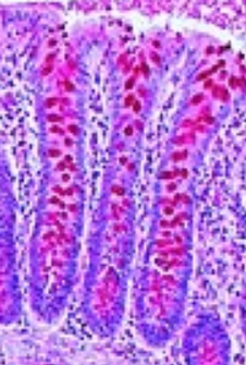
Figure 2: Goblet cells
The mucins (MUC2) are viscous, slime-forming substances consisting of a protein string bound to many sugar chains. Due to their oligosaccharide chain structure, they offer adhesion binding sites for intestinal commensal bacteria and enhance probiotic colonization (Liu et al, 2020). They have a high water-binding capacity, which is responsible for their slimy and protective characteristics. In the case of inflammation, mucin production can increase strongly.
By providing bicarbonate for proper mucin unfolding in the small intestine, goblet cells help maintain homeostasis and the intestinal barrier function. Furthermore, goblet cells can form goblet cell-associated passages (GAPs) and deliver luminal substances to the antigen-presenting cells in the underlying lamina propria that can start an adaptive immune response (Knoop and Newberry, 2018).
As with Paneth cells, the number of goblet cells also increases by feeding quinoa soluble fibers.
2.6 Neuroendocrine cells
Enterochromaffin cells are neuroendocrine cells found in the epithelium of the whole digestive tract, mainly in the small intestine, the colon, and the ceca. They belong to the enteric endocrine system, are part of the diffuse neuroendocrine system, and produce 95% of the serotonin in the organism. Enterochromaffin cells act as chemo- and mechanosensors. They react to free fatty acids, amino acids, and other chemicals as well as physical forces occurring during peristaltic activity in the gut, thus modulating the secretion of water and electrolytes as well as gut motility and visceral sensation of pain (Linan-Rico et al., 2016; Diwakarla et al., 2018).
Serotonin, on its side, has been shown to affect the composition of the gut microbiota (Kwon et al., 2019) and to modulate bacterial physiology (Knecht et al., 2016). Gut-derived serotonin is responsible for immune responses (Baganz and Blakely, 2012) but also for the regulation of other functions such as bone development (Chabbi-Achengli et al., 2012), gut motility, and platelet aggregation (Berger et al., 2009). A deficient serotonergic system can cause psychopathological behaviors such as feather pecking.
3. Last but not least – the microbiome
The poultry gut microbiome consists of bacteria, fungi, protozoa, and viruses. Beneficial microbes, such as Lactobacillus, Bifidobacterium, and Bacteroides, contribute to gut health and immunity.
On the one hand, microbes are involved in digestion and nutrient synthesis. They assist in breaking down fiber, producing short-chain fatty acids, and synthesizing essential vitamins. On the other hand, they contribute to immune defense:
Beneficial bacteria (BB) prevent the colonization of harmful microbes: The bacteria inhabiting the poultry gut act against pathogens by competing with them for nutrients and binding sites at the intestinal mucosa.
Beneficial bacteria prevent/reduce inflammation and stabilize the intestinal mucosa Abaidullah et al. (2019) showed in their review how beneficial bacteria influence the immune response to diverse viruses (AIV, IBDV, MDV, NDV). Bacteria such as Collinsella, Faecalibacterium, Oscillibacter, etc., increase the release of IFN-α, IFN-β, and IL-22. These substances control virus replication and repair mucosal tissue damage. Other bacteria, such as Clostridium XIVa or Firmicutes, provoke T-cells to produce anti-inflammatory cytokines to suppress inflammation. By promoting the antimicrobial peptides such as MUC, TFF, ZO, and tight junction proteins comprised of claudins, occludin, and zona occludens mRNA expression, Bacteroides, Candidatus, SMB53, Parabacteroides, Lactobacillus, Paenibacillus, Enterococcus, and Streptococcus spp. inhibit pathobiont colonization and translocation, and suppress inflammation. Butyrate succinate and lactate, produced by Faecalibacterium and Blautia spp., provide energy and reduce inflammation. Bacteroides fragilis produce bacterial polysaccharides that communicate with the immune system and influence the transformation of CD4+ (T-helper cells) and Foxp3+ cells (the master transcription factor of regulatory T cells in mammals, but also present in chicken (Burkhardt et al., 2022)).
“Negative” bacteria increase inflammation and enhance viral shedding Clostridium Cluster XI, Salmonella, and Shigella downregulate the anti-inflammatory and tight junction-stabilizing substances, which would be increased by the beneficial bacteria and increase IFN-γ and IF-17A to cause mucosal inflammation and tissue damage, as well as increased virus replication and fecal shedding. Further bacteria, which enhance mucosal and GIT inflammation, are Desulfovibrionaceae, producing hydrogen sulfides, Vampirovibrio, Clostridium cluster XIVb, and the genus Rumicoccus. They induce the pro-inflammatory cytokines IL-6 and IL-1β. The latter three bacteria also increase viral shedding. Salmonella typhimurium and Campylobacter jejuni also achieve higher viral shedding by decreasing viral-specific IgG and IgA production (Abaidullah et al., 2019)
Factors impairing intestinal immune defense
As the previous paragraph indicates, an imbalance of the intestinal microbiome called dysbiosis makes chickens more prone to diseases such as necrotic enteritis (Stanley et al., 2014). Several factors are disturbing the balance in the microbiome (Heinzl, 2020):
An abrupt change of feed
High contents of non-starch polysaccharides increase viscosity, decrease passage rate, lower the digestibility of other nutrients, and serve as nutrients for, e.g., Clostridium perfringens
High protein levels can also serve as a substrate for pathogens and cause a shift in the balance of the intestinal flora
Finely ground feed does not stimulate the gizzard muscles to do their work. pH increases, transit time decreases, and pathogenic microbes such as Salmonella, Campylobacter, and Clostridia proliferate.
Stress (heat or cold stress, re-assembling of groups, high stocking densities)
Mycotoxins
However, besides all these factors causing an overgrowth of commensal bacteria such as E. coli, ingested pathogens such as Marek’s or Newcastle Disease viruses can also cause this imbalance.
Immune defense in the gut – an interplay of different tools that must be protected
The first line of defense, the intestine, comprises different tools working together to fight pathogens and harmful substances. Besides the mucus layers and the specialized cells, the intestinal microbiome plays an essential role in immune defense by competing with pathogens for nutrients and binding sites, enhancing the secretion of anti-inflammatory substances, and stimulating the production of interferons, which fight the pathogens. However, several factors can impact the balance of the microbiome and cause dysbiosis. The best protection of this sensitive equilibrium can support the organism in defending against diseases and maintaining immunity and performance. Understanding the interplay between microbiota, immune function, and nutrition allows for effective strategies to enhance poultry health while reducing reliance on antibiotics. Future research will continue to provide insights into optimizing gut-immune interactions in poultry production.
References
Abaidullah, Muhammad, Shuwei Peng, Muhammad Kamran, Xu Song, and Zhongqiong Yin. “Current Findings on Gut Microbiota Mediated Immune Modulation against Viral Diseases in Chicken.” Viruses 11, no. 8 (July 25, 2019): 681. https://doi.org/10.3390/v11080681.
Baganz, Nicole L., and Randy D. Blakely. “A Dialogue between the Immune System and Brain, Spoken in the Language of Serotonin.” ACS Chemical Neuroscience 4, no. 1 (December 24, 2012): 48–63. https://doi.org/10.1021/cn300186b.
Berger, Miles, John A. Gray, and Bryan L. Roth. “The Expanded Biology of Serotonin.” Annual Review of Medicine 60, no. 1 (February 1, 2009): 355–66. https://doi.org/10.1146/annurev.med.60.042307.110802.
Broom, Leon J., and Michael H. Kogut. “The Role of the Gut Microbiome in Shaping the Immune System of Chickens.” Veterinary Immunology and Immunopathology 204 (October 2018): 44–51. https://doi.org/10.1016/j.vetimm.2018.10.002.
Burkhardt, Nina B, Daniel Elleder, Benjamin Schusser, Veronika Krchlíková, Thomas W Göbel, Sonja Härtle, and Bernd Kaspers. “The Discovery of Chicken Foxp3 Demands Redefinition of Avian Regulatory T Cells.” The Journal of Immunology 208, no. 5 (March 1, 2022): 1128–38. https://doi.org/10.4049/jimmunol.2000301.
Chabbi-Achengli, Yasmine, Amélie E. Coudert, Jacques Callebert, Valérie Geoffroy, Francine Côté, Corinne Collet, and Marie-Christine de Vernejoul. “Decreased Osteoclastogenesis in Serotonin-Deficient Mice.” Proceedings of the National Academy of Sciences 109, no. 7 (January 30, 2012): 2567–72. https://doi.org/10.1073/pnas.1117792109.
Clarke, G, S Grenham, P Scully, P Fitzgerald, R D Moloney, F Shanahan, T G Dinan, and J F Cryan. “The Microbiome-Gut-Brain Axis during Early Life Regulates the Hippocampal Serotonergic System in a Sex-Dependent Manner.” Molecular Psychiatry 18, no. 6 (June 2013): 666–73. https://doi.org/10.1038/mp.2012.77.
Diwakarla, S., L. J. Fothergill, J. Fakhry, B. Callaghan, and J. B. Furness. “Heterogeneity of Enterochromaffin Cells within the Gastrointestinal Tract.” Neurogastroenterology & Motility 29, no. 6 (May 9, 2017). https://doi.org/10.1111/nmo.13101.
Duangnumsawang, Yada, Jürgen Zentek, and Farshad Goodarzi Boroojeni. “Development and Functional Properties of Intestinal Mucus Layer in Poultry.” Frontiers in Immunology 12 (October 4, 2021). https://doi.org/10.3389/fimmu.2021.745849.
Heinzl, Inge. “Necrotic Enteritis: The Complete Overview.” EW Nutrition, August 8, 2023. https://ew-nutrition.com/necrotic-enteritis-complete-overview/.
Knecht, Leslie D., Gregory O’Connor, Rahul Mittal, Xue Z. Liu, Pirouz Daftarian, Sapna K. Deo, and Sylvia Daunert. “Serotonin Activates Bacterial Quorum Sensing and Enhances the Virulence of Pseudomonas Aeruginosa in the Host.” EBioMedicine 9 (July 2016): 161–69. https://doi.org/10.1016/j.ebiom.2016.05.037.
Kong, Shanshan, Yanhui H. Zhang, and Weiqiang Zhang. “Regulation of Intestinal Epithelial Cells Properties and Functions by Amino Acids.” BioMed Research International 2018 (2018): 1–10. https://doi.org/10.1155/2018/2819154.
Kwon, Yun Han, Huaqing Wang, Emmanuel Denou, Jean-Eric Ghia, Laura Rossi, Michelle E. Fontes, Steve P. Bernier, et al. “Modulation of Gut Microbiota Composition by Serotonin Signaling Influences Intestinal Immune Response and Susceptibility to Colitis.” Cellular and Molecular Gastroenterology and Hepatology 7, no. 4 (2019): 709–28. https://doi.org/10.1016/j.jcmgh.2019.01.004.
Linan-Rico, Andromeda, Fernando Ochoa-Cortes, Arthur Beyder, Suren Soghomonyan, Alix Zuleta-Alarcon, Vincenzo Coppola, and Fievos L. Christofi. “Mechanosensory Signaling in Enterochromaffin Cells and 5-HT Release: Potential Implications for Gut Inflammation.” Frontiers in Neuroscience 10 (December 19, 2016). https://doi.org/10.3389/fnins.2016.00564.
Liu, Yang, Xinjie Yu, Jianxin Zhao, Hao Zhang, Qixiao Zhai, and Wei Chen. “The Role of MUC2 Mucin in Intestinal Homeostasis and the Impact of Dietary Components on MUC2 Expression.” International Journal of Biological Macromolecules 164 (December 2020): 884–91. https://doi.org/10.1016/j.ijbiomac.2020.07.191.
Lyte, Mark. “Microbial Endocrinology in the Microbiome-Gut-Brain Axis: How Bacterial Production and Utilization of Neurochemicals Influence Behavior.” PLoS Pathogens 9, no. 11 (November 14, 2013). https://doi.org/10.1371/journal.ppat.1003726.
Marcobal, A., P. C. Kashyap, T. A. Nelson, P. A. Aronov, M. S. Donia, A. Spormann, M. A. Fischbach, and J. L. Sonnenburg. “A Metabolomic View of How the Human Gut Microbiota Impacts the Host Metabolome Using Humanized and Gnotobiotic Mice.” The ISME Journal 7, no. 10 (June 6, 2013): 1933–43. https://doi.org/10.1038/ismej.2013.89.
Stanley, Dragana, Shu-Biao Wu, Nicholas Rodgers, Robert A. Swick, and Robert J. Moore. “Differential Responses of Cecal Microbiota to Fishmeal, Eimeria and Clostridium Perfringens in a Necrotic Enteritis Challenge Model in Chickens.” PLoS ONE 9, no. 8 (August 28, 2014). https://doi.org/10.1371/journal.pone.0104739.
Wallaeys, Charlotte, Natalia Garcia‐Gonzalez, and Claude Libert. “Paneth Cells as the Cornerstones of Intestinal and Organismal Health: A Primer.” EMBO Molecular Medicine 15, no. 2 (December 27, 2022). https://doi.org/10.15252/emmm.202216427.
Yano, Jessica M., Kristie Yu, Gregory P. Donaldson, Gauri G. Shastri, Phoebe Ann, Liang Ma, Cathryn R. Nagler, Rustem F. Ismagilov, Sarkis K. Mazmanian, and Elaine Y. Hsiao. “Indigenous Bacteria from the Gut Microbiota Regulate Host Serotonin Biosynthesis.” Cell 163, no. 1 (September 2015): 258. https://doi.org/10.1016/j.cell.2015.09.017.
Health management of nursery piglets through nutrition
Conference Report
An optimized gut function is essential for pigs’ overall health and performance. When managed correctly, gut health can significantly enhance growth, immunity, and productivity. However, if gut health is compromised, it can lead to lifetime negative impacts on a pig’s performance.
Early feed intake enhances GIT development
Dr. Edwards emphasized that good health and performance in the nursery are closely linked to maintaining feed intake, which is essential for developing stomach capacity and function. A larger stomach capacity increases the exposure to digestive enzymes and prolongs stomach dwell time.
Acid output takes time to develop and develops in response to substrate. It directly influences stomach pH and is closely related to pepsin output, which, on its part, influences protein digestibility and the risk of diarrhea.
Protein and immunity
Protein is a double-edged sword, warned Dr. Edwards:
Excess or undigested protein can create inflammation and oxidative stress in the body. This occurs when the metabolism of surplus protein leads to the production of reactive oxygen species (ROS), which can damage cells and tissues, further exacerbating inflammatory responses. Chronic inflammation may impair immune responses, making pigs more susceptible to infections and diseases.
On the other hand, a deficiency in amino acids can limit immune response. Amino acids do more than build muscle – they are critical for synthesizing antibodies and other immune-related proteins. Without adequate levels, pigs may struggle to mount effective immune responses, increasing their vulnerability to pathogens.
Table 1: Effects of amino acids on pig gut health and functions (Yang & Liao, 2019)
Amino acid
Functions
Glutamine/glutamate
Metabolic fuel for rapidly dividing cells, including lymphocytes, enterocytes
maintains or enhances villus height/crypt depth
enhances microbial diversity
is utilized to synthesize GSH and protect against oxidative stress
stimulates both innate and adaptive immunity
Arginine
promotes intestinal healing and reverses intestinal dysfunction
has anti-inflammatory effects
Cysteine
is utilized to synthesize GSH (antioxidant)
utilized to synthesize taurine (antioxidant/cell membrane stabilizer)
utilized for mucin synthesis (physical protection)
Threonine
utilized for mucin synthesis
important component of immunoglobulins
enhances microbial diversity
Glycine
anti-inflammatory effects
utilized to synthesize GSH (antioxidant)
Methionine
acts as an antioxidant by protecting other proteins against oxidative damage
important for the proliferation of lymphocytes
Diets should be formulated to all ten essential amino acids (arginine, histidine, isoleucine, leucine, lysine, methionine, phenylalanine, threonine, tryptophan, and valine) while ensuring a ratio of about 50:50 for essential amino acids to non-essential amino acids is optimal for nitrogen retention and utilization in pigs.
During immune challenges, the pig’s amino acid requirements, including methionine, cysteine, tryptophan, threonine, and glutamine, increase relative to lysine. Well-known examples are threonine, a key component of mucin (and immunoglobulins), supporting gut health and integrity during stress, and glutamine, a major energy source for rapidly dividing cells in the immune system.
Microbiome evolution and modulation
The microbiota of the pig evolves from birth up to about 20 weeks of age. The alpha diversity (the number of species) and species richness increase with age. The pig microbiome consists of both permanent members that establish stable populations throughout life and transient members that may fluctuate based on dietary changes or environmental factors.
Microbiome modulation through the diet
Diet can influence the rate and maturity of microbiota evolution. For instance, diets rich in fiber and specific carbohydrates can promote the growth of beneficial bacteria such as Lactobacillus and Bifidobacterium. In contrast, diets high in protein can increase potentially harmful bacteria if not appropriately balanced.
Understanding these dynamics is critical for optimizing nutrition strategies that support gut health and overall performance in pigs. Proper management of dietary components can lead to healthier microbiomes, enhancing nutrient absorption and immune responses throughout the pig’s life.
The following strategies accelerate the maturation of the microbiome, the gut, and the immune system:
Promoting and maintaining feed intake: consistent feed intake is crucial for microbial development. Early access to solid feed helps establish a diverse microbiome.
Raw material continuity: variability in feed composition can disrupt microbial communities, leading to dysbiosis. A step-wise approach to diet changes, with a broad range of ingredients at low inclusion levels, is recommended.
Regulating digest transit time: the rate at which digesta moves through the gastrointestinal tract affects nutrient absorption and microbial colonization. Strategies to optimize transit time, such as increasing particle size and incorporating insoluble fibers, can enhance nutrient digestibility and promote a healthy microbiome by allowing beneficial microbes to thrive.
Feeder access: adequate access to feeders encourages regular feeding behavior, supporting consistent nutrient intake and microbial activity. Frequent feeding can help maintain stable gut conditions conducive to microbial growth.
Inert fiber: helps maintain gut motility and provides substrates for beneficial bacteria, contributing to a balanced microbiome.
Minimizing stress: stress can negatively impact gut integrity and microbial balance, increasing susceptibility to infections and other health issues.
Limiting the use of antibiotics helps preserve the natural gut microbiota, which is essential for maintaining health and preventing disease. The use of antibiotics can lead to dysbiosis, making pigs more vulnerable to infections and impairing immune responses.
Limitations in the use of AGPs, Zn, and Cu require rethinking in pig nutrition
Reduced access to in-feed antibiotics and pharmacological levels of zinc and copper have exposed nutritional shortcomings for nursery pigs. Preventive strategies through nutrition, carefully designed diets, and optimal use of eubiotics and functional ingredients are the keys to getting pigs off to a healthy and efficient start.
Nursery nutrition programs should be designed for long-term gut health, efficiency, and functionality. The level of investment will depend on the weaning age/weight, health status, labor quality, etc., noted Dr. Edwards.
EW Nutrition’s Swine Academy took place in Ho Chi Minh City and Bangkok in October 2024. Dr. Megan Edwards, an Australian animal nutrition consultant with global research and praxis experience and a keen interest in immuno-nutrition and functional nutrients, was an esteemed guest speaker at this event.