Intrinsically Heat-Stable Xylanase: A New Standard for Improving Performance under High-Temperature Pelleting
Author: Ajay Bhoyar, Senior Global Technical Manager, EW Nutrition
The global use of feed enzymes has become a central feature of efficient monogastric animal production systems. Rising feed ingredient costs, tighter margins, and increasing regulatory pressure to reduce environmental impact have all accelerated enzyme innovation. At the same time, feed mills have shifted toward higher conditioning temperatures and time in pursuit of improved pellet durability, pathogen control, and throughput. However, this creates a hostile environment for most exogenous feed enzymes, which can lose significant activity under the harsh conditions of feed processing.
Historically, enzyme manufacturers have attempted to overcome heat degradation of by coating, encapsulating, or post-pelleting liquid application (PPLA) of enzymes. While these approaches provide partial solutions, they can also have limitations, including delayed enzyme activity, uneven distribution, reduced mixing uniformity, and reliance on specialized liquid enzyme applicators.
These limitations prompted a novel direction: enzymes designed or selected to be intrinsically heat-stable, capable of surviving pelleting without protective matrices.
This article highlights recent advancements in intrinsically heat-stable xylanase technology, explains its advantages over coated and post-pelleting enzyme solutions, and outlines its practical benefits for feed manufacturers, integrators, and nutritionists operating under modern high-temperature feed pelleting conditions.
Intrinsically Thermostable Enzymes
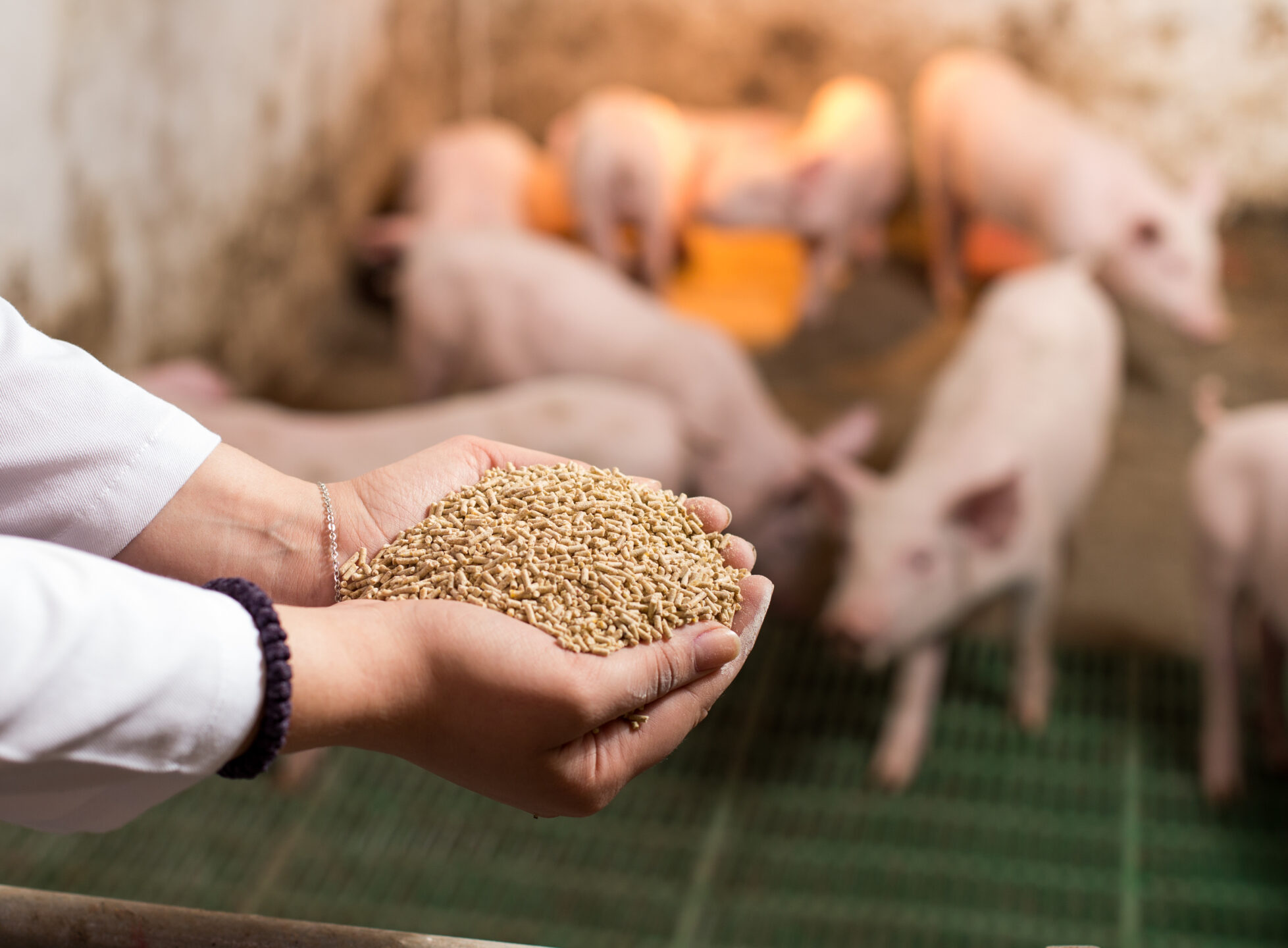
An enzyme is considered intrinsically heat-stable when its native protein structure resists unfolding and retains catalytic activity under high temperatures associated with feed processing—typically 80–95°C for 30–90 seconds. Unlike coated enzymes that rely on external protection, intrinsically thermostable enzymes depend on their internal protein architecture for heat tolerance. Enzymes from organisms living in compost, thermal springs, and geothermal soils naturally withstand temperatures of 80–100 °C or higher. Intrinsically thermostable enzymes are often sourced from thermophiles (organisms living in hot springs and deep-sea vents) or engineered for stability. They resist denaturation (loss of shape and function) at high-temperature processing.
Fig.1: Key benefits of intrinsically thermostable enzymes
Limitations of Current Thermostability Solutions
Coating / Encapsulation
A method of protecting enzymes from heat is to encapsulate or coat them with a protective coating. An ideal enzyme coating for animal feed needs to:
1. Protect the enzyme through steam conditioning (typically 85–90°C or higher) and through subsequent pelleting.
2. Release the enzyme from the coating quickly in the gastrointestinal tract of the target animal, to ensure optimum efficacy. (Gilbert and Cooney, 2007)
There is some evidence, however, suggesting that the coating of enzymes may reduce the efficacy of the product, compared to an uncoated version of the same product (Kwakkel et al., 2000).
Post-Pelleting Liquid Application (PPLA)
Post-pelleting liquid enzyme application requires sophisticated applicators to minimize the risk of uneven spraying or calibration errors, which is often not feasible in small or mid-size mills. Accurate application of the liquid enzyme, as with some other critical liquid micro-ingredients, requires specialized spraying equipment and, even then, consistency of accurate enzyme application can be an issue (Bedford and Cowieson, 2009). Research has shown that as much as 30% of the enzyme activity can be found in the pellet fines, and therefore, adding the enzyme before screening would result in a lower than expected dosage in the final feed and wastage of the enzyme product (Engelen, 1998). In some cases, adjusting the pelleting machines to the output of the PPLA’s spray nozzles to ensure a homogenous and even application of the enzyme on the pellets may reduce the overall pellet production rate, especially in big feed mills with very high throughput.
These limitations of the coated or PPLA technologies strengthen the value proposition of intrinsically heat-stable enzymes.
Nutritional and Commercial Benefits of Intrinsically Heat-Stable Xylanase
The use of intrinsically heat-stable xylanase delivers consistent nutritional benefits in poultry and swine feeds, including predictable non-starch polysaccharide (NSP) degradation, a significant increase in the metabolizable energy (ME) value of the feed, and enhanced gut health resilience supporting reduced antibiotic use.
From a commercial and operational perspective, this technology simplifies enzyme application, improves mixing uniformity, reduces formulation risk, and lowers feed cost per unit of meat or egg produced.
In-Vitro Thermal Stability Profile of Axxess XY
Axxess XY is a novel, intrinsically thermostable GH10 xylanase originating from Thermotoga maritima, a hyperthermophilic bacterium found in hydrothermal vents near volcanic grounds, and commercially it is produced by proprietary strain of Bacillus subtilis.
The superior heat stability of Axxess XY has been proven under various commercial pelleting conditions across different geographies. Axxess XY showed excellent post-pelleting recovery under commercial feed-milling conditions across varying temperatures and conditioning times (Fig. 2).
In one study, in addition to excellent post-pelleting recovery, Axxess XY also demonstrated high xylanase stability in pelleted feed over a 2-month feed storage period at>40°C, with humidity around 65%.
Fig.2: Demonstrated Intrinsic Thermostability of Axxess XY Across Geographies
Conclusions
As feed mills continue to operate at higher conditioning temperatures and longer retention times, enzyme heat stability has become a critical success factor in modern feed production. Intrinsically heat-stable xylanase offers a practical and reliable solution to this challenge by maintaining enzyme activity through pelleting without the need for coatings or post-pelleting liquid application systems.
By relying on its native protein structure rather than external protection, intrinsically thermostable xylanase delivers consistent post-pelleting recovery, uniform distribution in feed, and predictable nutritional performance across different feed mills and processing conditions. This reliability translates into improved nutrient utilization, better gut health support, and reduced cost per kilogram of meat or eggs produced.
From an operational standpoint, intrinsically heat-stable xylanase simplifies enzyme application, reduces dependence on specialized equipment, and minimizes the need for over-formulation or safety margins. These advantages help feed manufacturers and integrators improve efficiency, lower risk, and achieve more consistent results, especially under demanding commercial pelleting conditions.
In summary, intrinsically heat-stable xylanase aligns well with the evolving needs of today’s feed industry, offering a robust, cost-effective, and future-ready enzyme solution for high-performance animal production systems.
References:
Bedford, M. R., and A. J. Cowieson. 2009. “Phytate and Phytase Interactions.” In Proceedings of the 17th European Symposium on Poultry Nutrition, 7–13. Edinburgh, UK.
Eeckhout, M., M. De Schrijver, and E. Vanderbeke. 1995. “The Influence of Process Parameters on the Stability of Feed Enzymes during Steam Pelleting.” In Proceedings of the 2nd European Symposium on Feed Enzymes, 163–169. Noordwijkerhout, The Netherlands.
Engelen, G. M. A. 1998. Technology of Liquid Additives in Post-Pelleting Applications. Wageningen, The Netherlands: Wageningen Institute of Animal Science.
Gilbert, T. C., and G. Cooney. 2011. “Thermostability of Feed Enzymes and Their Practical Application in the Feed Mill.” In Enzymes in Farm Animal Nutrition, 2nd ed., edited by M. R. Bedford and G. G. Partridge, 249–259. Wallingford, UK: CABI.
Kwakkel, R. P., P. L. van der Togt, and K. A. B. M. Holkenborg. 2000. “Bio-Efficacy of Two Phytase Formulations Supplemented to a Corn–Soybean Broiler Diet.” In Proceedings of the 3rd European Symposium on Feed Enzymes, 63–64. Noordwijkerhout, The Netherlands.
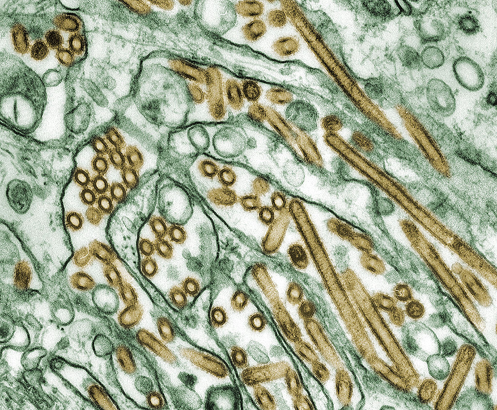
Europe – Disease Outbreak Report Summary, 6-12 November 2025
Widespread across Europe, indicating active transmission
Data Source: ADIS (Animal Disease Information System) Weekly Notification Created: November 14, 2025
Header image photo credit: Cynthia Goldsmith Content Providers: CDC/ Courtesy of Cynthia Goldsmith; Jacqueline Katz; Sherif R. Zaki
This media comes from the Centers for Disease Control and Prevention’s Public Health Image Library (PHIL), with identification number #1841
Energy Metabolism in Pigs: Disease and stress impact efficiency
By Dr. Inge Heinzl, Editor, and Predrag Persak, Regional Technical Manager North Europe
For profitable pig production, efficient energy metabolism is essential. Every kilojoule consumed must be wisely spent – on maintenance, growth, reproduction, or defense. An impacted energy metabolism due to disease or stress impacts animal performance and farm profitability.
Different faces of energy
Energy metabolism determines how efficiently pigs convert feed into body mass. The Gross energy (GE) of the diet, which the use of a calorimeter can determine, is progressively reduced by losses in feces (→digestible energy – DE), urine, gases (→metabolizable energy – ME), and heat, resulting in the →net energy (NE), which is then available for maintenance and performance (growth, milk…).
The requirements for maintenance include the minimum energy that an organism needs to maintain essential functions under standardized conditions and at complete rest. This includes respiration, thermoregulation, tissue turnover, and immune system activity. Only energy in excess of these needs is available for performance. The ratio between additional retained energy and additional energy intake defines the incremental efficiency of nutrient utilization. Under normal conditions, healthy, fast-growing pigs display high incremental efficiencies for both protein and energy deposition by channeling energy efficiently into lean tissue and approximately 25-30% of the metabolizable energy from the feed is used for maintenance, 20-25% for lean gain, and the rest for fat deposition, driving daily gain and carcass quality (Patience, 2019).
However, disease, immune stress, and suboptimal environmental conditions can disrupt this delicate balance, diverting nutrients from growth to survival processes (Obled, 2003). The activation of the immune system leads to reduced feed efficiency, slower growth, and inferior meat quality.
Disease generates costs
The health challenge of disease causes energy loss through several key mechanisms (Patience, 2019).
The activation of the immune system becomes an energetic priority. It consumes significant amounts of energy and nutrients, such as glucose and specific amino acids, to produce immune cells and acute-phase proteins, such as haptoglobin and CRP, and to combat pathogens. The nutrients are redirected away from performance toward immune defense, i.e., less energy available for growth performance or even a mobilization of body reserves (fat deposits). A study conducted by Huntley et al. (2017) showed a 23.6% higher requirement for metabolizable energy to activate and maintain the immune system, resulting in a 26% lower ADG.
Physiological responses to disease, such as fever (heat production), shivering, or increased physical activity due to discomfort or listlessness, require energy.
Additional lower feed intake due to reduced appetite, leading to less energy consumption and intensifying the problem of energy repartitioning.
Environmental challenges are energy-consuming
Besides environmental conditions that cause disease due to high pathogenic pressure, environmental challenges are often related to thermoregulation.
1. Cold stress
In the case of cold stress, the ambient temperature falls below the pig’s lower critical temperature. The animal must spend extra energy to produce heat and maintain a constant body temperature. Alternatively, it can achieve this through shivering (muscle friction generates heat) and the release of thyroid hormones, which increase the metabolic rate and boost body temperature. Another possibility is huddling with other pigs. If the pigs eat more to gain extra energy for warmth, they increase production costs.
2. Heat stress
Excessive temperature leads to heat stress, and the animals attempt to cope through several mechanisms. Increased respiratory evaporation by panting is energy-intensive. Other possibilities are lying spread out on cool surfaces (conduction), seeking shade, and reducing physical activity to minimize heat production. To reduce metabolic heat production, pigs decrease their feed intake; however, this results in an energy deficit and likely mobilizes body reserves, especially in lactating sows.
3. Poor housing and management
High ventilation rates, draughts, wet floors, high stocking densities, and, too often, mixing of pigs are other stressors that require adequate energy-consuming responses. Also, an environment that facilitates excessive heat loss, e.g., through cold concrete floors, constrains the pigs to expend more ME to compensate. Poor-quality air with high levels of harmful gases, such as ammonia or hydrogen sulfide, or dust can lead to respiratory issues and energy expenditure for immune defense.
What are the detailed consequences?
Energy required for immune defense cannot be used for the production of meat, milk, or eggs. Several energy-consuming processes are triggered during an immunological challenge.
Glucose, an important energy source
Several scientists (Spurlock, 1997; Rigobelo and Ávila, 2011) have stated that glucose is primarily used to meet the increased energy demands of an activated immune system. According to Kvidera et al. (2017), the reason might be that stimulated leucocytes change their metabolism from oxidative phosphorylation to aerobic glycolysis (Palsson-McDermott and O’Neill, 2013). A trial conducted by Kvidera et al. (2017) confirmed the high need for glucose. In their trial with E. coli LPS-challenged crossbred gilts, they measured the amount of glucose required to maintain normal blood glucose levels (euglycemia). They calculated that an acutely and intensely activated immune system requires 1.1 g of glucose/kg body weight0.75/h. As they obtained similar results in ruminants (Kvidera et al., 2016 and 2017), they regard this glucose requirement as conserved across species and physiological states. In a confirming study, McGilvray and coworkers (2018) observed a significant (P<0.01) decrease in blood glucose in pigs after injection of E. coli LPS.
A further energy-consuming process is the increase in body temperature (fever): To increase body temperature by 1°C, the metabolic rate must be raised by 10-12.5% (Evans et al., 2015).
Influence on protein metabolism
Stimulation of the immune system in growing pigs may lead to a redistribution of amino acids from protein retention to immune defense. Amino acids are needed as a ‘substrate’ to synthesize immune system metabolites, such as acute-phase proteins (e.g., haptoglobin, a-fibrinogen, antitrypsin, lipopolysaccharide-binding protein, C-reactive protein, and others (Rakhshandeh and De Lange, 2011)), immunoglobulins, and glutathione (Reeds and Jahoor, 2001). This impacts the requirements for amino acids quantitatively but also qualitatively, i.e., the amino acid profile. Various studies indicated an increased need for Methionine, cysteine, branched-chain amino acids (BCAAs), aromatic amino acids, Threonine, and Glutamine during immune system stimulation (Reeds et al., 1994; Melchior et al., 2004; Calder et al., 2006; Rakhshandeh and de Lange, 2011; Rakhshandeh et al., 2014).
If the required amino acids are not available, they must be either synthesized or obtained from body protein. This costs energy, leads to muscle mass degradation, and causes an imbalance in amino acid levels. Excess amino acids are catabolized, resulting in an increase in blood urea nitrogen (BUN). McGilvray et al. (2018), e.g., observed a 25% increase in BUN in their study, in which they stimulated pigs’ immune systems with LPS.
Another possibility is using amino acids as energy sources. L-Glutamine, for example, is a crucial energy source for immune cells and the primary energy substrate for mucosal cells (Mantwill, 2025).
Carcass and meat quality
As already mentioned, immune stimulation or disease leads to protein degradation. Plank and Hill (2000) reported a loss of up to 20% of body protein (mainly skeletal muscle) in critically ill humans over 3 weeks. This protein degradation influences carcass yield and quality by reducing the amount of muscle meat.
Another effect is a decrease in the muscle cross-sectional area of fibers and a significant shift from the myosin heavy chain (MHC)-II towards the MHC-I type (Gilvray et al, 2019)
How can feed additives support pigs in health challenges?
Health challenges can occur due to infections by bacteria, viruses, fungi, or protozoa, as well as due to myco-, exo-, or endotoxins. Phytomolecules-based and toxin-binding can help animals cope with these health challenges.
Phytomolecules have several health-supporting effects
Phytomolecules can support animals in the case of a health challenge by directly fighting bacteria – antimicrobial effect (Burt, 2004; Rowaiye et al., 2025), scavenging free radicals – antioxidant effect (Saravanan et al., 2025; Dhir, 2022), or mitigating infection – anti-inflammatory effect (Saravanan et al., 2025).
A trial with the phytomolecules-based product Ventar D demonstrated its antimicrobial and microbiome-modulating effects (Heinzl, 2022). The product clearly reduced the populations of Salmonella enterica, E. coli, and Clostridium perfringens but spared the beneficial lactobacilli.
The anti-inflammatory effects of phytomolecules inhibit the activity of pro-inflammatory cytokines and chemokines from endotoxin-stimulated immune cells and epithelial cells (Lang et al., 2004; Lee et al., 2005; Liu et al., 2020), and there is an indication that the anti-inflammatory effects might be mediated by blocking the NF-κB activation pathway (Lee et al., 2005). A trial confirmed this thesis by showing a dose-dependent reduction of NFκB activity in LPS-stimulated mouse cells (-11% & -54% with 50 & 200 ppm Ventar D, respectively) (Figure 1).
Figure 1: NFκB activity in LPS-stimulated mouse cells with different inclusion rates of Ventar D (light color: no LPS; dark color: 0.25 µg LPS/mL)
Additionally, Ventar D increases interleukin-10, a cytokine with anti-inflammatory properties, and decreases interleukin-6, a pro-inflammatory cytokine. The result is a dose-dependent decline in the ratio of IL-6 to IL-10 (Figure 2), indicating the effectiveness of the product.
Figure 2: IL-6/IL-10 ratio
The effects of Ventar D, which support the immune system and redirect energy to enhance growth performance, result in higher daily gains and improved feed conversion. This was observed in a trial conducted on a commercial farm in Germany, using, on average, 26-day-old weaned piglets with a mean body weight of approximately 8 kg. Just after weaning, young animals experience stress (new feed, new groups, and separation from the dam) and are more susceptible to disease.
Two groups of piglets were fed either the regular feed of the farm (Control) or the regular feed + 100 g Ventar per MT of feed. The results for final weight and FCR are shown in Figures 3 and 4
Figure 3: Final weight in weaned piglets with and without Ventar D
Figure 4: FCR in weaned piglets with and without Ventar D
Toxin-binding products support animals against health challenges caused by toxins
As mentioned, various toxins, including myco-, endo-, and exotoxins, can harm animals. The danger of mycotoxins lurks in many feeds, and exo- and endotoxins derive from bacteria. Toxin-binding products, possibly supplemented with phytomolecules that support health (e.g., liver protection), can help animals cope with these challenges.
Solis Max 2.0, a toxin solution containing bentonite and phytomolecules, showed excellent binding performance for myco- and endotoxins (Figures 5 and 6).
Trial with endotoxins
Two samples were prepared: one with only 25 EU (1 EU equivalent to approximately 100 pg or 10,000 cells) of LPS of E. coli O55:B5 LPS/mL solution, and one with the same concentration of LPS but also containing 700 mg Solis Max 2.0/mL.
Solis Max 2.0 bound about 80% of endotoxin.
Figure 5: Endotoxin-binding capacity of Solis Max
Trial with mycotoxins
In another in vitro trial, the binding capacity of Solis Max 2.0 for six different kinds of mycotoxins was evaluated. For that purpose, samples with 800 ppb AFB1, 400 ppb OTA, 800 ppb DON, 300 ppb T2, 2,000 ppb FB1, or 1,200 ppb ZEN were prepared, and Solis max was added at two inclusion rates, one corresponding to 1 kg/t, the other to 2 kg/t. The binding capacities ranged from 40.7% for OTA to 96% for AFB1, with the lower inclusion rate, and from 61.5% for OTA to 99% for AFB1, with the higher inclusion rate.
Figure 6: Mycotoxin-binding capacity of Solis Max
Health support by toxin-binding solutions improves performance
The mitigating effects of Solis Max concerning the negative impact of toxins are also reflected in performance. A trial involving 24 female weaned piglets was conducted to evaluate the mitigating effects of Solis Max in the event of a challenge with a naturally contaminated diet (3,400 ppb of DON and 700 ppb of ZEA). Solis Max was added to one half of the challenged piglets. The addition of Solis Max to the contaminated diet not only compensates for growth performance parameters, such as weight gain and feed conversion, but also for Vulva and tail necrosis scores. The results are shown in Figures 7-11.
Figure 7: Feed intake (g)
Figure 8: Body weight gain (g)
Figure 9: FCR
Figure 10: Vulva score
Figure 11: Tail necrosis score
Tools are available to prevent the unnecessary expenditure of energy for immune protection
As the various references in the article demonstrate, health challenges such as pathogens or toxins not only spoil the appetite of animals but also require energy due to the activation of the immune system. Products based on phytomolecules, as well as toxin solutions, can help animals cope with these challenges and conserve energy for improved performance.
References:
Balli, Swetha, Karlie R. Shumway, and Shweta Sharan. “Physiology, Fever.” StatPearls [Internet]., September 4, 2023. https://www.ncbi.nlm.nih.gov/books/NBK562334/.
Burt, Sara. “Essential Oils: Their Antibacterial Properties and Potential Applications in Foods—a Review.” International Journal of Food Microbiology 94, no. 3 (August 2004): 223–53. https://doi.org/10.1016/j.ijfoodmicro.2004.03.022.
Calder, Phillip C. “Branched-Chain Amino Acids and Immunity ,.” The Journal of Nutrition 136, no. 1 (January 2006). https://doi.org/10.1093/jn/136.1.288s.
Dhir, Vivek. “Emerging Prospective of Phytomolecules as Antioxidants against Chronic Diseases.” ECS Transactions 107, no. 1 (April 24, 2022): 9571–80. https://doi.org/10.1149/10701.9571ecst.
Evans, Sharon S., Elizabeth A. Repasky, and Daniel T. Fisher. “Fever and the Thermal Regulation of Immunity: The Immune System Feels the Heat.” Nature Reviews Immunology 15, no. 6 (May 15, 2015): 335–49. https://doi.org/10.1038/nri3843.
Heinzl, Inge. “Efficient Microbiome Modulation with Phytomolecules.” EW Nutrition, June 9, 2023. https://ew-nutrition.com/pushing-microbiome-in-right-direction-phytomolecules/.
Huntley, Nichole F., John F. Patience, and C. Martin Nyachoti. “Immune Stimulation UPS Maintenance Energy Requirements.” National Hog Farmer.com, September 28, 2017. https://www.nationalhogfarmer.com/hog-health/immune-stimulation-ups-maintenance-energy-requirements.
Kvidera, S. K., E. A. Horst, M. Abuajamieh, E. J. Mayorga, M. V. Sanz Fernandez, and L. H. Baumgard. “Technical Note: A Procedure to Estimate Glucose Requirements of an Activated Immune System in Steers.” Journal of Animal Science 94, no. 11 (November 1, 2016): 4591–99. https://doi.org/10.2527/jas.2016-0765.
Kvidera, S.K., E.A. Horst, M. Abuajamieh, E.J. Mayorga, M.V. Sanz Fernandez, and L.H. Baumgard. “Glucose Requirements of an Activated Immune System in Lactating Holstein Cows.” Journal of Dairy Science 100, no. 3 (March 2017): 2360–74. https://doi.org/10.3168/jds.2016-12001.
LANG, A. “Allicin Inhibits Spontaneous and Tnf-$alpha; Induced Secretion of Proinflammatory Cytokines and Chemokines from Intestinal Epithelial Cells.” Clinical Nutrition, May 2004. https://doi.org/10.1016/s0261-5614(04)00058-5.
Lee, Seung Ho, Sun Young Lee, Dong Ju Son, Heesoon Lee, Hwan Soo Yoo, Sukgil Song, Ki Wan Oh, Dong Cho Han, Byoung Mog Kwon, and Jin Tae Hong. “Inhibitory Effect of 2′-Hydroxycinnamaldehyde on Nitric Oxide Production through Inhibition of NF-ΚB Activation in RAW 264.7 Cells.” Biochemical Pharmacology 69, no. 5 (March 2005): 791–99. https://doi.org/10.1016/j.bcp.2004.11.013.
Liu, S. D., M. H. Song, W. Yun, J. H. Lee, H. B. Kim, and J. H. Cho. “Effect of Carvacrol Essential Oils on Growth Performance and Intestinal Barrier Function in Broilers with Lipopolysaccharide Challenge.” Animal Production Science 60, no. 4 (January 22, 2020): 545–52. https://doi.org/10.1071/an18326.
Liu, S. D., M. H. Song, W. Yun, J. H. Lee, H. B. Kim, and J. H. Cho. “Effect of Carvacrol Essential Oils on Growth Performance and Intestinal Barrier Function in Broilers with Lipopolysaccharide Challenge.” Animal Production Science 60, no. 4 (January 22, 2020): 545–52. https://doi.org/10.1071/an18326.
Mantwill, Elke. “Eiweiß & Immunsystem.” sportärztezeitung, April 10, 2025. https://sportaerztezeitung.com/rubriken/ernaehrung/9197/eiweiss-immunsystem/.
McGilvray, Whitney D, David Klein, Hailey Wooten, John A Dawson, Deltora Hewitt, Amanda R Rakhshandeh, Cornelius F de Lange, and Anoosh Rakhshandeh. “Immune System Stimulation Induced byEscherichia ColiLipopolysaccharide Alters Plasma Free Amino Acid Flux and Dietary Nitrogen Utilization in Growing Pigs1.” Journal of Animal Science 97, no. 1 (October 11, 2018): 315–26. https://doi.org/10.1093/jas/sky401.
Melchior, D., B. Sève, and N. Le Floc’h. “Chronic Lung Inflammation Affects Plasma Amino Acid Concentrations in Pigs.” Journal of Animal Science 82, no. 4 (April 1, 2004): 1091–99. https://doi.org/10.2527/2004.8241091x.
Obled, C. “Amino Acid Requirements in Inflammatory States.” Canadian Journal of Animal Science 83, no. 3 (September 1, 2003): 365–73. https://doi.org/10.4141/a03-021.
Palsson‐McDermott, Eva M., and Luke A. O’Neill. “The Warburg Effect Then and Now: From Cancer to Inflammatory Diseases.” BioEssays 35, no. 11 (September 20, 2013): 965–73. https://doi.org/10.1002/bies.201300084.
Pastorelli, H., J. van Milgen, P. Lovatto, and L. Montagne. “Meta-Analysis of Feed Intake and Growth Responses of Growing Pigs after a Sanitary Challenge.” Animal 6, no. 6 (2012): 952–61. https://doi.org/10.1017/s175173111100228x.
Patience, John. “One of the Most Important Decisions in Swine Production: Dietary Energy Level – Dr. John Patience by The Swine It Podcast Show.” Spotify for Creators, December 2, 2019. https://anchor.fm/swineitpodcast/episodes/One-of-the-most-important-decisions-in-swine-production-dietary-energy-level—Dr–John-Patience-e99j9u.
Plank, Lindsay D., and Graham L. Hill. “Sequential Metabolic Changes Following Induction of Systemic Inflammatory Response in Patients with Severe Sepsis or Major Blunt Trauma.” World Journal of Surgery 24, no. 6 (June 2000): 630–38. https://doi.org/10.1007/s002689910104.
Rakhshandeh, A., and C.F.M. de Lange. “Evaluation of Chronic Immune System Stimulation Models in Growing Pigs.” Animal 6, no. 2 (2012): 305–10. https://doi.org/10.1017/s1751731111001522.
Rakhshandeh, A., and C.F.M. De Lange. “Immune System Stimulation in the Pig: Effect on Performance and Implications for Amino Acid Nutrition.” Essay. In Manipulating Pig Production XIII, 31–46. Werribee, Victoria, Australia: Australasian Pig Science Association Incorporation, 2011.
Rakhshandeh, Anoosh, John K. Htoo, Neil Karrow, Stephen P. Miller, and Cornelis F. de Lange. “Impact of Immune System Stimulation on the Ileal Nutrient Digestibility and Utilisation of Methionine plus Cysteine Intake for Whole-Body Protein Deposition in Growing Pigs.” British Journal of Nutrition 111, no. 1 (January 14, 2014): 101–10. https://doi.org/10.1017/s0007114513001955.
Reeds, P., and F. Jahoor. “The Amino Acid Requirements of Disease.” Clinical Nutrition 20 (June 2001): 15–22. https://doi.org/10.1054/clnu.2001.0402.
Reeds, Peter J, Carla R Fjeld, and Farook Jahoor. “Do the Differences between the Amino Acid Compositions of Acute-Phase and Muscle Proteins Have a Bearing on Nitrogen Loss in Traumatic States?” The Journal of Nutrition 124, no. 6 (June 1994): 906–10. https://doi.org/10.1093/jn/124.6.906.
Rigobelo, E. Cid, and F. A. De Ávila. “Hypoglycemia Caused by Septicemia in Pigs.” Essay. In Hypoglycemia – Causes and Occurrences., 221–38. London, UK: InTechOpen, 2011.
Rowaiye, Adekunle, Gordon C. Ibeanu, Doofan Bur, Sandra Nnadi, Ugonna Morikwe, Akwoba Joseph Ogugua, and Chinwe Uzoma Chukwudi. “Phyto-Molecules Show Potentials to Combat Drug-Resistance in Bacterial Cell Membranes.” Microbial Pathogenesis 205 (August 2025): 107723. https://doi.org/10.1016/j.micpath.2025.107723.
Saravanan, Haribabu, Maida Engels SE, and Muthiah Ramanathan. “Phytomolecules Are Multi Targeted: Understanding the Interlinking Pathway of Antioxidant, Anti Inflammatory and Anti Cancer Response.” In Silico Research in Biomedicine 1 (2025): 100002. https://doi.org/10.1016/j.insi.2025.100002.
Spurlock, M E. “Regulation of Metabolism and Growth during Immune Challenge: An Overview of Cytokine Function.” Journal of Animal Science 75, no. 7 (1997): 1773–83. https://doi.org/10.2527/1997.7571773x.
Suchner, U., K. S. Kuhn, and P. Fürst. “The Scientific Basis of Immunonutrition.” Proceedings of the Nutrition Society 59, no. 4 (November 2000): 553–63. https://doi.org/10.1017/s0029665100000793.
Phytomolecules: Sustainability and Efficiency in Pig Production
Conference Report
By M. Rosenthal, Global Application Manager Swine, EW Nutrition GmbH
Sustainability is essential for the long-term survival of our planet. In pig production, sustainability involves maintaining economically viable outputs while simultaneously safeguarding animal health and welfare and minimizing environmental impact. The goal is to produce pork that is profitable, ethical, and has a minimal ecological footprint.
Phytomolecules, the bioactive constituents of plant-derived essential oils, play a promising role in advancing this goal. With multifunctional gut health benefits including antimicrobial, anti-inflammatory, antioxidant, and digestive-supportive properties, phytomolecules help maintain gut health and reduce the need for antibiotics. By improving feed efficiency, enhancing resilience, and supporting intestinal integrity, phytomolecules contribute to both sustainability and efficiency in pig production systems.
Targeting sustainability in pig production
Achieving sustainability in pig production requires a balanced approach that considers three key perspectives: those of the producer, the pig, and the environment.
For the producer, sustainable pig production must be profitable to ensure the long-term viability of the industry. This includes factors such as efficient feed conversion, optimized production practices, and fair market prices.
Another aspect is the maintenance of animal health and well-being, which is essential for optimal pig performance and can be achieved by providing appropriate housing, nutrition, and veterinary care, as well as minimizing stress and disease.
From an environmental perspective, minimizing negative impacts, such as greenhouse gas emissions, water pollution, and land degradation, is a key objective. Various strategies, such as improved manure management, efficient nutrient utilization, reuse of farm resources like manure and water, and the use of by-products from other industries as feed ingredients, can be applied.
Strategy for efficient pig production
Historically, pig production has relied heavily on the use of antibiotics to control enteric pathogens, promote gut health, and enhance growth. While effective in the short term, this practice led to unintended consequences, including the emergence of antimicrobial resistance (amr), disruption of microbiota across multiple organ systems, difficulties in manure management, and environmental contamination.
These outcomes triggered societal concern, regulatory interventions, and economic pressure, prompting a shift away from routine antibiotic use. The industry now faces increasing expectations for environmentally responsible practices, reduced dependence on antibiotics, and cost-effective, sustainable solutions.
Achieving both efficiency and sustainability in pig production requires a holistic, system-wide approach that includes an innovative, solution-oriented mindset, optimized management practices, and the adoption of effective gut health antibiotic alternatives.
The foundation of efficiency – the gut
The pigs gastrointestinal tract is the largest and most vulnerable interface between the pig and its external environment. It is a highly organized ecosystem comprised of epithelial cells, the mucosal immune system, and a diverse microbiome consisting of both beneficial commensal microbes and potentially harmful pathogens.
The functions of the gut include nutrient absorption, chemosensing of nutrients and other compounds, immune defence, and balancing the highly diverse microbiome within this complex environment (Furness et al. , 2013). Disruption of this ecosystems homeostasis can impair not only gut function and health but also negatively affect the overall well-being and growth efficiency of the pig.
When evaluating antibiotic alternatives to support this ecosystems homeostasis in the face of challenges, considerations include safety for humans, animals, and the environment, cost-effectiveness, antimicrobial efficacy, the ability to increase nutrient availability, and to modulate immune activation and inflammation.
Functional feed additives commonly utilized in pig nutrition, alone or in combination, include organic acids, probiotics, immunoglobulins, medium-chain fatty acids, and phytomolecules.
Phytomolecules: supporting gut health and performance
Phytomolecules are the bioactive components of plant-derived essential oils. Due to the variability in phytomolecule content and the presence of volatile and astringent components in essential oil extracts, utilizing commercial phytomolecule products is recommended. Proprietary formulations utilize encapsulation or matrix technology to protect the phytomolecules from damage or loss during storage, processing, and passage through the stomach.
Extensive research in humans and animals has identified phytomolecules as having antimicrobial, anti-inflammatory, antioxidative, and coccidiostatic properties. They enhance digestibility and immunity, promote gut health through differential modulation of bacterial populations, and reduce inflammation and oxidative stress (Brenes et al., 2010; Puvaca et al. , 2013; Chitprasert et al., 2014). Phytomolecules most researched and utilized in pig feed additives to date include terpenes (e. G., carvacrol and thymol) and phenylpropenes (e.g., cinnamaldehyde and eugenol).
1. Direct antimicrobial activity of phytomolecules
Phytomolecules such as carvacrol and thymol provide broad-spectrum antimicrobial activities against Gram- and Gram+ bacteria, fungi, and yeast and are regarded as promising alternatives to antibiotics in swine production systems (Lambert et al., 2001; Delaquis et al., 2002; Abbaszadeh et al., 2014).
Phytomolecules directly target bacterial cells through multiple mechanisms, with the cell wall and membrane being major sites of action. The lipophilic structure of phytomolecules enables their entry through bacterial membranes among the fatty acid chains, causing the cell wall and membranes to expand and become more fluid. This damage collapses the cell wall and cytoplasmic membrane, resulting in the destruction of membrane proteins, the coagulation of the cytoplasm, and a reduction in proton motive force. The result is leakage of vital intracellular contents and death of the bacterial cell (Cox et al., 1998; Faleiro, 2011; Nazzaro et al., 2013; Yap et al., 2014). For example, thymol and carvacrol can damage the outer membrane of Salmonellatyphimurium and Escherichia coli o157: h7 (Helander et al., 1998).
A further direct antimicrobial action involves phytomolecules acting as trans-membrane carriers, exchanging a hydroxyl proton for a potassium ion, resulting in dissipation of the ph gradient and electrical potential over the bacterial cytoplasmic membrane. The result is a reduced proton motive force and the depletion of the intracellular adenosine triphosphate (APT) pools. Loss of potassium further inhibits bacterial function as it is needed for the activation of cytoplasmic enzymes to maintain osmotic pressure and regulate intracellular pH. (Wendakoon et al., 1995).
In summary, the primary direct antimicrobial mechanism of action for terpene and phenylpropene phytomolecules is related to their effects on cell walls and cytoplasmic membranes, and energy metabolism of pathogenic bacteria.
2. Indirect antimicrobial activity of phytomolecules
Phytomolecules indirectly impact the physiological functioning and virulence capability of pathogenic bacteria through the interference of quorum-sensing (QS). QS involves pathogenic bacteria producing signaling molecules that are released based on cell numbers. The detection of these molecules regulates pathogen population behavior such as attachment, biofilm formation, and motility, i. e. , virulence (Greenberg, 2003; Joshi et al., 2016).
QS mechanisms require signal synthesis, signal accumulation, and signal detection, providing three opportunities for QS inhibitors to disrupt pathogenic bacteria from causing disease (Czajkowski and Jafra, 2009; Lasarre and Federle, 2013). Eugenol and carvacrol have been extensively studied for their QS inhibition activities (Zhou et al., 2013; Burt et al., 2014).
3. Combinations increase efficacy
Additional antimicrobial effects can be seen when different phytomolecules are combined, and/or applied with other functional additives such as organic acids (Souza et al., 2009; Hulankova and Borilova, 2011). Zhou et al. (2007) reported that carvacrol or thymol in combination with acetic or citric acid had a better efficacy against S. typhimurium when compared to the individual phytomolecule or organic acid. In recent studies, results have shown in vivo efficacy of such synergistic dietary strategies in pigs (Diao et al., 2015; Balasubramanian et al., 2016). The combined inclusion of phytomolecules and organic acids in pig diets before slaughter may hinder Salmonella shedding and seroprevalence (Walia et al., 2017; Noirrit et al., 2016).
4. Phytomolecules are more than antimicrobials
In addition to acting as antimicrobials, phytomolecules enhance production efficiency through multiple complementary mechanisms, including direct anti-inflammatory, antioxidative, digestive, and gut barrier-supportive effects.
Anti-inflammatory effects: Gut inflammation in pigs not only compromises intestinal function and barrier integrity but also has a direct negative impact on growth performance and overall health. Chronic or excessive immune activation diverts energy away from productive processes such as growth and feed efficiency.
Phytomolecules have demonstrated the ability to modulate immune responses by influencing key cell-signalling pathways involved in inflammation. For example, compounds such as cinnamaldehyde and carvacrol can modulate the activity of critical transcription factors, including nuclear factor erythroid 2 2-related factor 2 (Nrf2) and nuclear factor kappa B (NF-κB). Through this dual action, phytomolecules can simultaneously activate antioxidant defences and suppress pro-inflammatory signalling, thereby reducing intestinal inflammation and supporting improved performance outcomes (Krois-mayr et al., 2008; Wondrak et al., 2010; Zou et al., 2016).
Antioxidant effects: oxidative stress is a major biological challenge in modern swine production systems, where high-performance animals are frequently exposed to stressors such as weaning, disease challenges, heat stress, mycotoxin exposure, transport, and overcrowding. These stressors promote the generation of reactive oxygen species (ROS), and when ROS production exceeds the capacity of the pig’s antioxidant defence systems, oxidative stress occurs.
This imbalance can negatively affect growth, immunity, muscle integrity, feed intake, milk yield, and reproductive performance, including increased abortion rates in gestating sows (Zhou et al., 2013; Burt et al., 2014). As a result, there is growing interest in the use of natural antioxidant compounds, particularly phytomolecules, to counteract these detrimental effects. For example, carvacrol and thymol (1:1 ratio) at 100 mg/kg dietary supplementation reduced weaning-associated oxidative stress by decreasing TNF-α mRNA expression in the intestinal mucosa (Wei et al., 2017).
Additionally, carvacrol supplementation in the diets of late gestation and lactating sows under oxidative stress conditions significantly improved piglet performance (Tan et al., 2015).
Digestive function: The gastrointestinal tract functions not only as a site for nutrient absorption but also as a sensory organ. Specialized chemosensors in the gut monitor the concentration and composition of nutrients, playing a crucial role in the regulation of digestive enzyme secretion, gut peptide release, feed intake, and nutrient absorption and metabolism.
Studies in weaner piglets have shown that certain phytomolecules can stimulate the secretion of digestive enzymes and enhance gastrointestinal function (Maenner et al., 2011; Li et al., 2012).
Tight junctions and gut barrier integrity: The intestinal epithelium functions as a highly dynamic and selective barrier, facilitating the absorption of fluids and solutes while preventing the translocation of pathogens and toxins into underlying tissues. This barrier function occurs through intercellular tight junctions. During episodes of mucosal inflammation, the integrity of these junctions can be compromised, leading to increased intestinal permeability, reduced nutrient absorption, and systemic immune activation and inflammation.
Research has shown that phytomolecules can enhance transepithelial electrical resistance and upregulate the expression of tight junction proteins, reducing epithelial permeability and maintaining a functional barrier, even under inflammatory conditions (Yu et al., 2020; Kim and Kim, 2019).
Sustainable efficiency in pig production supported by in-feed phytomolecules
As the pig industry moves away from reliance on in-feed antibiotics, the need for sustainable, efficient, and health-focused production strategies has never been greater. Modern pig production systems must respond to societal expectations, regulatory mandates, and environmental pressures, while still maintaining profitability and high animal welfare standards.
Central to this transformation is a holistic approach-one that includes a shift in mindset among stakeholders, optimized management across all production domains, and the strategic use of effective antibiotic alternatives. The gastrointestinal tract, as the core of nutrient absorption and immune defence, is a critical control point for supporting health and performance.
Phytomolecules and other functional feed additives have demonstrated potential to enhance gut integrity, reduce inflammation, combat oxidative stress, and improve nutrient utilization. While no single solution can fully replace antibiotics, targeted combinations of these compounds have shown the most consistent success in promoting gut health and sustainable performance.
With continued innovation, collaboration, and science-based application of these alternatives, the industry is well-positioned to achieve its goals of profitable, ethical, and ecologically responsible pork production for the future.
References
Abbaszadeh, S., A. Sharifzadeh, H. Shokri, A. Khosravi, and A. Abbaszadeh. 2014. “Antifungal Efficacy of Thymol, Carvacrol, Eugenol and Menthol as Alternative Agents to Control the Growth of Food-Relevant Fungi.” Journal de Mycologie Médicale 24 (2): 51–56. Balasubramanian, B., J. W. Park, and I. H. Kim. 2016. “Evaluation of the Effectiveness of Supplementing Micro-Encapsulated Organic Acids and Essential Oils in Diets for Sows and Suckling Piglets.” Italian Journal of Animal Science 15 (4): 626–33. Baschieri, A., M. D. Ajvazi, J. L. F. Tonfack, L. Valgimigli, and R. Amorati. 2017. “Explaining the Antioxidant Activity of Some Common Non-Phenolic Components of Essential Oils.” Food Chemistry 232: 656–63. Berchieri-Ronchi, C., S. Kim, Y. Zhao, C. Correa, K.-J. Yeum, and A. Ferreira. 2011. “Oxidative Stress Status of Highly Prolific Sows During Gestation and Lactation.” Animal 5 (11): 1774–79. Brenes, A., and E. Roura. 2010. “Essential Oils in Poultry Nutrition: Main Effects and Modes of Action.” Animal Feed Science and Technology 158 (1): 1–14. Burt, S. A., V. T. Ojo-Fakunle, J. Woertman, and E. J. Veldhuizen. 2014. “The Natural Antimicrobial Carvacrol Inhibits Quorum Sensing in Chromobacterium violaceum and Reduces Bacterial Biofilm Formation at Sub-Lethal Concentrations.” PLoS One 9 (4): e93414. Chitprasert, P., and P. Sutaphanit. 2014. “Holy Basil (Ocimum sanctum Linn.) Essential Oil Delivery to Swine Gastrointestinal Tract Using Gelatine Microcapsules Coated with Aluminium Carboxymethyl Cellulose and Beeswax.” Journal of Agricultural and Food Chemistry 62 (52): 12641–48. Cox, S., J. Gustafson, C. Mann, J. Markham, Y. Liew, and R. Hartland, et al. 1998. “Tea Tree Oil Causes K⁺ Leakage and Inhibits Respiration in Escherichia coli.” Letters in Applied Microbiology 26 (5): 355–58. Czajkowski, R., and S. Jafra. 2009. “Quenching of Acyl-Homoserine Lactone-Dependent Quorum Sensing by Enzymatic Disruption of Signal Molecules.” Acta Biochimica Polonica 56 (1): 1–16. Delaquis, P. J., K. Stanich, B. Girard, and G. Mazza. 2002. “Antimicrobial Activity of Individual and Mixed Fractions of Dill, Cilantro, Coriander and Eucalyptus Essential Oils.” International Journal of Food Microbiology 74 (1): 101–9. Diao, H., P. Zheng, B. Yu, J. He, X. Mao, J. Yu, et al. 2015. “Effects of Benzoic Acid and Thymol on Growth Performance and Gut Characteristics of Weaned Piglets.” Asian-Australasian Journal of Animal Sciences 28 (6): 827–35. Faleiro, M. 2011. “The Mode of Antibacterial Action of Essential Oils.” In Science Against Microbial Pathogens: Communicating Current Research and Technological Advances, vol. 2, 1143–56. Badajoz, Spain: Formatex Research Center. Furness, J., L. Rivera, and H. J. Cho, et al. 2013. “The Gut as a Sensory Organ.” Nature Reviews Gastroenterology & Hepatology 10: 729–40. Greenberg, E. P. 2003. “Bacterial Communication and Group Behavior.” Journal of Clinical Investigation 112 (9): 1288–90. Helander, I. M., H.-L. Alakomi, K. Latva-Kala, T. Mattila-Sandholm, I. Pol, E. J. Smid, et al. 1998. “Characterization of the Action of Selected Essential Oil Components on Gram-Negative Bacteria.” Journal of Agricultural and Food Chemistry 46 (9): 3590–95. Hulankova, R., and G. Borilova. 2011. “In Vitro Combined Effect of Oregano Essential Oil and Caprylic Acid Against Salmonella Serovars, Escherichia coli O157:H7, Staphylococcus aureus and Listeria monocytogenes.” Acta Veterinaria Brno 80 (4): 343–48. Joshi, J. R., N. Khazanov, H. Senderowitz, S. Burdman, A. Lipsky, and I. Yedidia. 2016. “Plant Phenolic Volatiles Inhibit Quorum Sensing in Pectobacteria and Reduce Their Virulence by Potential Binding to ExpI and ExpR Proteins.” Scientific Reports 6: 38126. Kim, M. S., and J. Y. Kim. 2019. “Cinnamon Subcritical Water Extract Attenuates Intestinal Inflammation and Enhances Intestinal Tight Junction in a Caco-2 and RAW264.8 Co-Culture Model.” Food & Function 20: 4350–60. Kroismayr, A., J. Sehm, M. Pfaffl, K. Schedle, C. Plitzner, and W. Windisch.2008. “Effects of Avilamycin and Essential Oils on mRNA Expression of Apoptotic and Inflammatory Markers and Gut Morphology of Piglets.” Czech Journal of Animal Science 53: 377–87. Lambert, R., P. N. Skandamis, P. J. Coote, and G. J. Nychas. 2001. “A Study of the Minimum Inhibitory Concentration and Mode of Action of Oregano Essential Oil, Thymol and Carvacrol.” Journal of Applied Microbiology 91 (3): 453–62. LaSarre, B., and M. J. Federle. 2013. “Exploiting Quorum Sensing to Confuse Bacterial Pathogens.” Microbiology and Molecular Biology Reviews 77 (1): 73–111. Li, P., X. Piao, Y. Ru, X. Han, L. Xue, and H. Zhang. 2012. “Effects of Adding Essential Oil to the Diet of Weaned Pigs on Performance, Nutrient Utilization, Immune Response and Intestinal Health.” Asian-Australasian Journal of Animal Sciences 25 (11): 1617–26. Maenner, K., W. Vahjen, and O. Simon. 2011. “Studies on the Effects of Essential-Oil-Based Feed Additives on Performance, Ileal Nutrient Digestibility, and Selected Bacterial Groups in the Gastrointestinal Tract of Piglets.” Journal of Animal Science 89 (7): 2106–12. Nazzaro, F., F. Fratianni, L. De Martino, R. Coppola, and V. De Feo. 2013. “Effect of Essential Oils on Pathogenic Bacteria.” Pharmaceuticals 6 (12): 1451–74. Noirrit, M., and F. Philippe. 2016. “Reduction of Salmonella Prevalence on Sows and Finishing Pigs by Use of a Protected Mix of Organic Acids and Essential Oils in the Feed of Lactating Sows and Weaned Piglets.” Journées Recherche Porcine 48: 351–52. Puvaca, N., V. Stanacev, D. Glamocic, J. Levic, L. Peric, and D. Milic. 2013. “Beneficial Effects of Phytoadditives in Broiler Nutrition.” World’s Poultry Science Journal 69 (1): 27–34. Souza, E. L., J. C. Barros, M. L. Conceiçao, N. J. Gomes Neto, and A. C. V. Costa.2009. “Combined Application of Origanum vulgare L. Essential Oil and Acetic Acid for Controlling the Growth of Staphylococcus aureus in Foods.” Brazilian Journal of Microbiology 40 (2): 387–93. Tan, C., H. Wei, H. Sun, J. Ao, G. Long, S. Jiang, et al. 2015. “Effects of Dietary Supplementation of Oregano Essential Oil to Sows on Oxidative Stress Status, Lactation Feed Intake of Sows, and Piglet Performance.” BioMed Research International 2015: Article ID 941754. Walia, K., H. Argüello, H. Lynch, F. C. Leonard, J. Grant, D. Yearsley, et al. 2017. “Effect of Strategic Administration of an Encapsulated Blend of Formic Acid, Citric Acid, and Essential Oils on Salmonella Carriage, Seroprevalence, and Growth of Finishing Pigs.” Preventive Veterinary Medicine 137: 28–35. Wei, H. K., H. X. Xue, Z. Zhou, and J. Peng. 2017. “A Carvacrol-Thymol Blend Decreased Intestinal Oxidative Stress and Influenced Selected Microbes Without Changing the Messenger RNA Levels of Tight Junction Proteins in Jejunal Mucosa of Weaning Piglets.” Animal 11 (2): 193–201. Wendakoon, C. N., and M. Sakaguchi. 1995. “Inhibition of Amino Acid Decarboxylase Activity of Enterobacter aerogenes by Active Components in Spices.” Journal of Food Protection 58 (3): 280–83. Wondrak, G. T., N. F. Villeneuve, S. D. Lamore, A. S. Bause, T. Jiang, and D. D. Zhang. 2010. “The Cinnamon-Derived Dietary Factor Cinnamic Aldehyde Activates the Nrf2-Dependent Antioxidant Response in Human Epithelial Colon Cells.” Molecules 15 (5): 3338–55. Yap, P. S. X., B. C. Yiap, H. C. Ping, and S. H. E. Lim. 2014. “Essential Oils, a New Horizon in Combating Bacterial Antibiotic Resistance.” Open Microbiology Journal 8 (1). Yu, J., Y. Song, B. Yu, J. He, P. Zheng, X. Mao, Z. Huang, Y. Luo, J. Luo, H. Yan, Q. Wang, H. Wang, and D. Chen. 2020. “Tannic Acid Prevents Post-Weaning Diarrhea by Improving Intestinal Barrier Integrity and Function in Weaned Piglets.” Journal of Animal Science and Biotechnology 11: 87. Zhou, F., B. Ji, H. Zhang, H. Jiang, Z. Yang, J. Li, et al. 2007. “Synergistic Effect of Thymol and Carvacrol Combined with Chelators and Organic Acids Against Salmonella Typhimurium.” Journal of Food Protection 70 (7): 1704–9. Zhou, L., H. Zheng, Y. Tang, W. Yu, and Q. Gong. 2013. “Eugenol Inhibits Quorum Sensing at Subinhibitory Concentrations.” Biotechnology Letters 35 (4): 631–37. Zou, Y., J. Wang, J. Peng, and H. Wei. 2016. “Oregano Essential Oil Induces SOD1 and GSH Expression Through Nrf2 Activation and Alleviates Hydrogen Peroxide-Induced Oxidative Damage in IPEC-J2 Cells.” Oxidative Medicine and Cellular Longevity 2016: Article ID 5987183.
The big challenge: Keeping sows healthy and productive – Part 2 Nutritional interventions – Phytomolecules
Dr. Inge Heinzl – Editor of EW Nutrition, and Dr. Merideth Parke – Global Application Manager for Swine, EW Nutrition
The first of the two articles focused on general aspects to be observed to achieve a particular stock of healthy and well-performing sows, as well as high productivity on the farm. In addition to general measures, feed supplements can be used to further support the sows. Phytomolecules with characteristics supporting gut and overall health are effective for this purpose.
Phytomolecules – how can they help?
Phytogenics, also known as phytomolecules, are plant-derived, natural bioactive compounds that promote livestock health and well-being, as well as improve growth performance and production efficiency. Phytomolecules encompass a diverse range of compounds, including terpenes, phenols, glycosides, saccharides, aldehydes, esters, and alcohols.
The literature describes some of their effects, including stimulation of digestive secretions, immune stimulation and anti-inflammatory activity, intestinal microflora modulation, and antioxidant effects (Durmic and Blanche, 2012; Ehrlinger, 2007; Zhao et al., 2023), as well as estrogenic and hyperprolactinemic properties (Farmer, 2018) and effects on colostrum and milk porcine sensory profiles (Val-Laillet et al., 2018). They represent exciting antibiotic alternatives in swine production (Omonijo et al., 2018).
1. Phytomolecules modulate intestinal microbiota
Phytomolecules are microbiome modulators through different mechanisms. They can directly impact pathogenic bacteria by damaging the cell membrane, cell wall, or cytoplasm, interrupting the anion exchange, resulting in changes to cellular pH, and inhibiting the cell’s energy production system. Additionally, phytomolecules interfere with the virulence capacity of pathogenic bacteria through the indirect quorum quenching mechanism. (Rutherford and Bassler, 2012).
The favorable consequence of this differential microbial modulation is maintaining gut microbiome diversity, shifting it to a bacterial population with reduced pathogenic and increased beneficial microbes.
Proof of Ventar D’s pathogen-inhibiting effect
An in vitro study evaluated the effect of Ventar D on pathogenic Clostridium perfringens and beneficial Lactobacillus spp.
Process
To test the effect of Ventar D on four different beneficial Lactobacillus spp., and pathogenic Clostridium perfringens, the phytogenic formulation (Ventar D) was added to the respective culture medium in the following concentrations: 0 µg/mL – control, 500 µg/mL (only C. perfr.), 750 µg/mL, 1000 mg/mL (only C. perfr.), and 1250 µg/mL.
After cultivating the bacteria in the culture medium, the colony-forming units (CFU) were counted.
Results and discussion
The study demonstrated a dose-dependent decrease in the Clostridium perfringens population. At the lowest tested concentration (500 µg/mL), Ventar D’s antimicrobial effect was already detectable; at 750 µg/mL, scarce colonies were observed; and at 1000 µg/mL, C. perfringens could no longer grow.
Figure 1: Effect of Ventar D on Clostridium perfringens
In contrast, even at higher concentrations of Ventar D, the beneficial L. agilis S73 and L. agilis S1 populations were only mildly affected, and L. casei and L. plantarum were unaffected.
Figure 2: Effect of Ventar D on Lactobacillus spp.
These findings confirm the differential antimicrobial activity of Ventar D’s formulation, specifically a bactericidal effect on pathogenic C. perfringens populations and a mild to no inhibition of beneficial Lactobacillus spp.
2. Phytomolecules improve intestinal integrity
The gut barrier is semipermeable and is responsible for immune sensing and regulating the movement of nutrients and undesirable microbes and substances.
The “gatekeepers” are tight junctions (TJ), adherent junctions (AJ), and desmosomes situated between the intestinal enteric cells (IEC). The tight junctions regulate the transport of small molecules and ions. The adherent junctions and desmosomes maintain the integrity of the intestinal barrier by keeping the IECs together through cell-adhesion bonds.
Oxidative stress resulting from factors such as heat stress or fat oxidation in the feed, as well as dysbacteriosis caused by changes in diet, out-of-feed events, poor dietary formulation, or bacterial contamination, can compromise the integrity of these critical adhesions and junctions between enterocytes.
The support of these tight junctions prevents bacteria and toxins from passing into the organism. Besides reducing disease occurrence, it also reduces the activation of the immune system and inflammatory processes. Ingested nutrients can be used for growth and need not be spent for the defense of the organism.
Proof of Ventar D’s gut barrier-stabilizing effect
An experiment was conducted to determine the level of tight junction gene expression biomarkers closely related to gut integrity.
Process
The experiment was conducted in broilers. They were fed 100 g of Ventar D/ t of feed, and the gene expression of Claudin and Occludin was measured (the higher the gene expression, the higher the level of gut barrier damage).
Figure 3: Effect of Ventar D on gut barrier function
Results
The lower levels of both gut tight junction gene expression biomarkers, Claudin and Occludin, in Ventar D-supplemented birds support a lower level of damage and a more robust gut barrier function (Figure 3).
3. Phytomolecules act as antioxidants
As mentioned, oxidative stress can disrupt gut barrier function and negatively impact the health of sows and piglets. Therefore, it is vital to scavenge reactive oxygen species (ROS) to reduce the damage these free radicals can cause to enterocytes and tight junctions.
Proof of Ventar D’s antioxidant effect in vitro
In this case, an in vitro trial was conducted to show Ventar D’s antioxidant effects.
Process
Ventar D’s antioxidant activity was tested in vitro using the ORAC (Oxygen Radical Absorbent Capacity) test. The ORAC test measures the antioxidant activity of a compound compared to that of the Vitamin E analog Trolox.
Result
The components in Ventar D demonstrated its capacity as an antioxidant, with a more substantial effect than the Vitamin E analog Trolox (see Figure 4).
Figure 4: Antioxidant capacity of Ventar D compared to Vit. E analogue (AUC – Area under curve)
4. Phytomolecules decrease inflammation
In intensive production, animals face daily inflammation associated with various stressors, including gut incidents and intestinal dysbiosis, social hierarchy-associated fighting resulting in musculoskeletal or skin injuries, farrowing and lactation trauma to reproductive organs, and diseases affecting any system in the pig.
Animals with high-performance expectations, such as gestating, farrowing, and lactating sows, are particularly susceptible to high nutrient diversion, which can lead to inflammation and activation of the immune system. To mitigate the excessive continuation of inflammatory processes, phytomolecules with anti-inflammatory effects can be utilized.
Proof of Ventar D’s anti-inflammatory effect in vitro
The anti-inflammatory effect of Ventar D was shown in an in vitro trial conducted in the Netherlands.
Process
For the trial, cells from mice (Murine macrophages, RAW264.7) were stressed with lipopolysaccharides (LPS, Endotoxin) from E. coli O111:B4 (0.25 µg/ml) to provoke an immune response. To evaluate the effects of Ventar D, two different concentrations (50 and 200 ppm) were tested, and the levels of NF-κB, IL-6, and IL-10 were determined. IL-6 and IL-10 could be measured directly using specific ELISA kits, whereas, in the case of NF-κB activity, an enzyme induced by NF-κB (secreted embryonic alkaline phosphatase – SEAP) was used for measurement. The trial design was as follows (Figure 5):
Figure 5: Trial design
Results
The trial results showed a dose-dependent reduction of NF-κB activity in LPS-stimulated mouse cells, with 11% and 54% reductions at 50 and 200 ppm Ventar D, respectively. The pro-inflammatory cytokine IL-6 was downregulated, and the anti-inflammatory cytokine IL-10 was upregulated by 84% and 20%, respectively, resulting in a decrease in the IL-6 to IL-10 ratio. This ratio is essential in balancing the pro- and anti-inflammatory outcomes of cellular signaling.
Figure 6: Activity of NFκB
Figure 7: IL-6/IL-10 ratio
5. Phytomolecules improve production performance and efficiency
The intensive production systems of today encompass many factors that create stress in the animals. Phytomolecules exhibiting the positive characteristics mentioned in points 1 to 4 result in better performance in animals.
In pigs in suboptimal conditions, the antimicrobial effect of phytomolecules is the most important. However, in pigs held under optimal conditions and with extraordinary growth, the antioxidant and anti-inflammatory effects are most relevant. Anabolic processes, driven by strong growth, increase oxidative stress, while non-infectious inflammations burden the immune system.
Proof of Ventar D’s performance-promoting effect in pigs
To evaluate growth-promoting effects in pigs, a study was conducted on a commercial farm in the United States.
Process
A total of 532 approx. 24-day-old weaned piglets were housed in 28 pens, each containing 19 non-castrated males or gilts. Piglets were blocked by body weight and fed a three-phase feeding program (Table 1). Phase 1 and 2 diets were pellets, and phase 3 was mash. Diets were based on corn and soybeans, and a concentrate including soy protein concentrate, whey permeate, and fish meal was added in phases 1 and 2, at a ratio of 50% of the total feed in phase 1 and 25% in phase 2. No feed medication was used in this trial.
Table 1: Feeding scheme and product application
Trial groups
Feeding Phase 1 (day1 – day 14)
Feeding Phase 2 (day 15 – day 24)
Feeding Phase 3
Control
No additive
No additive
No additive
Ventar D
Ventar D 200 g/MT
Ventar D 200 g/MT
Ventar D 200 g/MT
Results
Adding Ventar D increased final body weight and improved FCR (see Figures 8 to 10). Furthermore, the addition of Ventar D to the feed reduced mortality.
Figures 8-10: Performance of piglets fed Ventar D in comparison to a negative control
Phytomolecules can help to keep sows healthy and productive
Intensive animal production places a significant strain on animal organisms. High stocking density often accompanies high pathogenic pressure and stress, and high growth performance can lead to increased oxidative stress and inflammation. It isn’t easy to keep all challenges under control. However, phytomolecules can be a solution as their modes of action cover different relevant topics.
References
Durmic, Z., and D. Blache. “Bioactive Plants and Plant Products: Effects on Animal Function, Health and Welfare.” Animal Feed Science and Technology 176, no. 1–4 (September 2012): 150–62. https://doi.org/10.1016/j.anifeedsci.2012.07.018.
Farmer, Chantal. “Nutritional Impact on Mammary Development in Pigs: A Review.” Journal of Animal Science 96, no. 9 (June 15, 2018): 3748–56. https://doi.org/10.1093/jas/sky243.
Omonijo, Faith A., Liju Ni, Joshua Gong, Qi Wang, Ludovic Lahaye, and Chengbo Yang. “Essential Oils as Alternatives to Antibiotics in Swine Production.” Animal Nutrition 4, no. 2 (June 2018): 126–36. https://doi.org/10.1016/j.aninu.2017.09.001.
Rutherford, S. T., and B. L. Bassler. “Bacterial Quorum Sensing: Its Role in Virulence and Possibilities for Its Control.” Cold Spring Harbor Perspectives in Medicine 2, no. 11 (November 1, 2012). https://doi.org/10.1101/cshperspect.a012427.
Val-Laillet, David, J Stephen Elmore, David Baines, Peter Naylor, and Robert Naylor. “Long-Term Exposure to Sensory Feed Additives during the Gestational and Postnatal Periods Affects Sows’ Colostrum and Milk Sensory Profiles, Piglets’ Growth, and Feed Intake1.” Journal of Animal Science, June 29, 2018. https://doi.org/10.1093/jas/sky171.
Zhao, Bi-Chen, Tian-Hao Wang, Jian Chen, Bai-Hao Qiu, Ya-Ru Xu, Qing Zhang, Jian-Jie Li, Chun-Jiang Wang, Qiu-Feng Nie, and Jin-Long Li. “Effects of Dietary Supplementation with a Carvacrol–Cinnamaldehyde–Thymol Blend on Growth Performance and Intestinal Health of Nursery Pigs.” Porcine Health Management 9, no. 24 (May 23, 2023). https://doi.org/10.1186/s40813-023-00317-x.
The big challenge: Keeping sows healthy and productive – Part 1 General aspects to be observed
Dr. Inge Heinzl – Editor of EW Nutrition and
Dr. Merideth Parke – Global Application Manager for Swine, EW Nutrition
Sow mortality critically impacts herd performance and efficiency in modern pig production. Keeping the sows healthy is, therefore, the best strategy to keep them alive and productive and the farm’s profitability high.
Rising mortality rates are alarming
In recent years, sow mortality has increased across pig-raising regions in many countries. Eckberg’s (2022) findings from the MetaFarms Ag Platform (including farms across the United States, Canada, Australia, and the Philippines) determined an increase of 66.2% between 2012 and 2021.
Figure 1: Sow mortality rates from 2012 to 2021 (Eckberg, 2022)
What can be done to decrease mortality rates?
Several measures can be taken to reach a particular stock of healthy and high-performing sows. In the following, the main remedial actions will be explained.
1. Determination of the cause of death
If a sow is dead, it must first be clarified why it has died. If the sow is culled, the reason for this decision is usually apparent. If the sow suddenly dies, investigations, including a thorough postmortem, are extremely valuable to determine the cause of death. Kikuti et al. (2022) provided a collection of the most-occurring causes of death in the years 2009 to 2018. As often, no necropsy is conducted, and the causes of death remain unclear, as shown by the high numbers of “other”. Locomotory (e.g., lameness) and reproductive (e.g., prolapse, endotoxemic shock from retained fetuses) incidents account for approximately half of the recorded sow mortalities (Kikuti et al., 2022), especially during the first three parities. (Marco, 2024).
Figure 2: Removal reasons and their frequency from 2009 to 2018 (Kikuti et al., 2022)
Evaluating detailed breeding history together with the cause of death will provide perspective and assist veterinary, nutritionist, and husbandry teams with interventions to prevent similar events and early sow mortality.
Selection of the gilts
After selecting the best genetics and rearing the gilts under the best conditions, further selection must focus on physical traits such as structure, weight, height, leg, and hoof integrity.
Additionally, as we have more and more group housing for sows, the selection for stress resilience can positively impact piglet performance (Luttmann and Ernst, 2024). The following table compares stress-resilient and stress-vulnerable sows concerning piglet performance and shows the piglets of the vulnerable sows with worse performance.
Least square means and standard error of stress resilient (SR) and stress vulnerable (SV) for each trait; significance threshold of p<0.05 with * indicating 0.01<p<0.05, ** indicating 0.001<p<0.01
How to manage the gilts best
The management of the gilts must consider the following:
Age at first estrus should be <195 days:
Gilts having their first estrus earlier show higher daily gain and usually higher lifetime productivity. In a study conducted by Roongsitthichai et al. (2013), sows culled at parity 0 or 1 exhibited first estrus at 204.4±0.7 days of age, while those culled at parity ≥5 exhibited first estrus at 198.9±2.1 days of age (P=0.015).
Age at first breeding should lay between 200 and 225 days:
If the sows are bred at a higher age, they have the risk of being overweight, leading to smaller second-parity litters, longer wean-to-service intervals, and shorter production life.
The body weight at first mating should be between 135 and 160 kg:
To reach this target within 200-225 days, the gilts must have 600-800 g of average daily gain. Breeding underweight gilts reduces first-litter size and lactation performance. Overweight gilts (>160 kg) face higher maintenance costs and locomotion issues.
The number of estruses at first mating should be 2 or 3:
Accurately track estrus and breed on the second estrus. Research shows that delaying breeding to the second estrus positively affects litter size. Only delay breeding to the third estrus to meet minimum weight targets.
Housing
Gestating sows are more and more held in groups. Understanding the process of group housing is essential for success. The following graphic shows factors impacting successful grouping.
Figure 3: Factors influencing group housing
If the groups are not well-established yet, the stress levels among sows are higher, leading to
More leg injuries due to aggressive behavior or fighting for resources
Higher rates of abortions and returns to service
Reduced sow performance, including decreased productivity, lower milk yield, and poor piglet growth due to compromised immune function and overall health
To mitigate stress in group housing, it is crucial to implement proper group management practices, which include gradual introductions, maintaining stable social structures, and ensuring adequate space and resources. This helps promote a calmer environment, improving animal welfare and herd performance.
Responsible on-farm pig care
Caregivers must be well-trained and equipped to provide high-quality care. Insufficient or unskilled pig caregivers can significantly affect the growth and development of prospective replacement gilts, ultimately influencing their suitability for the breeding herd:
Growth Rates: Suboptimal nutrition and health management result in slower growth rates and poor body condition.
Health Issues: Unskilled handling may increase the risk of disease transmission, injuries, and stress, all of which can adversely affect growth and overall development.
Behavioral Problems: Poorly managed environments can increase aggression and competition among animals, hindering growth and health.
Selection Criteria: Ineffective growth and health monitoring can result in misjudging the potential of gilts, leading to the selection of less suitable candidates for the breeding herd.
Table 2: Influence of handling on growth performance and corticosteroid concentration of female grower pigs from 7-13 weeks of age (Hemsworth et al., 1987)
Unpleasant
Pleasant
Inconsistent
Minimal
ADG (g)
404a
455b
420ab
4.58b
FCR (F:G)
2.62b
2.46a
2.56b
2.42a
Corticosteroid conc (ng/mL)
2.5a
1.6b
2.6a
1.7b
Responsible on-farm pig care is crucial to keep sows healthy and performing. Poor sow observations (e.g., failure to identify stressed, anorexic, or heat-stressed sows) or inappropriate farrowing interventions can directly influence sow health and potentially reduce subsequent performance or mortality. On the contrary, rapid and proactive identification of sows needing intervention can save many animals that would otherwise die or need to be culled.
Keeping sows healthy and performing is manageable
The maintenance of sows’ health is a challenge but manageable. Observing all the points mentioned, from selecting the right genetics over rearing the piglets under the best conditions to managing the young gilts, can help prevent disease and performance drops. For all these tasks, farmers and farm workers who do their jobs responsibly and passionately are needed. The following article will show nutritional interventions supporting the sow’s gut and overall health.
Hemsworth, P.H., J.L. Barnett, and C. Hansen. “The Influence of Inconsistent Handling by Humans on the Behaviour, Growth and Corticosteroids of Young Pigs.” Applied Animal Behaviour Science 17, no. 3–4 (June 1987): 245–52. https://doi.org/10.1016/0168-1591(87)90149-3.
Kikuti, Mariana, Guilherme Milanez Preis, John Deen, Juan Carlos Pinilla, and Cesar A. Corzo. “Sow Mortality in a Pig Production System in the Midwestern USA: Reasons for Removal and Factors Associated with Increased Mortality.” Veterinary Record 192, no. 7 (December 22, 2022). https://doi.org/10.1002/vetr.2539.
Roongsitthichai, A., P. Cheuchuchart, S. Chatwijitkul, O. Chantarothai, and P. Tummaruk. “Influence of Age at First Estrus, Body Weight, and Average Daily Gain of Replacement Gilts on Their Subsequent Reproductive Performance as Sows.” Livestock Science 151, no. 2–3 (February 2013): 238–45. https://doi.org/10.1016/j.livsci.2012.11.004.
Immunoglobulins – Novel solutions for swine health
Conference Report
Unlike humans and most mammals, piglets do not receive any maternal immunoglobulins (antibodies) via the placenta. Therefore, it is vital for piglets to receive maternal antibodies via the colostrum within 24 hours of birth. Otherwise, they are more vulnerable to illnesses in their early stages of life. In situations where piglets do not receive enough colostrum, such as due to large litter sizes or weak sows following a prolonged farrowing — supplemental colostrum or IgY products can provide essential immune protection.
In the following, Dr. Shofiqur Rahman describes the innovative role of IgY – yolk immunoglobulins in enhancing swine health.
IgY – modes of action
IgY is an antibody found in egg yolk. It is an entirely natural product; each egg contains approximately 100 mg of IgY. These egg-derived antibodies primarily function in the gut through several mechanisms:
Adherence inhibition – IgY antibodies bind to specific structures on the surface of pathogens (such as fimbriae, flagella, and lipopolysaccharides), preventing them from adhering to the intestinal mucosa and blocking the initial stages of infection. This is particularly significant for enterotoxigenic E. coli (ETEC), which causes piglet diarrhea by attaching to intestinal cells.
Neutralization – IgY can neutralize toxins produced by pathogens, preventing them from exerting harmful effects on host cells.
Agglutination – IgY promotes the clumping of pathogens by binding them together, effectively immobilizing them, and facilitating their removal from the animal’s gut.
Cell damage – IgY can damage the integrity of bacterial cell walls leading to cell lysis and reduced bacterial viability.
Furthermore, because these pathogens are bound in complexes with IgY and eliminated through feces in an inactivated form, IgY helps prevent environmental re-infection through manure.
IgY and IgG – functional differences
Both IgY and Immunoglobulin G (IgG) (IgG, the most abundant immunoglobulin in mammals) are antibodies. They, however, exhibit significant differences due to their distinct structural characteristics. “IgY, for instance, does not activate the complement system, a key function of IgG that enhances immune responses against infections. Additionally, IgY promotes more rapid phagocytosis and reduces inflammation compared to IgG. These effects contribute to energy conservation, thereby facilitating improved animal growth performance,” he explained.
IgY is more hydrophobic than IgG, which increases its stability and resistance to proteolytic degradation. This property is beneficial for maintaining its functionality in the gastrointestinal tract.
Production and quality control
IgY develops in hens in response to the pathogens they encounter, regardless of their relevance to the hens themselves. For instance, hens immunized with an infectious pathogen affecting pigs can produce IgY, effectively preventing the disease caused by that pathogen.
There are different methods of IgY production. One possibility is to hyperimmunize the hens simultaneously with multiple antigens. This method seems convenient, but it does not produce products with standardized levels of immunoglobulins for each antigen.
Another approach involves immunizing different groups of hens, each with a single antigen (e.g., transmissible gastroenteritis virus, rotavirus, E. coli) that commonly challenges piglets during the first weeks of life. The immunoglobulin content is then quantified, and the resulting egg powders are spray-dried, pasteurized, and mixed. This process yields an IgY product with standardized amounts of specific immunoglobulins that exhibit high affinity for the target pathogens.
One health application in swine
“The benefits of IgY have been demonstrated through extensive trials and commercial experiences, highlighting its potential for various applications not only in swine but also in other animals and humans,” said Dr. Rahman.
Due to concerns about antibiotic resistance, regulatory and consumer scrutiny increased over the use of in-feed antibiotics. IgY can serve as an effective and natural alternative for improving overall gut health, reducing the incidence and severity of diarrhea, reducing morbidity during the critical pre- and post-weaning periods, and, thereby, increasing performance.
Unlike antibiotics, which can indiscriminately kill both harmful and beneficial bacteria, IgY selectively targets specific pathogens. This selective action helps maintain a balanced gut microbiome, which is crucial for overall health and digestion in piglets. Disruption of the gut microbiota by antibiotics can lead to issues such as antibiotic-associated diarrhea and increased susceptibility to opportunistic infections due to the loss of beneficial microbes.
In contrast to antibiotics, IgY targets multiple antigenic sites on pathogens, requiring various genes for their protection, thereby avoiding resistance issues among pathogenic microorganisms. Additionally, IgY is effective not only against bacteria but also demonstrates significant efficacy against viruses and coccidia.
Conclusion
Dr. Rahman concluded that “the use of IgY as a passive immunization strategy, incorporated into a holistic approach to reducing piglet diarrhea, offers a safe and natural alternative to traditional antibiotics, particularly in the light of rising antibiotic resistance and the need for effective treatments also for viral diseases.”
EW Nutrition’s Swine Academy took place in Ho Chi Minh City and Bangkok in October 2024. Dr. Shofiqur Rahman, Senior Researcher at the Immunology Research Institute Gifu (IRIG) in Japan was one of the highly experienced speakers of EW Nutrition. Originally a microbiologist, Dr. Rahman focuses on researching and developing IgY products for Human, Animal, Pet, Fish, Plant, and Environmental health.
The Science Behind Phytogenics
Conference Report
Essential oils, secondary plant compounds, phytogenics – all these expressions can be found in the context of animal feed. In the following, Dr. Sabiha Kadari, Regional Technical Director Southeast Asia/Pacific at EW Nutrition, will show the difference between essential oils and phytomolecules and the science behind phytogenics.
Essential oils and phytomolecules– not the same
Let us first show what are essential oils using the example of oregano oil. Essential oils are extracted from plants and unpurified mixes of different phytomolecules. The raw oregano oil extract contains carvacrol, thymol, P-cymene, and several other phytomolecules. The concentration and composition of these phytomolecules can vary significantly, depending on factors such as geographical origin, seasonal variations, plant part, plant growth stage and harvest time, extraction methods, and post-harvest processing. As a result, there can be significant batch-to-batch variations, resulting in differences in animal performance. Furthermore, there is the potential for the presence of undesirable contaminants.
In contrast, phytomolecules are the active ingredients in essential oils or other plant materials. They are clearly defined as one active compound (IUPAC name/CAS number) by their unique chemical structures, such as carvacrol. By focusing on specific active compounds, standardized products don’t have batch-to-batch variation, enhancing consistent animal performance.
Stringent screening processes
To yield the best phytogenic formulations for animal production, a rigorous screening process is required:
The initial screening process consists of ensuring the bioactives are generally recognized as safe (GRAS) by the US Department of Agriculture and approved by the European Food Safety Authority (EFSA). This step is crucial to ensure that any compounds used in formulations do not pose health risks to animals or humans.
In addition to being selected for their chemical-physical properties, which play a significant role in determining how well the phytogenics will perform in various applications, and a thorough cost-benefit analysis, the phytogenics are mapped for their following biological activities.
Antioxidant
Phytomolecules exert their antioxidant effects through various mechanisms, including scavenging free radicals. The ORAC (Oxygen Radical Absorbance Capacity) test is widely regarded as a gold standard for measuring the antioxidant potential of phytomolecules. It quantitatively assesses the ability of compounds to scavenge free radicals, providing a reliable comparison against a known standard, specifically Trolox, a vitamin E analog. Trolox has well-documented antioxidant properties, making it a reliable benchmark for evaluating the effectiveness of other antioxidants.
Antimicrobial
Incorporating a comprehensive approach to testing the antibacterial properties of phytogenics is essential for developing effective feed additives. The antibacterial properties should not only be tested against harmful enteropathogenic bacteria, such as Clostridium perfringens, E. coli, and Salmonella. It should also be evaluated if beneficial species such as Lactobacilli, the proliferation of which is wanted, are preserved.
By evaluating both pathogenic and beneficial bacteria, researchers can ensure that phytogenic formulations support optimal gut health and reduce the reliance on antibiotics.
Anti-inflammatory
Anti-inflammatory properties also help to modulate the gut-associated immune system and mitigate excessive immune response so that animals can allocate more energy towards growth and production. This shift is vital for optimizing feed conversion ratios and overall performance.
Dr. Kadari noted that “EW Nutrition uses nuclear factor kappa beta (NFkß), which regulates the expression of various pro-inflammatory cytokines, and interleukin 6 (pro-inflammatory) and 10 (anti-inflammatory) cytokines as biomarkers, for measuring anti-inflammatory activity. A reduction in NFkß and the ratio of IL-6/ IL-10 indicates a decrease in inflammatory response.”
Anti-conjugation
Conjugation is a common mechanism of horizontal gene transfer that is instrumental in spreading antibiotic resistance between bacteria. “Most resistance genes are found on mobile genetic elements named plasmids and primarily spread by conjugation,” explained Dr. Kadari.
Cell stress of bacteria modulates the conjugation frequency. Among these stressors are antimicrobial phytogenics. The goal is to keep the conjugation frequency below the one that could occur under unchallenged conditions.
Figure 1: High throughput screening allows EW Nutrition researchers to quickly conduct millions of chemical, genetic, or pharmacological tests
Delivery mechanism
Lastly, to optimize the benefit of the selected phytogenics and deliver consistent results, the substances must be protected by, e.g., encapsulation to ensure homogenous distribution in feed and thermostability in pelleted feed. A special delivery system provides for the targeted release of the active ingredients within the organism, specifically ensuring that these compounds are effectively utilized within the body rather than eliminated through the feces. This is crucial for optimizing their benefits in animal production.
Phytomolecules are an essential support in antibiotic reduction
“Phytogenics are increasingly recognized as effective alternatives in antimicrobial reduction programs. The combination of stringent screening processes alongside rigorous in vitro and in vivo testing is essential for ensuring that phytogenics deliver optimal and consistent performance in animal production,” noted Dr. Kadari.
EW Nutrition’s Swine Academies took place in Ho Chi Minh City and Bangkok in October 2024. Dr. Sabiha Kadari, Regional Technical Director at EW Nutrition SEAP, was one of the highly experienced speakers of EW Nutrition. With expertise in feed cost optimization, feed additive management, audits, and lab support, she provides customized technical solutions and troubleshooting challenges for customers.
Consequences of genetic improvements and nutrient quality on production performance in swine
Conference Report
Achieving high performance and superior meat quality with preferably low investment – and here, we speak about feed costs, which account for up to 70% of the total costs – is a considerable challenge for pig producers. The following will focus on the effects of genetic enhancements and nutrient quality on overall pig performance.
Effect of body weight and gender on protein deposition
Based on Schothorst Feed Research recommendations for tailoring nutritional strategies to enhance feed efficiency and overall productivity, the following facts must be considered:
Castrates, boars, and gilts have significantly different nutritional requirements due to variations in growth rates, body composition, and hormonal influences. For instance, testosterone significantly impacts muscle development and protein metabolism, increasing muscle mass in males. In contrast, ovarian hormones may inhibit muscle protein synthesis in females, contributing to differences in overall protein deposition. Boars, therefore, require higher protein levels to support muscle growth. Castrates typically have a higher FCR compared to gilts and boars due to higher feed intake. Split-sex feeding allows for diet adjustments to optimize growth rates and reduce feed costs per kilogram gained.
Different body weight ranges: because puberty is delayed in modern genetics, we can produce heavier pigs without compromising carcass quality. Given that a finisher pig with 80-120 kg bodyweight consumes about half of the total feed of that pig, Dr. Fledderus concluded that extra profit could be realized with an extra feed phase diet for heavy pigs. Implementing multiple finisher diets can help reduce feed costs by allowing for lower nutrient concentrations, such as reducing the net energy and standardized ileal digestible lysine in later phases, without compromising performance.
Decision-making according to feedstuff prices
Least cost formulation is commonly used by nutritionists to formulate feeds for the lowest costs possible while meeting all nutrient requirements and feedstuff restrictions at the actual market prices of feedstuffs. However, diet optimization is more complex. The real question is, “How do you formulate diets for the lowest cost per kilogram of body weight gain?” You must always consider your specific situation, as economic results vary greatly and depend mainly on the prices of pork and feed and pig growth performance (e.g., feed efficiency, slaughter weight, and lean percentage).
How can you optimize your feeding strategy? Reducing net energy (NE) value will result in more fiber entering the diet. This makes sense if fiber by-products are cheaper than cereals. In contrast, an increase in the NE value will increase the inclusion of high-quality proteins and synthetic amino acids. It will use more energy from fat and less from carbohydrates.
The effects of diet composition on meat quality and fat composition also need to be considered.
How can nutrition improve meat quality?
Nutritional strategies not only improve the sensory attributes of pork but also enhance its shelf life, ultimately leading to higher consumer satisfaction and better marketability. Some of the factors Dr Fledderus considered included:
Improving fat quality
The source of dietary fat significantly impacts the quality of pork fat. Saturated fats tend to produce firmer fat, while unsaturated fats can lead to softer, less stable fat deposits. Diets high in unsaturated fats are more prone to lipid oxidation, negatively affecting shelf life and overall meat quality. The deposition of polyunsaturated fatty acids is only from dietary fat. Saturated fats in pork, partly originates from dietary fat and are also synthesized de novo. So, the amount of polyunsaturated fatty acids in pork depends on the content and composition of dietary fat, which can negatively affect the shelf life and perception of pork meat.
The iodine value (IV) is a measure of the degree of unsaturation in fats. A higher IV indicates a higher proportion of unsaturated fatty acids, leading to softer fat. Pork fat with an IV lower than 70 is considered high quality, as it tends to be firmer and more desirable for processing.
As per the American Oil Chemists Society, IV is calculated as:
Dr. Fledderus concluded that the pigs’ nutritional requirements are dynamic and influenced by factors such as required meat and fat quality, heat stress, slaughter weight, and genetic developments. Tailoring diets based on gender and body weight is crucial for optimizing protein deposition. Accurate information is essential to formulate diets that achieve optimum economic results, not just the least cost.
Continuous monitoring of feedstuff prices and nutritional content allows for timely adjustments in diet formulations, ensuring that producers capitalize on cost-effective ingredients while maintaining nutritional quality.
EW Nutrition’s Swine Academy took place in Ho Chi Minh City and Bangkok in October 2024. Dr. Jan Fledderus, Product Manager and Consultant at the S&C team at Schothorst Feed Research, with a strong focus on continuously improving the price/quality ratio of the diets for a competitive pig sector and one of the founders of the Advanced Feed Package, was a reputable guest speaker in these events.
Recent advances in energy evaluation in pigs
Conference Report
During the recent EW Nutrition Swine Academies in Ho Chi Minh City and Bangkok, Dr. Jan Fledderus, Product Manager and Consultant at Schothorst Feed Research, discussed that much money is involved in a correct energy evaluation system. “Net energy is 70% of feed costs, and feed is about 70% of total costs.” Therefore, an accurate energy evaluation system is important as it will give:
Flexibility to use different raw materials
Reduction of formulation costs
Best prediction of pig performance
Match the available dietary energy requirement of the feed to the pig’s requirement
Energy evaluation systems for pigs
The energy value of a raw material or complete feed can be expressed using different energy evaluation systems. Net energy (NE) in pigs refers to the amount of energy available for maintenance and production after accounting for energy losses during digestion, metabolism, and heat production. It is a crucial concept in swine nutrition as it provides a more accurate measure of the energy value of feed ingredients compared to other systems like digestible energy (DE) and metabolizable energy (ME). Diets formulated using NE are lower in crude protein than those using DE or ME because the heat lost during catabolism and excretion of excess nitrogen is considered in the NE system.
Effect of energy
Energy is derived from three nutrients: lipids (fats and oils), carbohydrates, and proteins. Using NE values instead of DE or ME values can lead to changes in ingredient ranking when formulating diets. For example:
Ingredients high in fat or starch may be undervalued in DE systems but receive appropriate recognition in NE evaluations.
Conversely, protein-rich or fibrous ingredients may be favored in DE systems.
Table 1: Energy values (kcal/kg) of nutrients
Nutrient
Energy
Starch
Protein
Fat
Gross energy
GE
4,486 (100)
5,489 (122)
9,283 (207)
Digestible energy
DE
4,176 (100)
4,916 (118)
8,424 (202)
Metabolizable energy
ME
4,176 (100)
4,295 (103)
8,424 (202)
Net energy
NE
3,436 (100)
2,434 (71)
7,517 (219)
Heat production (kcal/kg)
740
1,861
907
Heat production (% of NE)
22%
76%
12%
Calculation of net energy
Net energy (kcal/kg dry matter) is calculated as:
= 2,577 x digestible crude protein
+ 8,615 x digestible crude fat
+ 3,269 x ileal digestible starch
+ 2,959 x ileal digestible sugars
+ 2,291x fermentable carbohydrates
Factors affecting nutrient digestibility
This raises the obvious question, ‘What is the nutrient digestibility of your raw materials?’ Dr. Fledderus considered several factors that affect nutrient digestibility and, therefore, NE values, including
Age: as pigs grow, their digestive systems mature, leading to improved nutrient digestibility. Younger pigs typically have lower digestibility rates due to an underdeveloped gastrointestinal tract. Older pigs typically exhibit higher digestibility, especially for fibrous diets, as their digestive systems become more efficient at breaking down complex nutrients.
Physiological stage: the digestibility of diets can vary between pregnant and lactating sows. Digestibility is generally higher for gestating sows; lactating sows may have slightly lower digestibility due to higher feed intake. Also, lactating sows do not consume enough feed to meet their energy needs, leading to body tissue mobilization and weight loss.
Feed intake and number of meals per day: Increased feed intake and more frequent meals can enhance nutrient digestibility. Regular feeding helps maintain gut motility and reduces the risk of digestive disturbances. Studies indicate that pigs fed multiple smaller meals exhibit better nutrient absorption than those fed larger meals less frequently.
Use of antibiotics and feed additives: including exogenous enzymes and other additives can improve nutrient breakdown and overall digestibility of complex feed components, further influencing ingredient rankings within different energy evaluation systems. Antibiotics can lead to dysbiosis, negatively impacting overall gut health and digestion.
Feed processing: gelatinized starch is more easily broken down by digestive enzymes, resulting in higher and faster digestibility compared to raw or unprocessed starch. This increased digestibility leads to a greater proportion of energy being absorbed in the small intestine, contributing positively to the NE value of the feed. As the particle size of feed ingredients decreases, the NE increases. While smaller particles generally improve digestibility, excessively fine grinding can lead to adverse effects such as increased risk of gastric ulcers in pigs.
Intestinal health: a healthy gut is crucial for optimal nutrient absorption. Factors such as the presence of beneficial microbiota and the integrity of the intestinal barrier play significant roles in nutrient digestibility. Conditions like inflammation or dysbiosis can impair nutrient absorption and decrease overall performance.
NE system shows better the “true” energy of the diet
Dr. Fledderus concluded that the NE system offers a closer estimate of pigs’ “true” energy available for maintenance and production (growth, lactation, etc.). This leads to better ingredient rankings, reduced crude protein levels, which decreases nitrogen excretion, and enhanced nutrient utilization, contributing to more sustainable pig production practices. This aligns with increasing demands for environmentally responsible farming methods.
EW Nutrition’s Swine Academy took place in Ho Chi Minh City and Bangkok in October 2024. Dr. Jan Fledderus, Product Manager and Consultant at the S&C team at Schothorst Feed Research, one of the founders of the Advanced Feed Package and with a strong focus on continuously improving the price/quality ratio of the diets for a competitive pig sector, was a reputable guest speaker in these events.